P granule assembly and function in Caenorhabditis elegans germ cells - PubMed (original) (raw)
Review
P granule assembly and function in Caenorhabditis elegans germ cells
Dustin Updike et al. J Androl. 2010 Jan-Feb.
Abstract
Germ granules are large, non-membrane-bound, ribonucleoprotein (RNP) organelles found in the germ line cytoplasm of most, if not all, animals. The term germ granule is synonymous with the perinuclear nuage in mouse and human germ cells. These large RNPs are complexed with germ line-specific cytoplasmic structures such as the mitochondrial cloud, intermitochondrial cement, and chromatoid bodies. The widespread presence of germ granules across species and the associated germ line defects when germ granules are compromised suggest that germ granules are key determinants of the identity and special properties of germ cells. The nematode Caenorhabditis elegans has been a very fruitful model system for the study of germ granules, wherein they are referred to as P granules. P granules contain a heterogeneous mixture of RNAs and proteins. To date, most of the known germ granule proteins across species, and all of the known P granule components in C elegans, are associated with RNA metabolism, which suggests that a main function of germ granules is posttranscriptional regulation. Here we review P granule structure and localization, P granule composition, the genetic pathway of P granule assembly, and the consequences in the germ line when P granule components are lost. The findings in C elegans have important implications for the germ granule function during postnatal germ cell differentiation in mammals.
Figures
Figure 1
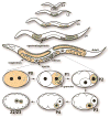
C elegans germ line development. The 2 primordial germ cells (Z2/Z3) begin to proliferate at the end of the first larval stage (L1). During the fourth larval stage (L4) approximately 40 meiotic germ cells undergo spermatogenesis. In the adult hermaphrodite germ line, germ cells switch from making sperm to making oocytes. After fertilization (P0 zygote), P granules are segregated to the germ line blastomeres P1 to P4. At the ~100-cell stage, the primordial germ cell P4 divides into the daughter cells (Z2/Z3). Germ line is in orange. P granules are in green. L2 indicates second larval stage; L3, third larval stage.
Figure 2
An oocyte and developing spermatocytes and sperm in C elegans. GLH-1 (green) is localized to P granules in the oocyte and in primary and secondary spermatocytes, dispersed throughout the cytoplasm of residual bodies, and undetectable in mature spermatids. PGL-3 (red) is localized to P granules in the oocyte and in primary spermatocytes, and then drops to undetectable at later stages of spermatogenesis. DAPI (4′,6-diamidino-2-phenylindole)-stained DNA is in blue.
Similar articles
- PGL proteins self associate and bind RNPs to mediate germ granule assembly in C. elegans.
Hanazawa M, Yonetani M, Sugimoto A. Hanazawa M, et al. J Cell Biol. 2011 Mar 21;192(6):929-37. doi: 10.1083/jcb.201010106. Epub 2011 Mar 14. J Cell Biol. 2011. PMID: 21402787 Free PMC article. - RNAi Screen Identifies Novel Regulators of RNP Granules in the Caenorhabditis elegans Germ Line.
Wood MP, Hollis A, Severance AL, Karrick ML, Schisa JA. Wood MP, et al. G3 (Bethesda). 2016 Aug 9;6(8):2643-54. doi: 10.1534/g3.116.031559. G3 (Bethesda). 2016. PMID: 27317775 Free PMC article. - Proximity labeling identifies LOTUS domain proteins that promote the formation of perinuclear germ granules in C. elegans.
Price IF, Hertz HL, Pastore B, Wagner J, Tang W. Price IF, et al. Elife. 2021 Nov 3;10:e72276. doi: 10.7554/eLife.72276. Elife. 2021. PMID: 34730513 Free PMC article. - Connecting the Dots: Linking Caenorhabditis elegans Small RNA Pathways and Germ Granules.
Sundby AE, Molnar RI, Claycomb JM. Sundby AE, et al. Trends Cell Biol. 2021 May;31(5):387-401. doi: 10.1016/j.tcb.2020.12.012. Epub 2021 Jan 29. Trends Cell Biol. 2021. PMID: 33526340 Review. - Germ granules and gene regulation in the Caenorhabditis elegans germline.
Phillips CM, Updike DL. Phillips CM, et al. Genetics. 2022 Mar 3;220(3):iyab195. doi: 10.1093/genetics/iyab195. Genetics. 2022. PMID: 35239965 Free PMC article. Review.
Cited by
- Complex formation of RNA silencing proteins in the perinuclear region of Neurospora crassa.
Decker LM, Boone EC, Xiao H, Shanker BS, Boone SF, Kingston SL, Lee SA, Hammond TM, Shiu PK. Decker LM, et al. Genetics. 2015 Apr;199(4):1017-21. doi: 10.1534/genetics.115.174623. Epub 2015 Feb 2. Genetics. 2015. PMID: 25644701 Free PMC article. - Germ cell specification.
Wang JT, Seydoux G. Wang JT, et al. Adv Exp Med Biol. 2013;757:17-39. doi: 10.1007/978-1-4614-4015-4_2. Adv Exp Med Biol. 2013. PMID: 22872473 Free PMC article. Review. - New Aspects of Magnesium Function: A Key Regulator in Nucleosome Self-Assembly, Chromatin Folding and Phase Separation.
Ohyama T. Ohyama T. Int J Mol Sci. 2019 Aug 29;20(17):4232. doi: 10.3390/ijms20174232. Int J Mol Sci. 2019. PMID: 31470631 Free PMC article. Review. - The role of RNA-binding proteins in orchestrating germline development in Caenorhabditis elegans.
Albarqi MMY, Ryder SP. Albarqi MMY, et al. Front Cell Dev Biol. 2023 Jan 4;10:1094295. doi: 10.3389/fcell.2022.1094295. eCollection 2022. Front Cell Dev Biol. 2023. PMID: 36684428 Free PMC article. Review. - Hypoxia induces transgenerational epigenetic inheritance of small RNAs.
Wang SY, Kim K, O'Brown ZK, Levan A, Dodson AE, Kennedy SG, Chernoff C, Greer EL. Wang SY, et al. Cell Rep. 2022 Dec 13;41(11):111800. doi: 10.1016/j.celrep.2022.111800. Cell Rep. 2022. PMID: 36516753 Free PMC article.
References
- Barbee SA, Lublin AL, Evans TC. A novel function for the Sm proteins in germ granule localization during C. elegans embryogenesis. Curr Biol. 2002;12:1502–1506. - PubMed
- Batista PJ, Ruby JG, Claycomb JM, Chiang R, Fahlgren N, Kasschau KD, Chaves DA, Gu W, Vasale JJ, Duan S, Conte D, Jr, Luo S, Schroth GP, Carrington JC, Bartel DP, Mello CC. PRG-1 and 21U-RNAs interact to form the piRNA complex required for fertility in C. elegans. Mol Cell. 2008;31:67–78. - PMC - PubMed
Publication types
MeSH terms
Substances
LinkOut - more resources
Full Text Sources

