Syk tyrosine kinase acts as a pancreatic adenocarcinoma tumor suppressor by regulating cellular growth and invasion - PubMed (original) (raw)
Syk tyrosine kinase acts as a pancreatic adenocarcinoma tumor suppressor by regulating cellular growth and invasion
Tracy Layton et al. Am J Pathol. 2009 Dec.
Abstract
We have identified the nonreceptor tyrosine kinase syk as a marker of differentiation/tumor suppressor in pancreatic ductal adenocarcinoma (PDAC). Syk expression is lost in poorly differentiated PDAC cells in vitro and in situ, and stable reexpression of syk in endogenously syk-negative Panc1 (Panc1/syk) cells retarded their growth in vitro and in vivo and reduced anchorage-independent growth in vitro. Panc1/syk cells exhibited a more differentiated morphology and down-regulated cyclin D1, akt, and CD171, which are overexpressed by Panc1 cells. Loss of PDAC syk expression in culture is due to promoter methylation, and reversal of promoter methylation caused reexpression of syk and concomitant down-regulation of CD171. Moreover, suppression of syk expression in BxPC3 cells caused de novo CD171 expression, consistent with the reciprocal expression of syk and CD171 we observe in situ. Importantly, Panc1/syk cells demonstrated dramatically reduced invasion in vitro. Affymetrix analysis identified statistically significant regulation of >2000 gene products by syk in Panc1 cells. Of these, matrix metalloproteinase-2 (MMP2) and tissue inhibitor of metalloproteinase-2 were down-regulated, suggesting that the MMP2 axis might mediate Panc1/mock invasion. Accordingly, MMP2 inhibition suppressed the in vitro invasion of Panc1/mock cells without effect on Panc1/syk cells. This study demonstrates a prominent role for syk in regulating the differentiation state and invasive phenotype of PDAC cells.
Figures
Figure 1
Syk expression correlates with PDAC grade in situ. Patient samples were obtained from the UCSD Department of Pathology archives. Patient demographics and tissue characteristics were described in detail previously., A: Tissue samples of varying PDAC grade were stained with anti-syk mAb 4D10, which recognizes both isoforms of syk but not Zap70 or src family members and is not affected by tyrosine phosphorylation of syk. Antibody binding was visualized with diaminobenzidine (brown). Top left: Solid arrowheads denote examples of discrete ducts. Open arrowheads denote examples of acinus-associated ductules. Top Right: Solid arrowhead denotes a poorly differentiated group of syk-negative cells invading away from syk-positive cells that maintain ductal morphology. Very strongly syk-positive stromal cells are lymphocytes. B: Summary of tissue samples by grade. Table shows the number of normal tissue samples and PDAC samples of each grade, as well as the corresponding syk expression within each group. Tissue differentiation grade was categorized as the highest grade present (ie, a patient whose tumor contained elements of G2 and G3 was classified as G3, and so on). IHC, immunohistochemical. C: Kaplan-Meier survival curve of syk-positive versus syk-negative patient samples using the log-rank test. Variables were coded as 1 for 100% syk-positive tumors and 0 for those that evinced heterogeneity. Median survivals were 18.8 and 6.4 months for syk-positive and syk-negative samples, respectively.
Figure 2
Syk expression is regulated by promoter methylation in vitro. A: Immunoblot analysis of syk expression in PDAC cell lines using the anti-syk mAb 4D10, which recognizes both isoforms of syk but not Zap70 or src family members and is not affected by tyrosine phosphorylation of syk. Erk2, control. B: Syk was immunoprecipitated from equal quantities of cell lysate with the anti-syk LR pAb, and samples were immunoblotted with the anti-syk mAb 4D10. C: RT-PCR was performed on PDAC cell lines using syk primers that amplify across the alternatively spliced sequence. HL60 cells, expression control. Actin, control. D: Extended SDS-polyacrylamide gel electrophoresis and immunoblotting of the BxPC3 and CFPAC1 lysates with anti-syk mAb 4D10 to detect syk isoforms. Erk2, control. Relative migrations of sykA (72) and sykB (69) are denoted in kDa. E: Syk-negative MIAPaCa2 and Panc1 cell lines were treated with 2 μmol/L 5AzaC for the indicated times (hours), and genomic DNA was analyzed by methylation-specific PCR with syk promoter-specific primers. BxPC3 cells, syk-expressing control. M, methylated; U, unmethylated; NT, no treatment. Part of a methylated product band in the adjacent right lane is shown to demonstrate where methylated product would be in the BxPC3 panel. F: Panc1 cells were plated to achieve confluent or subconfluent density as described in Materials and Methods and then were treated for 120 hours with 5AzaC in media containing the indicated amounts of FBS before syk promoter methylation-specific-PCR analysis. M, methylated; U, unmethylated. G: MIAPaCa2 or BxPC3 cells were treated with 5AzaC for the indicated times (hours), and then RNA was harvested and analyzed by RT-PCR with syk primers. NT, no treatment. Images are shown to achieve maximal exposure of negative lanes/bands.
Figure 3
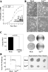
Syk exhibits the hallmarks of a tumor suppressor in PDAC. Panc1 cells were stably transfected with wild-type sykA (Sykwt) or empty vector (mock). A: Growth of Panc1/mock and Panc1/syk cells was measured daily after initial plating of equal numbers of cells (5 × 10²). Triplicate wells for each time point were stained with 1% crystal violet and quantitated at 550 nm after dye extraction. Inset: Expression of syk was assessed by immunoblot with the anti-syk mAb 4D10 in comparison with endogenously syk-positive BxPC3 cells. Actin, control. B: In vitro morphology of Panc1/mock and Panc1/syk cells at different densities. Note the more pronounced cell-cell contacts and lack of piling up of Panc1/syk cells, irrespective of density, in comparison with Panc1/mock cells. C: Anchorage-independent growth was determined as colonies present after 10 days of growth from 5 × 10⁴ single cells in soft agar. Images of plates are shown with quantitation points superimposed. D: Tumor growth was assessed subcutaneously after injection of 107 cells into the flanks of nude mice. After 4 weeks, tumors were harvested, fixed in formalin, and weighed wet.
Figure 4
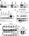
Syk regulates the expression of overexpressed proteins akt, cyclin D1, and CD171 in Panc1 cells. A: Immunoblot analysis of stable syk-reexpressing (Sykwt) or mock-transfected Panc1 cells with anti-cyclinD1, anti-pan-akt, and the anti-CD171 mAb UJ127. Actin, control. B: Immunoblot analysis of CD171 expression in PDAC cells. The membrane shown in Figure 2A was reprobed with the UJ127 anti-CD171 mAb. Erk2, control. C: BxPC3 cells were transiently transfected with pSUPER/syk6-siRNA and pEGFP as a reporter, and lysates were harvested on the indicated days for analysis of CD171, syk, and GFP expression by immunoblotting. Erk2, control. NT, no treatment. D: MIAPaCa2, Panc1, or BxPC3 cells were treated with 2 μmol/L 5AzaC for the indicated times (hours) and then lysates were harvested and immunoblotted with the anti-syk mAb 4D10 and the anti-CD171 mAb UJ127. Actin, control. NT, no treatment.
Figure 5
Syk and CD171 demonstrate reciprocal expression in situ. Patient samples were obtained from the UCSD Department of Pathology archives as described in the text and legend to Figure 1. Serial sections were stained with either the anti-syk mAb 4D10 (top panels) or the anti-CD171 mAb UJ127 (bottom panels). Twenty-two patients (of 92 total) demonstrated CD171 positivity; in all cases CD171-positive cells demonstrated syk-negative nuclei and dramatically reduced or completely absent cytoplasmic syk expression. Arrowheads denote syk−/CD171+ poorly differentiated malignant cells adjacent to syk+/CD171− well differentiated, normal or reactive ducts in the left and center panels. Highly anaplastic cells are essentially devoid of syk expression but strongly express CD171 in many cases (right panels). Very strongly syk-positive stromal cells are tumor-infiltrating lymphocytes.
Figure 6
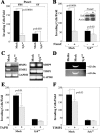
Syk mediates Panc1 invasion via regulation of the MMP2 axis. A and B: Cells from culture were seeded into the top chamber of BioCoat Growth Factor-Reduced Matrigel Invasion Chambers in serum-free medium and allowed to invade for 24 hours. A: Cells were provided FBS-containing (FBS) or serum-free (SF) medium in the lower chamber. B: Cells were pretreated for 120 hours with 5AzaC (or dimethyl sulfoxide) before seeding into the upper invasion chambers with FBS-containing media in the lower chamber. Inset: Immunoblot of syk expression in Panc1/syk cells at the time of application to invasion chambers. Leftover 5AzaC-treated cells were pelleted, lysed, and immunoblotted with the anti-syk mAb 4D10. Actin, control. C: RT-PCR was performed on RNA from Panc1/mock and Panc1/syk cells in normal culture using primers specific for MMP2 and its cognate partner TIMP2 or MMP9 and its cognate partner TIMP1. GAPDH control. D: Gelatin zymography was performed on equal volumes of serum-free media (media) or serum-free media conditioned by Panc1/mock or Panc1/syk cells. The relative migration of the active MMP2 species is indicated on the right. Lower panel: lighter zymogram image demonstrates the small amount of active MMP2 species in Panc1/syk conditioned media. E and F: Panc1/mock or Panc1/syk cells were pretreated for 15 minutes with 40 μmol/L TAPI1 (E) or 8 nmol/L TIMP2 (F) and then seeded into invasion chambers. Upper and lower chambers contained TAPI1 or TIMP2 for the 24-hour duration of the assay.
Similar articles
- Differential roles of cyclin D1 and D3 in pancreatic ductal adenocarcinoma.
Radulovich N, Pham NA, Strumpf D, Leung L, Xie W, Jurisica I, Tsao MS. Radulovich N, et al. Mol Cancer. 2010 Feb 1;9:24. doi: 10.1186/1476-4598-9-24. Mol Cancer. 2010. PMID: 20113529 Free PMC article. - Downregulated expression of IL‑28RA is involved in the pathogenesis of pancreatic ductal adenocarcinoma.
Liu L, Qi Y, Huang D, Xu Y, Li M, Hong Z, Gu Y, Jia B, Luo X, Zhang S, Zhang S. Liu L, et al. Int J Oncol. 2021 Aug;59(2):55. doi: 10.3892/ijo.2021.5235. Epub 2021 Jul 1. Int J Oncol. 2021. PMID: 34195850 - Analysis of protein expression regulated by lumican in PANC‑1 cells using shotgun proteomics.
Yamamoto T, Kudo M, Peng WX, Naito Z. Yamamoto T, et al. Oncol Rep. 2013 Oct;30(4):1609-21. doi: 10.3892/or.2013.2612. Epub 2013 Jul 11. Oncol Rep. 2013. PMID: 23846574 - Activated leukocyte cell adhesion molecule regulates the interaction between pancreatic cancer cells and stellate cells.
Zhang WW, Zhan SH, Geng CX, Sun X, Erkan M, Kleeff J, Xie XJ. Zhang WW, et al. Mol Med Rep. 2016 Oct;14(4):3627-33. doi: 10.3892/mmr.2016.5681. Epub 2016 Aug 26. Mol Med Rep. 2016. PMID: 27573419 Free PMC article. - KiSS‑1‑mediated suppression of the invasive ability of human pancreatic carcinoma cells is not dependent on the level of KiSS‑1 receptor GPR54.
Wang CH, Qiao C, Wang RC, Zhou WP. Wang CH, et al. Mol Med Rep. 2016 Jan;13(1):123-9. doi: 10.3892/mmr.2015.4535. Epub 2015 Nov 9. Mol Med Rep. 2016. PMID: 26572251 Free PMC article.
Cited by
- ADAM8 as a drug target in pancreatic cancer.
Schlomann U, Koller G, Conrad C, Ferdous T, Golfi P, Garcia AM, Höfling S, Parsons M, Costa P, Soper R, Bossard M, Hagemann T, Roshani R, Sewald N, Ketchem RR, Moss ML, Rasmussen FH, Miller MA, Lauffenburger DA, Tuveson DA, Nimsky C, Bartsch JW. Schlomann U, et al. Nat Commun. 2015 Jan 28;6:6175. doi: 10.1038/ncomms7175. Nat Commun. 2015. PMID: 25629724 Free PMC article. - Analysis of SYK Gene as a Prognostic Biomarker and Suggested Potential Bioactive Phytochemicals as an Alternative Therapeutic Option for Colorectal Cancer: An In-Silico Pharmaco-Informatics Investigation.
Biswas P, Dey D, Rahman A, Islam MA, Susmi TF, Kaium MA, Hasan MN, Rahman MDH, Mahmud S, Saleh MA, Paul P, Rahman MR, Al Saber M, Song H, Rahman MA, Kim B. Biswas P, et al. J Pers Med. 2021 Sep 6;11(9):888. doi: 10.3390/jpm11090888. J Pers Med. 2021. PMID: 34575665 Free PMC article. - Nanomechanical property maps of breast cancer cells as determined by multiharmonic atomic force microscopy reveal Syk-dependent changes in microtubule stability mediated by MAP1B.
Krisenko MO, Cartagena A, Raman A, Geahlen RL. Krisenko MO, et al. Biochemistry. 2015 Jan 13;54(1):60-8. doi: 10.1021/bi500325n. Epub 2014 Jun 18. Biochemistry. 2015. PMID: 24914616 Free PMC article. - Downregulation of spleen tyrosine kinase in hepatocellular carcinoma by promoter CpG island hypermethylation and its potential role in carcinogenesis.
Shin SH, Lee KH, Kim BH, Lee S, Lee HS, Jang JJ, Kang GH. Shin SH, et al. Lab Invest. 2014 Dec;94(12):1396-405. doi: 10.1038/labinvest.2014.118. Epub 2014 Oct 13. Lab Invest. 2014. PMID: 25310533 - Inhibition of ovarian tumor cell invasiveness by targeting SYK in the tyrosine kinase signaling pathway.
Yu Y, Suryo Rahmanto Y, Lee MH, Wu PH, Phillip JM, Huang CH, Vitolo MI, Gaillard S, Martin SS, Wirtz D, Shih IM, Wang TL. Yu Y, et al. Oncogene. 2018 Jul;37(28):3778-3789. doi: 10.1038/s41388-018-0241-0. Epub 2018 Apr 11. Oncogene. 2018. PMID: 29643476 Free PMC article.
References
- National Cancer Institute Bethesda, MD: National Cancer Institute,; Strategic plan for addressing the recommendations of the Pancreatic Cancer Progress Review Group. 2002
- Bardeesy N, DePinho RA. Pancreatic cancer biology and genetics. Nat Rev. 2002;2:897–909. - PubMed
- Sada K, Takano T, Yanagi S, Yamamura H. Structure and function of syk protein-tyrosine kinase. J Biochem. 2001;130:177–186. - PubMed
- Yanagi S, Inatome R, Takano T, Yamamura H. Syk expression and novel function in a wide variety of tissues. Biochem Biophys Res Commun. 2001;288:495–498. - PubMed
- Coopman PJ, Do MTH, Barth M, Bowden ET, Hayes AJ, Basyuk E, Blancato JK, Vezza PR, McLeskey SW, Mangeat PH, Mueller SC. The syk tyrosine kinase suppresses malignant growth of human breast cancer cells. Nature. 2000;406:742–747. - PubMed
Publication types
MeSH terms
Substances
Grants and funding
- CA109956/CA/NCI NIH HHS/United States
- CA130104/CA/NCI NIH HHS/United States
- R01 CA108597/CA/NCI NIH HHS/United States
- CA108597/CA/NCI NIH HHS/United States
- R03 CA109956/CA/NCI NIH HHS/United States
- R03 CA130104/CA/NCI NIH HHS/United States
LinkOut - more resources
Full Text Sources
Other Literature Sources
Medical
Molecular Biology Databases
Research Materials
Miscellaneous


