Astrocytes: biology and pathology - PubMed (original) (raw)
Review
Astrocytes: biology and pathology
Michael V Sofroniew et al. Acta Neuropathol. 2010 Jan.
Abstract
Astrocytes are specialized glial cells that outnumber neurons by over fivefold. They contiguously tile the entire central nervous system (CNS) and exert many essential complex functions in the healthy CNS. Astrocytes respond to all forms of CNS insults through a process referred to as reactive astrogliosis, which has become a pathological hallmark of CNS structural lesions. Substantial progress has been made recently in determining functions and mechanisms of reactive astrogliosis and in identifying roles of astrocytes in CNS disorders and pathologies. A vast molecular arsenal at the disposal of reactive astrocytes is being defined. Transgenic mouse models are dissecting specific aspects of reactive astrocytosis and glial scar formation in vivo. Astrocyte involvement in specific clinicopathological entities is being defined. It is now clear that reactive astrogliosis is not a simple all-or-none phenomenon but is a finely gradated continuum of changes that occur in context-dependent manners regulated by specific signaling events. These changes range from reversible alterations in gene expression and cell hypertrophy with preservation of cellular domains and tissue structure, to long-lasting scar formation with rearrangement of tissue structure. Increasing evidence points towards the potential of reactive astrogliosis to play either primary or contributing roles in CNS disorders via loss of normal astrocyte functions or gain of abnormal effects. This article reviews (1) astrocyte functions in healthy CNS, (2) mechanisms and functions of reactive astrogliosis and glial scar formation, and (3) ways in which reactive astrocytes may cause or contribute to specific CNS disorders and lesions.
Figures
Fig. 1
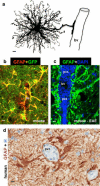
Astrocyte morphology and interactions with blood vessels in healthy and diseased tissue. a Protoplasmic astrocyte giving rise to a dense network of finely branching processes throughout its local gray matter neuropil, as well as to a large stem branch that extends foot processes along a blood vessel (bv). b Two color fluorescence showing astrocytes in healthy mouse gray matter stained immunohistochemically for GFAP (red) as well as the transgene-derived reporter molecule GFP (green). Note that in these transgenic mice [95], GFP reporter is present in all of the fine processes of the protoplasmic astrocytes throughout the neuropil, whereas the GFAP is present only in the large stem astrocyte processes and endfeet (which appear yellow where green and red staining overlap). Note that endfeet from many astrocytes contact and envelop bv. c Two color fluorescence showing dense accumulations of GFAP-positive (green) endfeet and processes of reactive astrocytes lining up along perivascular cuffs or clusters (pvc) of inflammatory cells stained with DAPI (blue) in a mouse with experimental autoimmune encephalomyelitis (EAE). Transgenic disruption of this reactive astrocyte barrier leads to widespread invasion of inflammatory cells away from perivascular clusters into CNS parenchyma during EAE [251]. d Two color brightfield staining showing human autopsy specimen with reactive astrocytes lining their processes along perivascular cuffs of inflammatory cells as if forming perivascular scar-like barriers similar to those observed in experimental animal models [251]. Scale bars a 3 μm, b 7.5 μm, c 15 μm, d 5 μm
Fig. 2
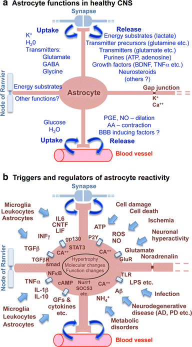
Schematic representations that summarize, a astrocyte functions in healthy CNS, and b triggers and molecular regulators of reactive astrogliosis
Fig. 3
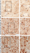
Astrocyte morphology remote from CNS lesions and with different gradations of reactive astrogliosis. Brightfield immunohistochemistry for GFAP counterstained with haematoxylin (H) in human autopsy specimens, surveys with details (boxed areas). a Appearance of astrocytes in tissue remote from a lesion and presumed healthy. Note that the territories of astrocyte processes do not overlap and that many astrocytes do not express detectable levels of GFAP. b Moderately reactive astrogliosis in which most (if not all) astrocytes have up regulated expression of GFAP and exhibit cellular hypertrophy, but with preservation of individual astrocyte domains and without pronounced overlap of astrocyte processes. c Severe diffuse reactive astrogliosis with pronounced up regulation of GFAP expression, astrocyte hypertrophy, astrocyte proliferation and pronounced overlap of astrocyte processes resulting in disruption of individual astrocyte domains. Scale bars surveys = 25 μm, details = 10 μm
Fig. 4
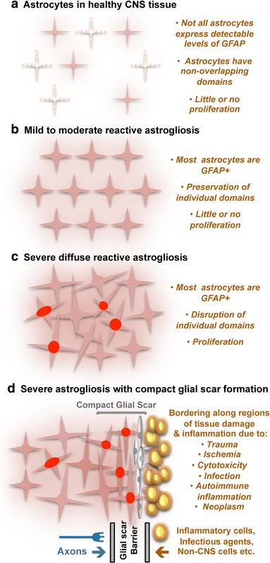
Schematic representations that summarize different gradations of reactive astrogliosis. a Astrocytes in healthy CNS tissue. b Mild to moderate reactive astrogliosis comprises variable changes in molecular expression and functional activity together with variable degrees of cellular hypertrophy. Such changes occur after mild trauma or at sites distant from a more severe injury, or after moderate metabolic or molecular insults or milder infections or inflammatory activation. These changes vary with insult severity, involve little anatomical overlap of the processes of neighboring astrocytes and exhibit the potential for structural resolution if the triggering insult is removed or resolves. c Severe diffuse reactive astrogliosis includes changes in molecular expression, functional activity and cellular hypertrophy, as well newly proliferated astrocytes (with red nuclei in figure), disrupting astrocyte domains and causing long-lasting reorganization of tissue architecture. Such changes are found in areas surrounding severe focal lesions, infections or areas responding to chronic neurodegenerative triggers. d Severe reactive astrogliosis with compact glial scar formation occurs along borders to areas of overt tissue damage and inflammation, and includes newly proliferated astrocytes (with red nuclei in figure) and other cell types (gray in figure) such as fibromeningeal cells and other glia, as well as deposition of dense collagenous extracellular matrix. In the compact glial scar, astrocytes have densely overlapping processes. Mature glial scars tend to persist for long periods and act as barriers not only to axon regeneration but also to inflammatory cells, infectious agents, and non-CNS cells in a manner that protects healthy tissue from nearby areas of intense inflammation
Fig. 5
Reactive astrogliosis demarcates cerebral microinfarcts. Survey image of cerebral cortex of an elderly individual showing microinfarcts (arrows) highlighted by dense clusters of prominently reactive astrocytes that stain intensely for GFAP. Fibrous astrocytes within subcortical white matter (wm) exhibit GFAP staining, whereas GFAP is not detectable in most protoplasmic gray matter astrocytes remote from the lesions in this specimen. H haematoxylin counterstain. Scale bar 180 μm
Fig. 6
Reactive astrogliosis in two disorders with seizures. a High magnification image of cerebral cortex from an individual with Rasmussen encephalitis (RE). Immunohistochemical staining for GFAP shows moderate reactive astrogliosis that is especially prominent around small blood vessels. Note the intensely stained and hypertrophic astrocytic foot processes extending to and lining the adventitia of microvessels. b, c High magnification images of a resection specimen from an individual with severe seizures associated with severe focal cortical dysplasia stained for H&E (b) or GFAP (c). b In this unique case, abundant Rosenthal fibers were found throughout the resection specimen, especially in the subpial regions (arrows) and around blood vessels (arrowheads). The density of the Rosenthal fibers suggested the diagnosis of ‘focal’ Alexander’s disease. However, testing of the GFAP gene revealed no mutations (for details, see [111]). c Comparable section to b showing abundant GFAP immunoreactive astrocytes and associated Rosenthal fibers (arrows). Scale bars a 10 μm, b 20 μm, c 15 μm
Fig. 7
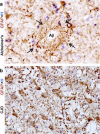
Reactive astrogliosis in two degenerative diseases. a High magnification image of autopsy specimen from a person with longstanding Alzheimer’s disease immunohistochemically stained for GFAP. Section of cerebral cortex shows an amyloid senile plaque with a pale unstained center (Aβ) ringed by dense layers of reactive astrocytic processes (arrows) that circumferentially surround the plaque as if forming a scar-like barrier around it. b High magnification image of autopsy specimen from a person with Creutzfeldt–Jakob disease (CJD) transmissible spongiform encephalopathy. Section of cerebral cortex shows pronounced neuron loss and severe diffuse reactive astrogliosis. Most of the cortex is packed with gemistocytic astrocytes, while spongiform change is relatively minimal in this area. Scale bars a, b 10 μm
Fig. 8
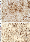
GFAP immunoreactive cells in primary CNS neoplasms. a High magnification image of a subependymal giant cell astrocytoma (SEGA) immunohistochemically stained for GFAP. Note variable staining intensity within tumor cells, including some that show uniform cytoplasmic staining. b High magnification image of a high-grade glioma (glioblastoma, WHO grade IV) immunohistochemically stained for GFAP. Note the variable staining of tumor cells as well as the pronounced nuclear and cytological atypia. Scale bars a, b 10 μm
Fig. 9
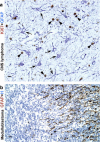
Reactive astrogliosis in response to CNS tumors. a High magnification image of autopsy specimen from a man with widely infiltrating primary CNS lymphoma. Two color immunohistochemistry for GFAP and the cell cycle marker Ki67, shows the cytoplasm of reactive astrocytes stained blue (arrows), with nuclei of infiltrating, malignant and highly proliferative lymphoid elements stained brown (arrowheads). Note that essentially none of the GFAP-immunoreactive astrocytes show Ki-67-immunoreactive nuclei and are thus not proliferative and exhibit only a moderate degree of reactive astrogliosis with cell hypertrophy and up regulation of GFAP, in spite of the presence of numerous infiltrating lymphoma cells. b High magnification image of a medulloblastoma immunohistochemically stained for GFAP and showing many processes of reactive astrocytes along one edge of the tumor (at right of figure). Scale bars a 15 μm, b 25 μm
Similar articles
- Reactive astrocytes as therapeutic targets for CNS disorders.
Hamby ME, Sofroniew MV. Hamby ME, et al. Neurotherapeutics. 2010 Oct;7(4):494-506. doi: 10.1016/j.nurt.2010.07.003. Neurotherapeutics. 2010. PMID: 20880511 Free PMC article. Review. - Heterogeneity of reactive astrocytes.
Anderson MA, Ao Y, Sofroniew MV. Anderson MA, et al. Neurosci Lett. 2014 Apr 17;565:23-9. doi: 10.1016/j.neulet.2013.12.030. Epub 2013 Dec 19. Neurosci Lett. 2014. PMID: 24361547 Free PMC article. Review. - Molecular dissection of reactive astrogliosis and glial scar formation.
Sofroniew MV. Sofroniew MV. Trends Neurosci. 2009 Dec;32(12):638-47. doi: 10.1016/j.tins.2009.08.002. Epub 2009 Sep 24. Trends Neurosci. 2009. PMID: 19782411 Free PMC article. Review. - Astrogliosis.
Sofroniew MV. Sofroniew MV. Cold Spring Harb Perspect Biol. 2014 Nov 7;7(2):a020420. doi: 10.1101/cshperspect.a020420. Cold Spring Harb Perspect Biol. 2014. PMID: 25380660 Free PMC article. Review. - Recent insights on astrocyte mechanisms in CNS homeostasis, pathology, and repair.
Hart CG, Karimi-Abdolrezaee S. Hart CG, et al. J Neurosci Res. 2021 Oct;99(10):2427-2462. doi: 10.1002/jnr.24922. Epub 2021 Jul 14. J Neurosci Res. 2021. PMID: 34259342 Review.
Cited by
- The Role of Inflammatory Cascade and Reactive Astrogliosis in Glial Scar Formation Post-spinal Cord Injury.
Bhatt M, Sharma M, Das B. Bhatt M, et al. Cell Mol Neurobiol. 2024 Nov 23;44(1):78. doi: 10.1007/s10571-024-01519-9. Cell Mol Neurobiol. 2024. PMID: 39579235 Free PMC article. Review. - Immunohistochemical Expression of GFAP in the Brain Astrocytes of Deceased Newborns Depending on the Postmortem Interval.
Shchegolev AI, Tumanova UN, Savva OV, Sukhikh GT. Shchegolev AI, et al. Bull Exp Biol Med. 2024 Nov;178(1):105-109. doi: 10.1007/s10517-024-06291-w. Epub 2024 Nov 23. Bull Exp Biol Med. 2024. PMID: 39578276 - The relevance of combined testing of cerebrospinal fluid glial fibrillary acidic protein and ubiquitin C-terminal hydrolase L1 in multiple sclerosis and peripheral neuropathy.
Csecsei P, Acs P, Gottschal M, Imre P, Miklos E, Simon D, Erdo-Bonyar S, Berki T, Zavori L, Varnai R. Csecsei P, et al. Neurol Sci. 2024 Nov 20. doi: 10.1007/s10072-024-07790-4. Online ahead of print. Neurol Sci. 2024. PMID: 39565457 - Glial fibrillary acidic protein in Alzheimer's disease: a narrative review.
Leipp F, Vialaret J, Mohaupt P, Coppens S, Jaffuel A, Niehoff AC, Lehmann S, Hirtz C. Leipp F, et al. Brain Commun. 2024 Nov 7;6(6):fcae396. doi: 10.1093/braincomms/fcae396. eCollection 2024. Brain Commun. 2024. PMID: 39554381 Free PMC article. Review. - The Development of Epilepsy Following CNS Viral Infections: Mechanisms.
Savoca G, Gianfredi A, Bartolini L. Savoca G, et al. Curr Neurol Neurosci Rep. 2024 Nov 16;25(1):2. doi: 10.1007/s11910-024-01393-4. Curr Neurol Neurosci Rep. 2024. PMID: 39549124 Review.
References
- Abbott NJ, Ronnback L, Hansson E. Astrocyte-endothelial interactions at the blood-brain barrier. Nat Rev Neurosci. 2006;7:41–53. - PubMed
- Acosta MT, Gioia GA, Silva AJ. Neurofibromatosis type 1: new insights into neurocognitive issues. Curr Neurol Neurosci Rep. 2006;6:136–143. - PubMed
- Akaoka H, Szymocha R, Beurton-Marduel P, Bernard A, Belin MF, Giraudon P. Functional changes in astrocytes by human T-lymphotropic virus type-1 T-lymphocytes. Virus Res. 2001;78:57–66. - PubMed
- Andreiuolo F, Junier MP, Hol EM, Miquel C, Chimelli L, Leonard N, Chneiweiss H, Daumas-Duport C, Varlet P. GFAPdelta immunostaining improves visualization of normal and pathologic astrocytic heterogeneity. Neuropathology. 2009;29:31–39. - PubMed
- Araque A, Parpura V, Sanzgiri RP, Haydon PG. Tripartite synapses: glia, the unacknowledged partner. Trends Neurosci. 1999;22:208–215. - PubMed
Publication types
MeSH terms
Grants and funding
- P50 AG16570/AG/NIA NIH HHS/United States
- P01 AG12435/AG/NIA NIH HHS/United States
- R01 NS057624/NS/NINDS NIH HHS/United States
- P50 AG016570/AG/NIA NIH HHS/United States
- P01 AG012435/AG/NIA NIH HHS/United States
LinkOut - more resources
Full Text Sources
Other Literature Sources
