Cortical thickness reduction in individuals at ultra-high-risk for psychosis - PubMed (original) (raw)
Cortical thickness reduction in individuals at ultra-high-risk for psychosis
Wi Hoon Jung et al. Schizophr Bull. 2011 Jul.
Abstract
Although schizophrenia is characterized by gray matter (GM) abnormalities, particularly in the prefrontal and temporal cortices, it is unclear whether cerebral cortical GM is abnormal in individuals at ultra-high-risk (UHR) for psychosis. We addressed this issue by studying cortical thickness in this group with magnetic resonance imaging (MRI). We measured cortical thickness of 29 individuals with no family history of psychosis at UHR, 31 patients with schizophrenia, and 29 healthy matched control subjects using automated surface-based analysis of structural MRI data. Hemispheric mean and regional cortical thickness were significantly different according to the stage of the disease. Significant cortical differences across these 3 groups were found in the distributed area of cerebral cortices. UHR group showed significant cortical thinning in the prefrontal cortex, anterior cingulate cortex, inferior parietal cortex, parahippocampal cortex, and superior temporal gyrus compared with healthy control subjects. Significant cortical thinning in schizophrenia group relative to UHR group was found in all the regions described above in addition with posterior cingulate cortex, insular cortex, and precentral cortex. These changes were more pronounced in the schizophrenia group compared with the control subjects. These findings suggest that UHR is associated with cortical thinning in regions that correspond to the structural abnormalities found in schizophrenia. These structural abnormalities might reflect functional decline at the prodromal stage of schizophrenia, and there may be progressive thinning of GM cortex over time.
Figures
Fig. 1.
Mean Cortical Thickness in 3 Groups for Each Hemisphere. The plots show gradual decreases in the mean cortical thickness according to the psychotic stages. UHR, ultra-high-risk (n = 29); schizophrenia (n = 31); HC subjects, healthy control subjects (n = 29).
Fig. 2.
Regional Maps of the Differences in the Mean Cortical Thickness (A) and Statistical Maps Between Groups: HC Subjects vs UHR Subjects, HC Subjects vs Schizophrenia Subjects, and UHR Subjects vs Schizophrenia Subjects (B). UHR, ultra-high-risk (n = 29); schizophrenia (n = 31); HC subjects, healthy control subjects (n = 29).
Fig. 3.
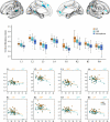
Brain Regions with Significant Reduced Cortical Thickness in UHR Subjects Compared with HC Subjects. In the left hemisphere, the cortical thickness was decreased in superior temporal gyrus (L1), anterior cingulate cortex (ACC, L2), parahippocampal cortex (L3), and medial superior frontal cortex (L4). In the right hemisphere, the cortical thickness was reduced in dorsal ACC (R1), rostral ACC (R2), inferior frontal cortex (R3), and inferior parietal cortex (R4). All these regions showed statistically significant differences among 3 groups. In the boxplot, the x-axis shows brain regions with significant reduction of cortical thickness, and the y-axis indicates cortical thickness (millimeters). The horizontal thick line inside each box indicates the median value. The upper and lower boundaries of each box mean lower quartile and upper quartile values, respectively. The whiskers represent smallest and largest nonoutlier observations. The circle depicts a mild outlier, and asterisk indicates an extreme outlier. Scatterplots and age correlation slopes for mean cortical thickness (millimeters) with increasing age within regions for UHR and HC subjects. The x-axis indicates age (y), and the y-axis indicates cortical thickness (millimeters). Significant correlations between age and mean cortical thickness within each region are marked (asterisk). UHR, ultra-high-risk (n = 29); schizophrenia (n = 31); HC subjects, healthy control subjects (n = 29); ACC, anterior cingulate cortex.
Similar articles
- Insular cortex gray matter changes in individuals at ultra-high-risk of developing psychosis.
Takahashi T, Wood SJ, Yung AR, Phillips LJ, Soulsby B, McGorry PD, Tanino R, Zhou SY, Suzuki M, Velakoulis D, Pantelis C. Takahashi T, et al. Schizophr Res. 2009 Jun;111(1-3):94-102. doi: 10.1016/j.schres.2009.03.024. Epub 2009 Apr 5. Schizophr Res. 2009. PMID: 19349150 - Differences and similarities in insular and temporal pole MRI gray matter volume abnormalities in first-episode schizophrenia and affective psychosis.
Kasai K, Shenton ME, Salisbury DF, Onitsuka T, Toner SK, Yurgelun-Todd D, Kikinis R, Jolesz FA, McCarley RW. Kasai K, et al. Arch Gen Psychiatry. 2003 Nov;60(11):1069-77. doi: 10.1001/archpsyc.60.11.1069. Arch Gen Psychiatry. 2003. PMID: 14609882 - Cigarette smoking is associated with thinner cingulate and insular cortices in patients with severe mental illness.
Jørgensen KN, Skjærvø I, Mørch-Johnsen L, Haukvik UK, Lange EH, Melle I, Andreassen OA, Agartz I. Jørgensen KN, et al. J Psychiatry Neurosci. 2015 Jul;40(4):241-9. doi: 10.1503/jpn.140163. J Psychiatry Neurosci. 2015. PMID: 25672482 Free PMC article. - Structural brain alterations in individuals at ultra-high risk for psychosis: a review of magnetic resonance imaging studies and future directions.
Jung WH, Jang JH, Byun MS, An SK, Kwon JS. Jung WH, et al. J Korean Med Sci. 2010 Dec;25(12):1700-9. doi: 10.3346/jkms.2010.25.12.1700. Epub 2010 Nov 24. J Korean Med Sci. 2010. PMID: 21165282 Free PMC article. Review. - Progressive loss of cortical gray matter in schizophrenia: a meta-analysis and meta-regression of longitudinal MRI studies.
Vita A, De Peri L, Deste G, Sacchetti E. Vita A, et al. Transl Psychiatry. 2012 Nov 20;2(11):e190. doi: 10.1038/tp.2012.116. Transl Psychiatry. 2012. PMID: 23168990 Free PMC article. Review.
Cited by
- Investigation of Schizophrenia with Human Induced Pluripotent Stem Cells.
Powell SK, O'Shea CP, Shannon SR, Akbarian S, Brennand KJ. Powell SK, et al. Adv Neurobiol. 2020;25:155-206. doi: 10.1007/978-3-030-45493-7_6. Adv Neurobiol. 2020. PMID: 32578147 Free PMC article. Review. - Using structural neuroimaging to make quantitative predictions of symptom progression in individuals at ultra-high risk for psychosis.
Tognin S, Pettersson-Yeo W, Valli I, Hutton C, Woolley J, Allen P, McGuire P, Mechelli A. Tognin S, et al. Front Psychiatry. 2014 Jan 29;4:187. doi: 10.3389/fpsyt.2013.00187. eCollection 2013. Front Psychiatry. 2014. PMID: 24523700 Free PMC article. - Fronto-temporal cortical grey matter thickness and surface area in the at-risk mental state and recent-onset schizophrenia: a magnetic resonance imaging study.
Rasser PE, Ehlkes T, Schall U. Rasser PE, et al. BMC Psychiatry. 2024 Jan 9;24(1):33. doi: 10.1186/s12888-024-05494-9. BMC Psychiatry. 2024. PMID: 38191320 Free PMC article. - Associating Psychotic Symptoms with Altered Brain Anatomy in Psychotic Disorders Using Multidimensional Item Response Theory Models.
Stan AD, Tamminga CA, Han K, Kim JB, Padmanabhan J, Tandon N, Hudgens-Haney ME, Keshavan MS, Clementz BA, Pearlson GD, Sweeney JA, Gibbons RD. Stan AD, et al. Cereb Cortex. 2020 May 14;30(5):2939-2947. doi: 10.1093/cercor/bhz285. Cereb Cortex. 2020. PMID: 31813988 Free PMC article. - Cortical Morphology Differences in Subjects at Increased Vulnerability for Developing a Psychotic Disorder: A Comparison between Subjects with Ultra-High Risk and 22q11.2 Deletion Syndrome.
Bakker G, Caan MW, Vingerhoets WA, da Silva-Alves F, de Koning M, Boot E, Nieman DH, de Haan L, Bloemen OJ, Booij J, van Amelsvoort TA. Bakker G, et al. PLoS One. 2016 Nov 9;11(11):e0159928. doi: 10.1371/journal.pone.0159928. eCollection 2016. PLoS One. 2016. PMID: 27828960 Free PMC article.
References
- Shim G, Kang DH, Chung YS, Yoo SY, Shin NY, Kwon JS. Social functioning deficits in young people at risk for schizophrenia. Aust N Z J Psychiatry. 2008;42:678–685. - PubMed
- Cannon TD. Clinical and genetic high-risk strategies in understanding vulnerability to psychosis. Schizophr Res. 2005;79:35–44. - PubMed
- Lencz T, Smith CW, McLaughlin D, et al. Generalized and specific neurocognitive deficits in prodromal schizophrenia. Biol Psychiatry. 2006;59:863–871. - PubMed
- Bramon E, Shaikh M, Broome M, et al. Abnormal P300 in people with high risk of developing psychosis. NeuroImage. 2008;41:553–560. - PubMed


