Cryomesh: a new substrate for cryo-electron microscopy - PubMed (original) (raw)
Cryomesh: a new substrate for cryo-electron microscopy
Craig Yoshioka et al. Microsc Microanal. 2010 Feb.
Abstract
Here we evaluate a new grid substrate developed by ProtoChips Inc. (Raleigh, NC) for cryo-transmission electron microscopy. The new grids are fabricated from doped silicon carbide using processes adapted from the semiconductor industry. A major motivating purpose in the development of these grids was to increase the low-temperature conductivity of the substrate, a characteristic that is thought to affect the appearance of beam-induced movement (BIM) in transmission electron microscope (TEM) images of biological specimens. BIM degrades the quality of data and is especially severe when frozen biological specimens are tilted in the microscope. Our results show that this new substrate does indeed have a significant impact on reducing the appearance and severity of beam-induced movement in TEM images of tilted cryo-preserved samples. Furthermore, while we have not been able to ascertain the exact causes underlying the BIM phenomenon, we have evidence that the rigidity and flatness of these grids may play a major role in its reduction. This improvement in the reliability of imaging at tilt has a significant impact on using data collection methods such as random conical tilt or orthogonal tilt reconstruction with cryo-preserved samples. Reduction in BIM also has the potential for improving the resolution of three-dimensional cryo-reconstructions in general.
Figures
Figure 1
Schematic diagram of Cryomesh construction. The holey ceramic film is bonded to the Si support layer and sits above it in the side-on view. Image is adapted from material provided courtesy of Protochips Inc.
Figure 2
A visual characterization of Cryomesh grids. A) Cryomesh grids as imaged under standard fluorescent lighting with the ceramic silicon layer on the side of the grid facing the viewer. The colors of the grids are related to the thickness of this film layer (see Figure 1) and extend over the outside support ring since the film layer is above the support layer when viewed from this side. B) Another Cryomesh grid that has been flipped over so that the film layer is facing away from the viewer and is on the other side of the support layer. In this case, the outside support ring now has neutral, rather than colored appearance. The outside support ring of all of the grids in panel A would have a similar appearance if they were flipped over. C,D,E) low magnification (49X) and higher magnification (500x) images of different Cryomesh grids with different mesh sizes. All have samples preserved in vitreous ice. Most interesting is the grid in panel E, which has a very large mesh size, with each square containing thousands of holes and even ice distribution.
Figure 3
A) High magnification, 29,000X of a Cryomesh hole with vitreous ice at 45° tilt. Along the edges of the hole ridges can be seen that are left behind from the manufacturing process. By measuring the length of these ridges in projection we can estimate the thickness of the support substrate. B) An image taken over the ceramic support substrate, with examples of the features that are artifacts of the ceramic material. These would shift and disappear/reappear if the stage where tilted or the beam-tilt changed. C) The power spectrum of the image in panel B.
Figure 4
Example of a 40962 pixel CCD image at 50,000X and 45° tilt exhibiting BIM. This image would have been marked as bad in the manual evaluation even though only a small region in the lower left corner is clearly showing BIM. In this image the power spectrum peaks that were used in the computer evaluation are shown in red, centered over the 10242 pixel areas from which they were calculated. Next to each peak in white text is the computed score assigned to that region. The yellow contour lines were generated using Matlab, each encloses an image area with BIM greater than or equal to the shown score (found using interpolation). On the right, the lowest and highest scoring regions from the image are expanded for clearer visibility.
Figure 5
At the top is a table with datasets that were evaluated by eye and their scores. Below is a graph with histograms of the computed scores for several different datasets. In black are 0° vitreous ice datasets on C-Flats as controls. Note the very large peak in the ~1.1 range. In blue are the 45° Cryomesh datasets, and in red are the 45° C-Flat datasets. The dashed lines represent the datasets that had an additional layer of thin carbon placed on the grids. Note that while the C-Flat w/ carbon dataset has improved slightly, the majority of the improvement comes at a level where there is still noticeable BIM, ~1.6. Likewise the Cryomesh w/carbon dataset has a much smaller peak in the low score range ~1.15 compared to the unaltered Cryomesh datasets. For comparison to the manual evaluation in the Table above, the green line denotes the score that most closely corresponds to where BIM becomes visually discernible.
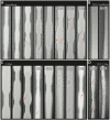
Figure 6
A gallery of channels that have been cut through the ice layers on different grid types. For the images in panels A and C, 45° and 0° images of the same area were aligned so that the thickness and variations in the ice layer could be easily observed. Further details can be found in Methods 2.4. A) Cryomesh grids B) carbon-coated Cryomesh C) C-Flat and D) carbon-coated C-Flat. Areas with subtle thinning or curvatures in the ice are denoted with red arrows. The red asterisk marks locations where the bilateral symmetry has been broken, likely due to ice movement. Channels that belong to images that exhibited BIM are marked by an upper-case ‘BIM’. Note that overall, the Cryomesh ice is more regular in flatness and thickness than the C-Flat ice. Also note that additional carbon film makes ice more irregular in thickness, and even more in flatness. The thin carbon coating also allows one to visualize the channel thickness without having to align the 0° images with the 45° images (though we did this anyways to confirm what we saw). Presumably this is because the ice/carbon interface creates contrast in addition to the ice/vacuum interface.
Similar articles
- Deformed grids for single-particle cryo-electron microscopy of specimens exhibiting a preferred orientation.
Liu Y, Meng X, Liu Z. Liu Y, et al. J Struct Biol. 2013 Jun;182(3):255-8. doi: 10.1016/j.jsb.2013.03.005. Epub 2013 Mar 26. J Struct Biol. 2013. PMID: 23537848 Free PMC article. - Minimizing Crinkling of Soft Specimens Using Holey Gold Films on Molybdenum Grids for Cryogenic Electron Microscopy.
Jiang X, Xuan S, Zuckermann RN, Glaeser RM, Downing KH, Balsara NP. Jiang X, et al. Microsc Microanal. 2021 Aug;27(4):767-775. doi: 10.1017/S1431927621000520. Microsc Microanal. 2021. PMID: 34085628 - Parameters affecting specimen flatness of two-dimensional crystals for electron crystallography.
Vonck J. Vonck J. Ultramicroscopy. 2000 Nov;85(3):123-9. doi: 10.1016/s0304-3991(00)00052-8. Ultramicroscopy. 2000. PMID: 11071349 - Cryo-focused-ion-beam applications in structural biology.
Rigort A, Plitzko JM. Rigort A, et al. Arch Biochem Biophys. 2015 Sep 1;581:122-30. doi: 10.1016/j.abb.2015.02.009. Epub 2015 Feb 20. Arch Biochem Biophys. 2015. PMID: 25703192 Review. - Cryo-electron microscopy and cryo-electron tomography of nanoparticles.
Stewart PL. Stewart PL. Wiley Interdiscip Rev Nanomed Nanobiotechnol. 2017 Mar;9(2). doi: 10.1002/wnan.1417. Epub 2016 Jun 23. Wiley Interdiscip Rev Nanomed Nanobiotechnol. 2017. PMID: 27339510 Review.
Cited by
- Low-cooling-rate freezing in biomolecular cryo-electron microscopy for recovery of initial frames.
Wu C, Shi H, Zhu D, Fan K, Zhang X. Wu C, et al. QRB Discov. 2021 Sep 6;2:e11. doi: 10.1017/qrd.2021.8. eCollection 2021. QRB Discov. 2021. PMID: 37529673 Free PMC article. - Current Microscopy Strategies to Image Fungal Vesicles: From the Intracellular Trafficking and Secretion to the Inner Structure of Isolated Vesicles.
Wendt C, Vieira V, Lima A, Augusto I, de Almeida FP, Gadelha APR, Nimrichter L, Rodrigues ML, Miranda K. Wendt C, et al. Curr Top Microbiol Immunol. 2021;432:139-159. doi: 10.1007/978-3-030-83391-6_11. Curr Top Microbiol Immunol. 2021. PMID: 34972883 - A primer to single-particle cryo-electron microscopy.
Cheng Y, Grigorieff N, Penczek PA, Walz T. Cheng Y, et al. Cell. 2015 Apr 23;161(3):438-449. doi: 10.1016/j.cell.2015.03.050. Cell. 2015. PMID: 25910204 Free PMC article. Review. - Biological Applications at the Cutting Edge of Cryo-Electron Microscopy.
Dillard RS, Hampton CM, Strauss JD, Ke Z, Altomara D, Guerrero-Ferreira RC, Kiss G, Wright ER. Dillard RS, et al. Microsc Microanal. 2018 Aug;24(4):406-419. doi: 10.1017/S1431927618012382. Microsc Microanal. 2018. PMID: 30175702 Free PMC article. Review. - Non-rigid image registration to reduce beam-induced blurring of cryo-electron microscopy images.
Karimi Nejadasl F, Karuppasamy M, Newman ER, McGeehan JE, Ravelli RB. Karimi Nejadasl F, et al. J Synchrotron Radiat. 2013 Jan;20(Pt 1):58-66. doi: 10.1107/S0909049512044408. Epub 2012 Nov 29. J Synchrotron Radiat. 2013. PMID: 23254656 Free PMC article.
References
- Booy FP, Pawley JB. Cryo-crinkling: what happens to carbon films on copper grids at low temperature. Ultramicroscopy. 1993;48:273–280. - PubMed
- Bottcher B. Electron cryo-microscopy of graphite in amorphous ice. Ultramicroscopy. 1995;58:417–424.
- Brink J, Sherman MB, Berriman J, Chiu W. Evaluation of charging on macromolecules in electron cryomicroscopy. Ultramicroscopy. 1998;72:41–52. - PubMed
- Downing K, Sui H, Auer M. Electron Tomography: A 3D View of the Subcellular World. Analytical Chemistry. 2007;79:7949–7957. - PubMed
Publication types
Grants and funding
- R01 RR023093/RR/NCRR NIH HHS/United States
- R01 RR023093-09/RR/NCRR NIH HHS/United States
- P41 RR017573-07/RR/NCRR NIH HHS/United States
- RR17573/RR/NCRR NIH HHS/United States
- P41 RR017573/RR/NCRR NIH HHS/United States
- RR23093/RR/NCRR NIH HHS/United States
LinkOut - more resources
Full Text Sources
Other Literature Sources


