Exercise protects against MPTP-induced neurotoxicity in mice - PubMed (original) (raw)
Exercise protects against MPTP-induced neurotoxicity in mice
Kimberly M Gerecke et al. Brain Res. 2010.
Abstract
Exercise has been shown to be potently neuroprotective in several neurodegenerative models, including 1-methyl-4-phenyl-1, 2, 3, 6-tetrahydropyridine (MPTP) model of Parkinson's disease (PD). In order to determine the critical duration of exercise necessary for DA neuroprotection, mice were allowed to run for either 1, 2 or 3months prior to treatment with saline or MPTP. Quantification of DA neurons in the SNpc show that mice allowed to run unrestricted for 1 or 2months lost significant numbers of neurons following MPTP administration as compared to saline treated mice; however, 3months of exercise provided complete protection against MPTP-induced neurotoxicity. To determine the critical intensity of exercise for DA neuroprotection, mice were restricted in their running to either 1/3 or 2/3 that of the full running group for 3months prior to treatment with saline or MPTP. Quantification of DA neurons in the SNpc show that mice whose running was restricted lost significant numbers of DA neurons due to MPTP toxicity; however, the 2/3 running group demonstrated partial protection. Neurochemical analyses of DA and its metabolites DOPAC and HVA show that exercise also functionally protects neurons from MPTP-induced neurotoxicity. Proteomic analysis of SN and STR tissues indicates that 3months of exercise induces changes in proteins related to energy regulation, cellular metabolism, the cytoskeleton, and intracellular signaling events. Taken together, these data indicate that exercise potently protects DA neurons from acute MPTP toxicity, suggesting that this simple lifestyle element may also confer significant protection against developing PD in humans.
Copyright 2010 Elsevier B.V. All rights reserved.
Figures
Figure 1
Pattern of average running activity for mice over the 90-day experimental period. Breaks in the Daytime Running data indicate days on which cages were changed; breaks in the nighttime running data are exclusions based on significant periods of missing values (>4h). Points represent the average running for mice (n=8) monitored either during the day or night period ± SEM.
Figure 2
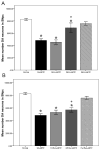
(A) Exercise protects DA neurons in the SNpc against MPTP neurotoxicity. Mice kept in SH or that exercised for 1 month prior to MPTP administration had significantly fewer DA neurons in the SNpc as compared to SH+Sal. Mice that ran for at least 3 months were not significantly different in terms of neuron number as compared to SH+Sal. While mice that ran for 2 months lost a significant number of neurons compared to SH+Sal, they still lost significantly fewer DA neurons in the SNpc than those that had not been allowed to exercise. (B) Exercise-mediated protection against MPTP-induced cell death requires full running. Mice were allowed to run the average number of revolutions/24 hours (18,000), or 1/3 (6,000) or 2/3 (12,000) the amount of distance, for 3 months total. Mice that were kept in SH or whose running was restricted prior to MPTP administration lost significantly more neurons in the SNpc as compared to SH Sal (p<0.001); however, animals allowed to run unrestricted were not significantly different in terms of neuron number as compared to SH+Sal. Bars represent the average ± SEM; *p<0.05 as compared to SH+Sal; + = p<0.05 as compared to SH+MPTP.
Figure 3
Exercise functionally protects DA neurons in the SNpc against MPTP neurotoxicity. HPLC/ED analysis of DA and the metabolites DOPAC and HVAC in the STR of SH or Ex mice administered either Sal or MPTP. Exercise significantly increased the measures of DA and its metabolites in the STR. Mice that exercised for 3 months administered MPTP had significantly lower measures of DA and its metabolites in the STR as compared to SH+Sal. However, Ex+MPTP mice lost significantly less DA in the STR compared to those kept in SH+MPTP. Bars represent the average ± SEM; *p<0.05 as compared to SH Sal; + = p<0.05 as compared to SH+MPTP.
Similar articles
- Intervention with exercise restores motor deficits but not nigrostriatal loss in a progressive MPTP mouse model of Parkinson's disease.
Sconce MD, Churchill MJ, Greene RE, Meshul CK. Sconce MD, et al. Neuroscience. 2015 Jul 23;299:156-74. doi: 10.1016/j.neuroscience.2015.04.069. Epub 2015 May 2. Neuroscience. 2015. PMID: 25943481 - Cannabinoid receptor type 1 protects nigrostriatal dopaminergic neurons against MPTP neurotoxicity by inhibiting microglial activation.
Chung YC, Bok E, Huh SH, Park JY, Yoon SH, Kim SR, Kim YS, Maeng S, Park SH, Jin BK. Chung YC, et al. J Immunol. 2011 Dec 15;187(12):6508-17. doi: 10.4049/jimmunol.1102435. Epub 2011 Nov 11. J Immunol. 2011. PMID: 22079984 - Neuroprotection in Parkinson models varies with toxin administration protocol.
Anderson DW, Bradbury KA, Schneider JS. Anderson DW, et al. Eur J Neurosci. 2006 Dec;24(11):3174-82. doi: 10.1111/j.1460-9568.2006.05192.x. Eur J Neurosci. 2006. PMID: 17156378 - MPTP: insights into parkinsonian neurodegeneration.
Speciale SG. Speciale SG. Neurotoxicol Teratol. 2002 Sep-Oct;24(5):607-20. doi: 10.1016/s0892-0362(02)00222-2. Neurotoxicol Teratol. 2002. PMID: 12200192 Review. - Neurotoxicity and Underlying Mechanisms of Endogenous Neurotoxins.
Cao Y, Li B, Ismail N, Smith K, Li T, Dai R, Deng Y. Cao Y, et al. Int J Mol Sci. 2021 Nov 26;22(23):12805. doi: 10.3390/ijms222312805. Int J Mol Sci. 2021. PMID: 34884606 Free PMC article. Review.
Cited by
- Inhibition of Nigral Microglial Activation Reduces Age-Related Loss of Dopaminergic Neurons and Motor Deficits.
Wang TF, Wu SY, Pan BS, Tsai SF, Kuo YM. Wang TF, et al. Cells. 2022 Jan 30;11(3):481. doi: 10.3390/cells11030481. Cells. 2022. PMID: 35159290 Free PMC article. - Delayed exercise-induced functional and neurochemical partial restoration following MPTP.
Archer T, Fredriksson A. Archer T, et al. Neurotox Res. 2012 Feb;21(2):210-21. doi: 10.1007/s12640-011-9261-z. Epub 2011 Aug 10. Neurotox Res. 2012. PMID: 21830164 - Study in Parkinson disease of exercise (SPARX): translating high-intensity exercise from animals to humans.
Moore CG, Schenkman M, Kohrt WM, Delitto A, Hall DA, Corcos D. Moore CG, et al. Contemp Clin Trials. 2013 Sep;36(1):90-8. doi: 10.1016/j.cct.2013.06.002. Epub 2013 Jun 14. Contemp Clin Trials. 2013. PMID: 23770108 Free PMC article. Clinical Trial. - BDNF alleviates Parkinson's disease by promoting STAT3 phosphorylation and regulating neuronal autophagy.
Geng X, Zou Y, Li J, Li S, Qi R, Yu H, Zhong L. Geng X, et al. Cell Tissue Res. 2023 Sep;393(3):455-470. doi: 10.1007/s00441-023-03806-1. Epub 2023 Jul 14. Cell Tissue Res. 2023. PMID: 37450039 Free PMC article. - Effects of a formal exercise program on Parkinson's disease: a pilot study using a delayed start design.
Park A, Zid D, Russell J, Malone A, Rendon A, Wehr A, Li X. Park A, et al. Parkinsonism Relat Disord. 2014 Jan;20(1):106-11. doi: 10.1016/j.parkreldis.2013.10.003. Epub 2013 Oct 15. Parkinsonism Relat Disord. 2014. PMID: 24209458 Free PMC article. Clinical Trial.
References
- Andreeva AV, Kutuzov MA, Voyno-Yasenetskaya TA. A ubiquitous membrane fusion protein alpha SNAP: a potential therapeutic target for cancer, diabetes and neurological disorders? Expert Opin Ther Targets. 2006;10:723–33. - PubMed
- Anstrom KK, Schallert T, Woodlee MT, Shattuck A, Roberts DC. Repetitive vibrissae-elicited forelimb placing before and immediately after unilateral 6-hydroxydopamine improves outcome in a model of Parkinson’s disease. Behav Brain Res. 2007;179:183–91. - PubMed
- Bender A, Krishnan KJ, Morris CM, Taylor GA, Reeve AK, Perry RH, Jaros E, Hersheson JS, Betts J, Klopstock T, Taylor RW, Turnbull DM. High levels of mitochondrial DNA deletions in substantia nigra neurons in aging and Parkinson disease. Nat Genet. 2006;38:515–7. - PubMed
MeSH terms
Substances
Grants and funding
- R01 NS039006-08/NS/NINDS NIH HHS/United States
- R01 NS070825/NS/NINDS NIH HHS/United States
- R21 NS045906-01A2/NS/NINDS NIH HHS/United States
- R21 NS045906/NS/NINDS NIH HHS/United States
- R01 NS039006/NS/NINDS NIH HHS/United States
LinkOut - more resources
Full Text Sources


