The clinical use of structural MRI in Alzheimer disease - PubMed (original) (raw)
Review
The clinical use of structural MRI in Alzheimer disease
Giovanni B Frisoni et al. Nat Rev Neurol. 2010 Feb.
Abstract
Structural imaging based on magnetic resonance is an integral part of the clinical assessment of patients with suspected Alzheimer dementia. Prospective data on the natural history of change in structural markers from preclinical to overt stages of Alzheimer disease are radically changing how the disease is conceptualized, and will influence its future diagnosis and treatment. Atrophy of medial temporal structures is now considered to be a valid diagnostic marker at the mild cognitive impairment stage. Structural imaging is also included in diagnostic criteria for the most prevalent non-Alzheimer dementias, reflecting its value in differential diagnosis. In addition, rates of whole-brain and hippocampal atrophy are sensitive markers of neurodegeneration, and are increasingly used as outcome measures in trials of potentially disease-modifying therapies. Large multicenter studies are currently investigating the value of other imaging and nonimaging markers as adjuncts to clinical assessment in diagnosis and monitoring of progression. The utility of structural imaging and other markers will be increased by standardization of acquisition and analysis methods, and by development of robust algorithms for automated assessment.
Conflict of interest statement
Competing interests: N. C. Fox declares associations with the following companies: Abbott Laboratories, Elan Pharmaceuticals, Eisai, Eli Lilly, GE Healthcare, IXICO, Lundbeck, Pfizer, Sanofi-Aventis, Wyeth Pharmaceuticals. See the article online for full details of the relationships. The other authors declare no competing interests.
Figures
Figure 1
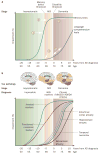
Natural progression of cognitive and biological markers of Alzheimer disease: a theoretical model. Some markers are sensitive to disease state and useful for diagnosis; others are more sensitive to disease progression and useful as surrogate markers in clinical trials. a | Known natural history of cognitive markers implies that memory tests, which change relatively early in the disease course (1) and soon reach the maximal level of impairment (2), are useful for diagnosis at the MCI stage, but are less useful for tracking later disease progression (3). Verbal comprehension tests start to change later in the disease course: during MCI they show mild or no impairment (4), and are of limited use in diagnosis. These markers become more sensitive at the dementia stage, when the slope of change steepens (5). b | Amyloid markers (cerebrospinal fluid amyloid-β42 and PET amyloid tracer uptake) represent the earliest detectable changes in the Alzheimer disease course, but have already plateaued by the MCI stage. Functional and metabolic markers detected by task-dependent activation on functional MRI and 18F-fluorodeoxyglucose PET are abnormal by the MCI stage, and continue to change well into the dementia stage. Structural changes come later,, following a temporal pattern mirroring tau pathology deposition., Abbreviations: AD, Alzheimer disease; MCI, mild cognitive impairment; NINCDS–ADRDA, National Institute of Neurological and Communicative Disorders and Stroke–Alzheimer's Disease and Related Disorders Association.
Figure 2
Progressive enrichment of a mild cognitive impairment cohort with future converters to Alzheimer dementia by screening for low hippocampal volume. Figures are computed from 339 patients with mild cognitive impairment from the North American Alzheimer's Disease Neuroimaging Initiative study with known conversion status at 12 month follow-up. The threshold for screening refers to the percentile of the distribution of hippocampal volume (average of right and left) in healthy elderly individuals. With an increasingly restrictive threshold, the ratio between true positives and false positives increases from 0.7 to 1.8, but the ratio of screened negatives to screened positives increases from 0.0 to 3.0.
Figure 3
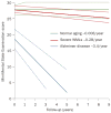
Effect of severe WMLs on the progression of cognitive deterioration. The rate of global cognitive decline in elderly individuals with severe WMLs is only marginally greater than that in healthy elderly people. The rate of decline in patients with Alzheimer dementia is about 12-fold greater than that in patients with severe WMLs. The confidence areas indicated by the dotted lines denote 95% confidence limits of the slope and the limits of the interquartile range of the intercept. Abbreviation: WMLs, white matter lesions. Permission obtained from Nature Publishing Group © Frisoni, G. B. et al. Nat. Clin. Pract. Neurol. 3, 620–627 (2007).
Figure 4
Cortical thinning in patients with mild cognitive impairment. Measurements were performed between baseline and 12 months in the left hemisphere of eight patients with mild cognitive impairment (mean age 72 years, Mini-Mental State Examination score 27.4) taken from the Alzheimer's Disease Neuroimaging Initiative data set who would develop Alzheimer dementia 12–24 months after the baseline measurement. The difference map shows thinning in the range of 0.5 mm in the medial temporal cortex and frontal, parietal and temporal neocortices, with relative sparing of the sensorimotor strip and visual cortex. The thinning maps closely with the known progression of neurofibrillary tangles and neurodegeneration at autopsy. Maps were obtained with the CIVET algorithm.
Similar articles
- Structural magnetic resonance imaging for the early diagnosis of dementia due to Alzheimer's disease in people with mild cognitive impairment.
Lombardi G, Crescioli G, Cavedo E, Lucenteforte E, Casazza G, Bellatorre AG, Lista C, Costantino G, Frisoni G, Virgili G, Filippini G. Lombardi G, et al. Cochrane Database Syst Rev. 2020 Mar 2;3(3):CD009628. doi: 10.1002/14651858.CD009628.pub2. Cochrane Database Syst Rev. 2020. PMID: 32119112 Free PMC article. - Relevance of magnetic resonance imaging for early detection and diagnosis of Alzheimer disease.
Teipel SJ, Grothe M, Lista S, Toschi N, Garaci FG, Hampel H. Teipel SJ, et al. Med Clin North Am. 2013 May;97(3):399-424. doi: 10.1016/j.mcna.2012.12.013. Epub 2013 Feb 1. Med Clin North Am. 2013. PMID: 23642578 Review. - Volumetric MRI and cognitive measures in Alzheimer disease : comparison of markers of progression.
Ridha BH, Anderson VM, Barnes J, Boyes RG, Price SL, Rossor MN, Whitwell JL, Jenkins L, Black RS, Grundman M, Fox NC. Ridha BH, et al. J Neurol. 2008 Apr;255(4):567-74. doi: 10.1007/s00415-008-0750-9. Epub 2008 Feb 18. J Neurol. 2008. PMID: 18274807 Clinical Trial. - Volumetric MRI vs clinical predictors of Alzheimer disease in mild cognitive impairment.
Fleisher AS, Sun S, Taylor C, Ward CP, Gamst AC, Petersen RC, Jack CR Jr, Aisen PS, Thal LJ. Fleisher AS, et al. Neurology. 2008 Jan 15;70(3):191-9. doi: 10.1212/01.wnl.0000287091.57376.65. Neurology. 2008. PMID: 18195264 Clinical Trial. - Quantitative structural MRI for early detection of Alzheimer's disease.
McEvoy LK, Brewer JB. McEvoy LK, et al. Expert Rev Neurother. 2010 Nov;10(11):1675-88. doi: 10.1586/ern.10.162. Expert Rev Neurother. 2010. PMID: 20977326 Free PMC article. Review.
Cited by
- Predicting future cognitive decline with hyperbolic stochastic coding.
Zhang J, Dong Q, Shi J, Li Q, Stonnington CM, Gutman BA, Chen K, Reiman EM, Caselli RJ, Thompson PM, Ye J, Wang Y. Zhang J, et al. Med Image Anal. 2021 May;70:102009. doi: 10.1016/j.media.2021.102009. Epub 2021 Feb 24. Med Image Anal. 2021. PMID: 33711742 Free PMC article. - BrainAGE in Mild Cognitive Impaired Patients: Predicting the Conversion to Alzheimer's Disease.
Gaser C, Franke K, Klöppel S, Koutsouleris N, Sauer H; Alzheimer's Disease Neuroimaging Initiative. Gaser C, et al. PLoS One. 2013 Jun 27;8(6):e67346. doi: 10.1371/journal.pone.0067346. Print 2013. PLoS One. 2013. PMID: 23826273 Free PMC article. - Diffusion tensor imaging reveals visual pathway damage in patients with mild cognitive impairment and Alzheimer's disease.
Nishioka C, Poh C, Sun SW. Nishioka C, et al. J Alzheimers Dis. 2015;45(1):97-107. doi: 10.3233/JAD-141239. J Alzheimers Dis. 2015. PMID: 25537012 Free PMC article. - New Drug Design Avenues Targeting Alzheimer's Disease by Pharmacoinformatics-Aided Tools.
Arrué L, Cigna-Méndez A, Barbosa T, Borrego-Muñoz P, Struve-Villalobos S, Oviedo V, Martínez-García C, Sepúlveda-Lara A, Millán N, Márquez Montesinos JCE, Muñoz J, Santana PA, Peña-Varas C, Barreto GE, González J, Ramírez D. Arrué L, et al. Pharmaceutics. 2022 Sep 9;14(9):1914. doi: 10.3390/pharmaceutics14091914. Pharmaceutics. 2022. PMID: 36145662 Free PMC article. Review. - Automated methods for hippocampus segmentation: the evolution and a review of the state of the art.
Dill V, Franco AR, Pinho MS. Dill V, et al. Neuroinformatics. 2015 Apr;13(2):133-50. doi: 10.1007/s12021-014-9243-4. Neuroinformatics. 2015. PMID: 26022748 Review.
References
- Braak H, Braak E. Neuropathological staging of Alzheimer-related changes. Acta Neuropathol. 1991;82:239–259. - PubMed
- Delacourte A, et al. The biochemical pathway of neurofibrillary degeneration in aging and Alzheimer's disease. Neurology. 1999;52:1158–1165. - PubMed
- McKhann G, et al. Clinical diagnosis of Alzheimer's disease: report of the NINCDS–ADRDA Work Group under the auspices of Department of Health and Human Services Task Force on Alzheimer's Disease. Neurology. 1984;34:939–944. - PubMed
- Bosscher L, Scheltens P. In: Evidence-Based Dementia Practice. Qizilbash N, et al., editors. Blackwell; Oxford: 2002. pp. 154–162.
Publication types
MeSH terms
Grants and funding
- EB007813/EB/NIBIB NIH HHS/United States
- R01 EB007813/EB/NIBIB NIH HHS/United States
- P41 RR013642-12/RR/NCRR NIH HHS/United States
- R56 AG020098/AG/NIA NIH HHS/United States
- HD050735/HD/NICHD NIH HHS/United States
- M01 RR000865-36/RR/NCRR NIH HHS/United States
- M01 RR000865-33/RR/NCRR NIH HHS/United States
- R01 HD050735-04/HD/NICHD NIH HHS/United States
- R01 AG011378-10/AG/NIA NIH HHS/United States
- R01 EB008432-04/EB/NIBIB NIH HHS/United States
- R37 AG011378/AG/NIA NIH HHS/United States
- R01 EB008281/EB/NIBIB NIH HHS/United States
- R01 EB007813-04/EB/NIBIB NIH HHS/United States
- G0601846/MRC_/Medical Research Council/United Kingdom
- G0801306/MRC_/Medical Research Council/United Kingdom
- U01 AG024904/AG/NIA NIH HHS/United States
- AG020098/AG/NIA NIH HHS/United States
- M01 RR000865/RR/NCRR NIH HHS/United States
- R01 EB008432-02/EB/NIBIB NIH HHS/United States
- R01 AG011378/AG/NIA NIH HHS/United States
- EB008432/EB/NIBIB NIH HHS/United States
- U01 AG024904-06/AG/NIA NIH HHS/United States
- P41 RR013642-10/RR/NCRR NIH HHS/United States
- R01 HD050735-05/HD/NICHD NIH HHS/United States
- R01 EB008281-14/EB/NIBIB NIH HHS/United States
- R01 HD050735/HD/NICHD NIH HHS/United States
- R01 EB008432/EB/NIBIB NIH HHS/United States
- R01 AG011378-18/AG/NIA NIH HHS/United States
- AG11378/AG/NIA NIH HHS/United States
- R01 AG020098-09/AG/NIA NIH HHS/United States
- P41 RR013642/RR/NCRR NIH HHS/United States
- R01 AG011378-17/AG/NIA NIH HHS/United States
- EB008281/EB/NIBIB NIH HHS/United States
- R01 AG020098/AG/NIA NIH HHS/United States
LinkOut - more resources
Full Text Sources
Other Literature Sources
Medical


