Mediator head module structure and functional interactions - PubMed (original) (raw)
Mediator head module structure and functional interactions
Gang Cai et al. Nat Struct Mol Biol. 2010 Mar.
Abstract
We used single-particle electron microscopy to characterize the structure and subunit organization of the Mediator Head module that controls Mediator-RNA polymerase II (RNAPII) and Mediator-promoter interactions. The Head module adopts several conformations differing in the position of a movable jaw formed by the Med18-Med20 subcomplex. We also characterized, by structural, biochemical and genetic means, the interactions of the Head module with TATA-binding protein (TBP) and RNAPII subunits Rpb4 and Rpb7. TBP binds near the Med18-Med20 attachment point and stabilizes an open conformation of the Head module. Rpb4 and Rpb7 bind between the Head jaws, establishing contacts essential for yeast-cell viability. These results, and consideration of the structure of the Mediator-RNAPII holoenzyme, shed light on the stabilization of the pre-initiation complex by Mediator and suggest how Mediator might influence initiation by modulating polymerase conformation and interaction with promoter DNA.
Conflict of interest statement
COMPETING INTERESTS STATEMENT
The authors declare no competing financial interests.
Figures
Figure 1
Mediator and Head module structure. (a) A cryo-EM reconstruction of Mediator shows the overall structure of the complex at ~25-Å resolution. Previous biochemical, functional and structural analyses suggest a modular organization of Mediator. The Head, Middle/Arm and Tail structural modules have been identified by comparing structures of Mediator in different conformations. A portion of the structurally defined Head module (dashed in green) comprises density corresponding to subunits biochemically identified with the Middle module. (b) A micrograph showing single Head module particles preserved in uranyl acetate. Scale bar, 200 Å. (c) Three different conformations of the Head module were identified through reference-free alignment and classification of EM images. Head module particles were nearly evenly distributed among the three different conformations that differ in the position of a smaller corner-shaped domain on the bottom-right of the structure. The angle (α) between the larger and smaller portions of the Head module structure (see diagram) is <90° in the collapsed conformation, ~90° in the closed conformation and >90° in the open conformation. Information from images of tilted particles was used to obtain 3D reconstruction of the Head module in all three different conformations. Scale bar, 100 Å. (d) Different views of Head module volumes in the collapsed (left column), closed (middle column) and open (right column) conformations. Scale bar, 100 Å.
Figure 2
EM analysis of Head module and subcomplexes. (a) Comparison of class averages obtained from images of Head and Core subcomplexes (corresponding to approximately the same projection direction) and difference mapping establishes that the extended domain flexibly attached to the rest of the Head module corresponds to subunits Med18 and Med20, with Med18 directly connected to the rest of the Head module structure and Med20 forming the distal end of the mobile domain. The above-mentioned comparison also indicates that the Mini complex corresponds to the left portion of the Core complex structure. All class averages are also shown as contour plots to facilitate visual comparison. (b) Contour plots calculated from 2D class averages of the Head and Core complexes were color-coded to highlight the approximate boundaries between different sets of Head module subunits. Two views (marked 1 and 2) of the 3D Head module structure matching the two projections of the Core (also marked 1 and 2) used for difference mapping indicate that the two 2D maps arise from roughly perpendicular orientations of the Head and Core particles in the EM samples (the Med18–Med20 portion of the Head structure is shown as a green mesh in orientation 2). (c) The EM results can be used to derive a description of the overall organization of subunits in the Head module and its interface to the rest of the Mediator complex (see Supplementary Fig. 4 for docking of Med7–Med21 and Med31 into the Mediator cryo-EM structure).
Figure 3
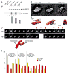
Head–TBP interactions. (a) A GST pulldown assay was used to measure the interaction between TBP and the Head, Core and Mini complexes. GST fusion complexes (as indicated) were immobilized on a glutathione-agarose resin incubated in the presence (lanes 4, 6 and 8) or absence (lanes 3, 5 and 7) of TBP. Controls are shown in lanes 1 and 2. Values for relative TBP binding to the different complexes are represented by the bars below the immunoblot. (b) Class averages obtained after incubation of the Head module with (top) and without TBP (bottom). Interaction with TBP had no effect on the range of conformations adopted by the Head module. (c) TBP interaction influences the distribution of particles among the different conformations of the Head module. The order of groups in the histogram is derived from the order of class averages in b. (d) Class averages calculated from images of Core particles alone (middle) and after incubation with TBP (left), and the difference between them (right). (e) An approximate 3D model of the Head–TBP complex based on the Core + TBP 2D class average and the 3D structure of the Head module. The Core portion of the Head module is shown in red, density corresponding to subunits Med18 and Med20 is represented as a gray mesh, and TBP is shown as a purple surface calculated by low-pass filtering the X-ray structure of TBP.
Figure 4
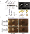
Head module–Rpb4–Rpb7 interaction. (a) A GST pulldown assay was used to measure the interactions between Rpb4–Rpb7 and the Head, Core and Mini complexes. GST fusion complexes (as indicated) were immobilized on glutathione-agarose resin incubated in the presence (lanes 4, 6 and 8) or absence (lanes 3, 5 and 7) of recombinant 6×His–Rpb4–Rpb7. Controls are shown in lanes 1 and 2. Values for relative binding to the different complexes are represented by the bars below the immunoblot. (b) Comparison of class averages obtained after alignment of Head alone (left, 1,748 images) and Head–Rpb4–Rpb7 particles (right, 1,332 images) shows the presence of additional density in the region corresponding to the jaws of the Head module. A 3D reconstruction of the Head–Rpb4–Rpb7 complex (solid yellow surface, right panel top) shows density (semitransparent yellow surface, right panel bottom) matching the size and shape of a low-resolution model calculated from the Rpb4–Rpb7 X-ray structure (purple surface, right panel bottom). (c) Genetic interaction between Rpb4 and Mediator subunits in the Head (Med18 and Med20), Middle (Med1, Med9 and Med31) and Tail (Med16 and Med5) modules was tested by assessing the viability of the synthetic double mutant strains as described in Online Methods. The wild-type (WT) and mutant strains were grown in SC –Leu medium, spotted in five-fold dilutions onto SC –Ura-Leu and SC +5-FOA plates and incubated at 30 °C for 3 (SC –Ura-Leu plates) or 5 d (SC +5-FOA plates).
Figure 5
Interaction of the Head module with components of the mPIC and a possible mechanism for initiation regulation. (a) Positioning TBP (shown as a purple surface calculated by low-pass filtering the X-ray structure of TBP) in its approximate binding location to the Head module (adjacent to the Med8 subunit) places the transcription factor in a position matching that predicted by current models of the minimal preinitiation complex structure. This positioning suggests how interaction of RNAPII (shown in orange) with Mediator in the Mediator–RNAPII holoenzyme structure (shown in gray) might help stabilize the preinitiation complex. The magenta circle denotes the approximate position of the RNAPII active site. (b) Interaction of the Head module with the Rpb4–Rpb7 polymerase subunit complex (shown in ruby) documented in this study could be important for enabling Mediator and the general transcription factors to affect the conformation of the polymerase clamp domain (shown in blue), possibly facilitating opening (as indicated by the yellow arrow) of the RNA polymerase II active-site cleft (outlined in black) to allow access of double-stranded promoter DNA to the polymerase active site.
Similar articles
- Interaction of the mediator head module with RNA polymerase II.
Cai G, Chaban YL, Imasaki T, Kovacs JA, Calero G, Penczek PA, Takagi Y, Asturias FJ. Cai G, et al. Structure. 2012 May 9;20(5):899-910. doi: 10.1016/j.str.2012.02.023. Structure. 2012. PMID: 22579255 Free PMC article. - Architecture of the RNA polymerase II-Mediator core initiation complex.
Plaschka C, Larivière L, Wenzeck L, Seizl M, Hemann M, Tegunov D, Petrotchenko EV, Borchers CH, Baumeister W, Herzog F, Villa E, Cramer P. Plaschka C, et al. Nature. 2015 Feb 19;518(7539):376-80. doi: 10.1038/nature14229. Epub 2015 Feb 4. Nature. 2015. PMID: 25652824 - Structure and TBP binding of the Mediator head subcomplex Med8-Med18-Med20.
Larivière L, Geiger S, Hoeppner S, Röther S, Strässer K, Cramer P. Larivière L, et al. Nat Struct Mol Biol. 2006 Oct;13(10):895-901. doi: 10.1038/nsmb1143. Epub 2006 Sep 10. Nat Struct Mol Biol. 2006. PMID: 16964259 - More pieces to the puzzle: recent structural insights into class II transcription initiation.
Kandiah E, Trowitzsch S, Gupta K, Haffke M, Berger I. Kandiah E, et al. Curr Opin Struct Biol. 2014 Feb;24:91-7. doi: 10.1016/j.sbi.2013.12.005. Epub 2014 Jan 16. Curr Opin Struct Biol. 2014. PMID: 24440461 Review. - Origins and activity of the Mediator complex.
Conaway RC, Conaway JW. Conaway RC, et al. Semin Cell Dev Biol. 2011 Sep;22(7):729-34. doi: 10.1016/j.semcdb.2011.07.021. Epub 2011 Jul 28. Semin Cell Dev Biol. 2011. PMID: 21821140 Free PMC article. Review.
Cited by
- Interaction of the mediator head module with RNA polymerase II.
Cai G, Chaban YL, Imasaki T, Kovacs JA, Calero G, Penczek PA, Takagi Y, Asturias FJ. Cai G, et al. Structure. 2012 May 9;20(5):899-910. doi: 10.1016/j.str.2012.02.023. Structure. 2012. PMID: 22579255 Free PMC article. - Crystal and EM structures of human phosphoribosyl pyrophosphate synthase I (PRS1) provide novel insights into the disease-associated mutations.
Chen P, Liu Z, Wang X, Peng J, Sun Q, Li J, Wang M, Niu L, Zhang Z, Cai G, Teng M, Li X. Chen P, et al. PLoS One. 2015 Mar 17;10(3):e0120304. doi: 10.1371/journal.pone.0120304. eCollection 2015. PLoS One. 2015. PMID: 25781187 Free PMC article. - Evidence for Multiple Mediator Complexes in Yeast Independently Recruited by Activated Heat Shock Factor.
Anandhakumar J, Moustafa YW, Chowdhary S, Kainth AS, Gross DS. Anandhakumar J, et al. Mol Cell Biol. 2016 Jun 29;36(14):1943-60. doi: 10.1128/MCB.00005-16. Print 2016 Jul 15. Mol Cell Biol. 2016. PMID: 27185874 Free PMC article. - Titer estimation for quality control (TEQC) method: A practical approach for optimal production of protein complexes using the baculovirus expression vector system.
Imasaki T, Wenzel S, Yamada K, Bryant ML, Takagi Y. Imasaki T, et al. PLoS One. 2018 Apr 3;13(4):e0195356. doi: 10.1371/journal.pone.0195356. eCollection 2018. PLoS One. 2018. PMID: 29614134 Free PMC article. - A functional portrait of Med7 and the mediator complex in Candida albicans.
Tebbji F, Chen Y, Richard Albert J, Gunsalus KT, Kumamoto CA, Nantel A, Sellam A, Whiteway M. Tebbji F, et al. PLoS Genet. 2014 Nov 6;10(11):e1004770. doi: 10.1371/journal.pgen.1004770. eCollection 2014 Nov. PLoS Genet. 2014. PMID: 25375174 Free PMC article.
References
- Flanagan PM, et al. Resolution of factors required for the initiation of transcription by yeast RNA polymerase II. J Biol Chem. 1990;265:11105–11107. - PubMed
- Flanagan PM, Kelleher RJ, III, Sayre MH, Tschochner H, Kornberg RD. A mediator required for activation of RNA polymerase II transcription in vitro. Nature. 1991;350:436–438. - PubMed
- Naar AM, et al. Composite co-activator ARC mediates chromatin-directed transcriptional activation. Nature. 1999;398:828–832. - PubMed
- Takagi Y, Kornberg RD. Mediator as a general transcription factor. J Biol Chem. 2006;281:80–89. - PubMed
Publication types
MeSH terms
Substances
Grants and funding
- R01 GM067167/GM/NIGMS NIH HHS/United States
- R01 GM067167-04/GM/NIGMS NIH HHS/United States
- R01 GM067167-05A2/GM/NIGMS NIH HHS/United States
- R01 GM67167/GM/NIGMS NIH HHS/United States
LinkOut - more resources
Full Text Sources
Molecular Biology Databases


