Perinuclear P granules are the principal sites of mRNA export in adult C. elegans germ cells - PubMed (original) (raw)
Perinuclear P granules are the principal sites of mRNA export in adult C. elegans germ cells
Ujwal Sheth et al. Development. 2010 Apr.
Abstract
Germline-specific granules of unknown function are found in a wide variety of organisms, including C. elegans, where they are called P granules. P granules are cytoplasmic bodies in oocytes and early embryos. Throughout most of the C. elegans life cycle, however, P granules are associated with clusters of nuclear pore complexes (NPCs) on germ cell nuclei. We show that perinuclear P granules differ from cytoplasmic P granules in many respects, including structure, stability and response to metabolic changes. Our results suggest that nuclear-associated P granules provide a perinuclear compartment where newly exported mRNAs are collected prior to their release to the general cytoplasm. First, we show that mRNA export factors are highly enriched at the NPCs associated with P granules. Second, we discovered that the expression of high-copy transgenes could be induced in a subset of germ cells, and used this system to demonstrate that nascent mRNA traffics directly to P granules. P granules appear to sequester large amounts of mRNA in quiescent germ cells, presumably preventing translation of that mRNA. However, we did not find evidence that P granules normally sequester aberrant mRNAs, or mRNAs targeted for destruction by the RNAi pathway.
Figures
Fig. 1.
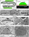
Perinuclear P granules are asymmetric. (A) One arm of the adult hermaphrodite gonad showing nuclei (black) and P granules (green). (B) A single P granule as described in the text; compare with F. env, nuclear envelope; Pg-NPCs, P granule-associated nuclear pore complexes. (C-E) Examples of small, possibly newly formed, P granules in the mitotic zone showing nuclear pores (small black arrows), presumptive crests (white arrows) and the base (black arrowhead). (F-H) Perinuclear P granules in the pachytene zone (F,G) and in a late oogonium (H); note that NPCs are not clustered in late oogonia and oocytes. (I) Detached, cytoplasmic P granule in a young oocyte. Arrows and arrowheads are the same as in C-E. Scale bars: 500 nm.
Fig. 2.
Perinuclear P granules are persistent structures with dynamic components. (A,A′) P granules (numbered) on three pachytene germ nuclei filmed for 20 minutes. (B-B′) Photobleaching of a single perinuclear P granule (arrow); the elapsed time shown is 79 seconds. (C) Quantified fluorescence recovery curve for the P granule photobleached in B (black diamonds) compared with a single-exponential best-fit curve (white circles). (D) Dissected, normal gonad showing PGL-1::GFP in P granules and in the cytoplasm. (E) Gonad 5 hours after injection with α-amanitin showing PGL-1::GFP; clumps of PGL-1 persist in the distal mitotic germ cells but PGL-1 is lost from pachytene P granules. (F) High magnification of germ nuclei from the gonad in panel E; inset shows the same magnification of pachytene germ nuclei in an α-amanitin-injected ama-1(m188) control gonad. (G) Pachytene nuclei 5 hours after injection of cycloheximide. (H) The left panel shows pachytene-specific loss of PGL-1::GFP in an nxf-1(RNAi) gonad; the right panel shows the nuclear envelope staining (NPP-9) in the same gonad. Asterisk indicates the distal tip of the gonad. Scale bars: 2.5 μm in A,B; 25 μm in D,E,H; 5 μm in F,G.
Fig. 3.
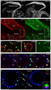
DDX-19 is enriched at the base of P granules. (A) Gonad and intestine co-stained for NPP-9 and DDX-19 as indicated. (B) Eight-plane projection of gonad co-stained for NPP-9 and PGL-1 as indicated. (C-E) Single-plane merged images of dashed boxes in c, d and e are shown at high magnification in C (rotated), D and E, respectively. (F) Germ nucleus from a fourth larval stage (L4) gonad co-stained for DDX-19, PGL-1 and NPP-9 as indicated. (G) Adult oogonia showing detached P granules (green), NPP-9 (blue) and DDX-19 (red); inset shows a detached P granule at high magnification. Arrows indicate examples of detached P granules. Scale bars: 25 μm in A,B; 5 μm in C-E,G; 2.5 μm in F.
Fig. 4.
DDX-19 is concentrated at Pg-NPCs. (A) Surface view of germ nuclei in a gonad stained for NPP-9, PGL-1 and DDX-19 as indicated. (B) Merged images as indicated of a single germ nucleus (outlined) from A, stained as indicated. In all panels, arrows indicate large patches of NPP-9 associated with P granules (Pg-NPCs) and arrowheads indicate small foci of NPP-9 (NPCs). Scale bars: 2.5 μm.
Fig. 5.
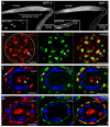
NXF-1 is concentrated at Pg-NPCs. (A) Gonad and intestine (inset) co-stained for NPP-9 and NXF-1 as indicated. (B) Surface view of a single pachytene germ nucleus (outlined) stained for NXF-1 and PGL-1 as indicated; note extensive association of the two proteins (arrow). (C) Optical cross-section of a single pachytene nucleus stained for NXF-1, PGL-1 and DNA as indicated. Note NXF-1 localization to the base of P granules (arrow) and in the nucleolus (arrowhead). (D) Optical cross-section of a single pachytene nucleus stained for NXF-1, NPP-9 and DNA as indicated. Scale bars: 25 μm in A; 2.5 μm in B-D.
Fig. 6.
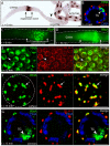
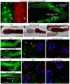
Newly exported mRNA traffics through P granules. (A-G) Panels show in situ hybridization detection with either alkaline phosphatase (A) or fluorescence (all others) of tissues/nuclei after recovery from heat shock for the times (t) indicated. Worms contained a high-copy array of a hsp16.2::gfp::unc-54 3′UTR transgene; nascent mRNA was visualized with an in situ probe against gfp mRNA. (A) Gonad and intestine. The transgene is expressed in all intestinal nuclei, somatic nuclei in the gonad (arrowhead) and germ nuclei within the expression zone (bracket). (B) High magnification of a pachytene nucleus in the expression zone showing P granules, mRNA and DNA as indicated; the arrowhead indicates the presumptive site of transgene (TG) expression. (C) Surface view of the gonad immediately after heat shock. (D) Optical section through the gonad core 30 minutes after heat shock; transgene-derived mRNA is abundant in the core underlying the expression zone. (E) Germ nuclei 15 minutes after heat shock showing nascent mRNA and the P granule marker GLH-1 as indicated. The arrow points to one example of perinuclear foci of mRNA coinciding with P granules. (F) High-magnification surface view of a single pachytene nucleus stained for the induced mRNA and GLH-1 as indicated. Arrows indicate the same as in E. (G) Optical cross-section through a pachytene nucleus showing mRNA (green) in P granules (red). Merge is in yellow. Scale bars: 25 μm in A,C,D; 2.5 μm in B,E-G.
Fig. 7.
Perinuclear mRNA localization requires P granules. (A) Intestinal cell 15 minutes after heat shock, showing little or no mRNA (green) adjacent to the NPP-9 zone (red). (B) Germ cell in the same animals showing perinuclear foci of RNA (green, left panel) adjacent to the NPP-9 zone (red) and coincident with P granules (green, right panel). (C,C′) Single pachytene germ cell from a heat-shocked, glh-1(RNAi) animal. Staining as indicated for transgenic mRNA, NPP-9 and DNA. Note the very low level of perinuclear mRNA (arrow; shown at high magnification in C′). (D,D′) Germ cell from mock-treated control animal stained as indicated and showing a high level of perinuclear mRNA (arrow; shown at high magnification in D′). Scale bars: 25 μm in A,C,D; 2.5 μm in B,E-G.
Fig. 8.
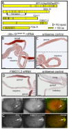
mRNAs targeted for degradation are not enriched in P granules. (A,B) FISH analysis of hsp16.2::gfp::pos-1 3′UTR expression on worms exposed to dsRNA for the pos-1 3′UTR. (A) Pachytene nuclei; double-headed arrow indicates an example of colocalized nascent mRNA (green) and P granules (red). (B) Low magnification view of the same gonad in A, showing that the induced mRNA has entered the gonad core and moved downstream to oogonia. (C-E) Panels show wild-type (WT) or mutant mex-3 mRNA detected by in situ hybridization in the strains indicated. mex-3(zu155) mRNA with a premature stop codon is degraded in the most mature oocytes, where it normally is translated (D, arrowhead), but persists when the NMD pathway is inhibited [smg-2(RNAi)] (E). (F-I′) FISH of the indicated mRNA in WT or mutant gonads (F-I); high magnification of the respective regions indicated by arrowheads are shown in F′-I′. Adjacent mRNA (green) and PGL-1 (red) signals are indicated in F′ for perinuclear P granules (arrowhead) and cytoplasmic P granules (arrows). Scale bars: 2.5 μm in A; 25 μm in B-I; 5 μm in F′-I′.
Fig. 9.
FG-repeat proteins in P granules. (A) Diagram showing FG repeats (vertical bars) in the nucleoporin NPP-9 and in four additional C. elegans proteins encoded by gonad-enriched mRNAs. (B,C) In situ hybridization for sense (B) and antisense (C) ddx-19(exon4) mRNA. (D,E) In situ hybridization for sense (D) and antisense (E) f58g11.2 mRNA. (F)Two-cell embryo showing colocalization of F58G11.2::GFP with cytoplasmic P granules (arrow). (G) Fifty-cell embryo showing colocalization of F58G11.2::GFP with P granules (arrow). Scale bars: 25 μm.
Similar articles
- RNAi Screen Identifies Novel Regulators of RNP Granules in the Caenorhabditis elegans Germ Line.
Wood MP, Hollis A, Severance AL, Karrick ML, Schisa JA. Wood MP, et al. G3 (Bethesda). 2016 Aug 9;6(8):2643-54. doi: 10.1534/g3.116.031559. G3 (Bethesda). 2016. PMID: 27317775 Free PMC article. - A genomewide RNAi screen for genes that affect the stability, distribution and function of P granules in Caenorhabditis elegans.
Updike DL, Strome S. Updike DL, et al. Genetics. 2009 Dec;183(4):1397-419. doi: 10.1534/genetics.109.110171. Epub 2009 Oct 5. Genetics. 2009. PMID: 19805813 Free PMC article. - P granules extend the nuclear pore complex environment in the C. elegans germ line.
Updike DL, Hachey SJ, Kreher J, Strome S. Updike DL, et al. J Cell Biol. 2011 Mar 21;192(6):939-48. doi: 10.1083/jcb.201010104. Epub 2011 Mar 14. J Cell Biol. 2011. PMID: 21402789 Free PMC article. - Germ granules and the control of mRNA translation.
Sengupta MS, Boag PR. Sengupta MS, et al. IUBMB Life. 2012 Jul;64(7):586-94. doi: 10.1002/iub.1039. Epub 2012 May 28. IUBMB Life. 2012. PMID: 22639345 Review. - Germ granules and gene regulation in the Caenorhabditis elegans germline.
Phillips CM, Updike DL. Phillips CM, et al. Genetics. 2022 Mar 3;220(3):iyab195. doi: 10.1093/genetics/iyab195. Genetics. 2022. PMID: 35239965 Free PMC article. Review.
Cited by
- PLP-1 is essential for germ cell development and germline gene silencing in Caenorhabditis elegans.
Vishnupriya R, Thomas L, Wahba L, Fire A, Subramaniam K. Vishnupriya R, et al. Development. 2020 Nov 27;147(22):dev195578. doi: 10.1242/dev.195578. Development. 2020. PMID: 33051256 Free PMC article. - C. elegans germ cells show temperature and age-dependent expression of Cer1, a Gypsy/Ty3-related retrotransposon.
Dennis S, Sheth U, Feldman JL, English KA, Priess JR. Dennis S, et al. PLoS Pathog. 2012;8(3):e1002591. doi: 10.1371/journal.ppat.1002591. Epub 2012 Mar 29. PLoS Pathog. 2012. PMID: 22479180 Free PMC article. - Small RNAs and chromatin in the multigenerational epigenetic landscape of Caenorhabditis elegans.
Frolows N, Ashe A. Frolows N, et al. Philos Trans R Soc Lond B Biol Sci. 2021 Jun 7;376(1826):20200112. doi: 10.1098/rstb.2020.0112. Epub 2021 Apr 19. Philos Trans R Soc Lond B Biol Sci. 2021. PMID: 33866817 Free PMC article. Review. - RNA Granules: A View from the RNA Perspective.
Tian S, Curnutte HA, Trcek T. Tian S, et al. Molecules. 2020 Jul 8;25(14):3130. doi: 10.3390/molecules25143130. Molecules. 2020. PMID: 32650583 Free PMC article. Review. - Reduction of mRNA export unmasks different tissue sensitivities to low mRNA levels during Caenorhabditis elegans development.
Zheleva A, Gómez-Orte E, Sáenz-Narciso B, Ezcurra B, Kassahun H, de Toro M, Miranda-Vizuete A, Schnabel R, Nilsen H, Cabello J. Zheleva A, et al. PLoS Genet. 2019 Sep 16;15(9):e1008338. doi: 10.1371/journal.pgen.1008338. eCollection 2019 Sep. PLoS Genet. 2019. PMID: 31525188 Free PMC article.
References
- Anderson P., Kedersha N. (2009). RNA granules: post-transcriptional and epigenetic modulators of gene expression. Nat. Rev. Mol. Cell Biol. 10, 430-436 - PubMed
- Brangwynne C. P., Eckmann C. R., Courson D. S., Rybarska A., Hoege C., Gharakhani J., Julicher F., Hyman A. A. (2009). Germline P granules are liquid droplets that localize by controlled dissolution/condensation. Science 324, 1729-1732 - PubMed
Publication types
MeSH terms
Substances
LinkOut - more resources
Full Text Sources
Molecular Biology Databases
