Multi-organ distribution of phosphorylated alpha-synuclein histopathology in subjects with Lewy body disorders - PubMed (original) (raw)
Multi-organ distribution of phosphorylated alpha-synuclein histopathology in subjects with Lewy body disorders
Thomas G Beach et al. Acta Neuropathol. 2010 Jun.
Abstract
A sensitive immunohistochemical method for phosphorylated alpha-synuclein was used to stain sets of sections of spinal cord and tissue from 41 different sites in the bodies of 92 subjects, including 23 normal elderly, 7 with incidental Lewy body disease (ILBD), 17 with Parkinson's disease (PD), 9 with dementia with Lewy bodies (DLB), 19 with Alzheimer's disease with Lewy bodies (ADLB) and 17 with Alzheimer's disease with no Lewy bodies (ADNLB). The relative densities and frequencies of occurrence of phosphorylated alpha-synuclein histopathology (PASH) were tabulated and correlated with diagnostic category. The greatest densities and frequencies of PASH occurred in the spinal cord, followed by the paraspinal sympathetic ganglia, the vagus nerve, the gastrointestinal tract and endocrine organs. The frequency of PASH within other organs and tissue types was much lower. Spinal cord and peripheral PASH was most common in subjects with PD and DLB, where it appears likely that it is universally widespread. Subjects with ILBD had lesser densities of PASH within all regions, but had frequent involvement of the spinal cord and paraspinal sympathetic ganglia, with less-frequent involvement of end-organs. Subjects with ADLB had infrequent involvement of the spinal cord and paraspinal sympathetic ganglia with rare involvement of end-organs. Within the gastrointestinal tract, there was a rostrocaudal gradient of decreasing PASH frequency and density, with the lower esophagus and submandibular gland having the greatest involvement and the colon and rectum the lowest.
Figures
Fig. 1
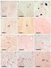
Photomicrographs of slides of immunohistochemical staining for phosphorylated α-synuclein histopathology (PASH) in paraffin sections of the spinal cord, sympathetic ganglia and vagus nerve. Positive staining is black, the counterstain is neutral red. a The intermediolateral horn of the thoracic spinal cord of a subject with PD, showing immunoreactive fibers, puncta and perikaryal cytoplasmic inclusions. Calibration bar 80 µm. b, c Low and higher magnification images of the posterior root entry into the sacral spinal cord of a subject with DLB, showing immunoreactive neuronal perikaryal staining. Calibration bar in b 0.3 mm; in c 80 µm. d, e Low and higher magnification images of the anterior horn of the sacral spinal cord in a subject with PD, showing diffuse cytoplasmic perikaryal immunoreactivity of a large motorneuron. Calibration bar in d 0.5 mm; in e 60 µm. f Sacral spinal cord adjacent to the central canal of a subject with PD, showing immunoreactive swollen degenerating neurons or neurites. Calibration bar 80 µm. g, h Low and higher magnification images of a middle cervical sympathetic ganglion of a subject with DLB, showing frequent immunoreactive dystrophic neurites, puncta and neuronal perikaryal cytoplasmic inclusions. Calibration bar in g 0.1 mm; calibration bar in h 100 µm. i Small branch of the vagus nerve in a subject with PD, showing several immunoreactive nerve fibers. Calibration bar 60 µm
Fig. 2
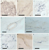
Photomicrographs of immunohistochemical staining for phosphorylated α-synuclein in paraffin sections of bodily organs and tissue. Positive staining is black, the counterstain is neutral red. a, b Low and higher magnification images of the submandibular gland in two subjects with PD, showing immunoreactive nerve fibers within the stroma of the gland. Calibration bar in a 0.2 mm; in b 100 µm. c The submucosa of the lower esophagus of a subject with PD, showing immunoreactive puncta, fibers and perikaryal cytoplasmic inclusions in ganglion cells. d Immunostaining of fibers in the duodenal submucosa of a subjects with DLB. Calibration bar 20 µm. e A single immunoreactive fiber in the stroma of the pancreas from a subject with PD. Calibration bar 40 µm. f A single immunoreactive fiber in the submucosa of a primary bronchus of a subject with PD. Calibration bar 40 µm. g A few immunoreactive fibers in the submucosa of the larynx of a subject with PD. Calibration bar 20 µm. h Several immunoreactive fibers in an epicardial nerve twig entering the myocardium in a subject with DLB. Calibration bar 100 µm. i A single immunoreactive fiber in the intermyenteric plexus of the urinary bladder of a subject with DLB. Calibration bar 20 µm. j Frequent immunoreactive fibers, puncta as well as cells with diffusely stained perikaryal cytoplasm in the adrenal medulla of a subject with PD. Calibration bar 100 µm. k A single immunoreactive fiber in the stroma of the parathyroid gland of a subject with PD. Calibration bar 10 µm. l The ovary of a woman with PD showing diffuse perikaryal immunostaining of a neuron-like cell with adjacent immunoreactive fibers and puncta. Calibration bar 10 µm
Fig. 3
Photomicrographs of immunohistochemical staining for phosphorylated α-synuclein in 80-µm thick frozen sections of formalin-fixed, cryoprotected tissue blocks of submandibular gland and lower esophagus. Positive staining is black. There is no counterstain. Submandibular gland from subjects with DLB (a–c) and PD (d) showing frequent immunoreactive fibers in the gland parenchyma (a), stroma (b) and around a small artery (d), while frequent immunoreactive puncta are seen in c. Lower esophagus from subjects with PD (e, g, h) and DLB (f) showing immunoreactive nerve fibers in e, nerve fibers and puncta in f, a thickened nerve fiber adherent to a bundle of smooth muscle fibers in g and a ganglion cell with diffuse perikaryal cytoplasmic staining in h
Fig. 4
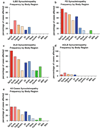
Relative frequency of PASH by diagnostic group for phosphorylated α-synuclein in different body regions including spinal cord, sympathetic ganglia, vagus nerve, sciatic nerve and multiple organs and tissues. Relative frequency is the percentage of subjects that showed immunoreactive tissue elements of any kind (fibers, puncta, perikaryal diffuse staining, perikaryal cytoplasmic inclusions) in single slides from each of the sites evaluated. For list of individual sites within each body region or organ system, see Table 1 and supplementary online table. Frequency was investigated further for some sites (see text)
Fig. 5
Relative frequency and density of PASH by diagnostic group in different spinal cord regions and sympathetic nervous system subdivisions. Relative frequency is the percentage of subjects that showed immunoreactive tissue elements of any kind (fibers, puncta, perikaryal diffuse staining, perikaryal cytoplasmic inclusions) in single slides taken from each of the sites evaluated. Density was calculated only for sites with immunoreactivity, negative or “zero” scores were not included
Similar articles
- [Clinical and pathological study on early diagnosis of Parkinson's disease and dementia with Lewy bodies].
Orimo S. Orimo S. Rinsho Shinkeigaku. 2008 Jan;48(1):11-24. doi: 10.5692/clinicalneurol.48.11. Rinsho Shinkeigaku. 2008. PMID: 18386627 Review. Japanese. - Multiple organ involvement by alpha-synuclein pathology in Lewy body disorders.
Gelpi E, Navarro-Otano J, Tolosa E, Gaig C, Compta Y, Rey MJ, Martí MJ, Hernández I, Valldeoriola F, Reñé R, Ribalta T. Gelpi E, et al. Mov Disord. 2014 Jul;29(8):1010-8. doi: 10.1002/mds.25776. Epub 2014 Jan 2. Mov Disord. 2014. PMID: 24395122 - Spectrum of abnormalities of sympathetic tyrosine hydroxylase and alpha-synuclein in chronic autonomic failure.
Isonaka R, Sullivan P, Jinsmaa Y, Corrales A, Goldstein DS. Isonaka R, et al. Clin Auton Res. 2018 Apr;28(2):223-230. doi: 10.1007/s10286-017-0495-6. Epub 2018 Feb 2. Clin Auton Res. 2018. PMID: 29396794 - Axonal alpha-synuclein aggregates herald centripetal degeneration of cardiac sympathetic nerve in Parkinson's disease.
Orimo S, Uchihara T, Nakamura A, Mori F, Kakita A, Wakabayashi K, Takahashi H. Orimo S, et al. Brain. 2008 Mar;131(Pt 3):642-50. doi: 10.1093/brain/awm302. Epub 2007 Dec 13. Brain. 2008. PMID: 18079166 - A critical evaluation of current staging of alpha-synuclein pathology in Lewy body disorders.
Jellinger KA. Jellinger KA. Biochim Biophys Acta. 2009 Jul;1792(7):730-40. doi: 10.1016/j.bbadis.2008.07.006. Epub 2008 Aug 5. Biochim Biophys Acta. 2009. PMID: 18718530 Review.
Cited by
- Neuronal hyperactivity-induced oxidant stress promotes in vivo α-synuclein brain spreading.
Helwig M, Ulusoy A, Rollar A, O'Sullivan SA, Lee SSL, Aboutalebi H, Pinto-Costa R, Jevans B, Klinkenberg M, Di Monte DA. Helwig M, et al. Sci Adv. 2022 Sep 2;8(35):eabn0356. doi: 10.1126/sciadv.abn0356. Epub 2022 Aug 31. Sci Adv. 2022. PMID: 36044566 Free PMC article. - Mitochondrial dysfunction in Parkinson's disease - a key disease hallmark with therapeutic potential.
Henrich MT, Oertel WH, Surmeier DJ, Geibl FF. Henrich MT, et al. Mol Neurodegener. 2023 Nov 11;18(1):83. doi: 10.1186/s13024-023-00676-7. Mol Neurodegener. 2023. PMID: 37951933 Free PMC article. Review. - Salivary acetylcholinesterase activity is increased in Parkinson's disease: a potential marker of parasympathetic dysfunction.
Fedorova T, Knudsen CS, Mouridsen K, Nexo E, Borghammer P. Fedorova T, et al. Parkinsons Dis. 2015;2015:156479. doi: 10.1155/2015/156479. Epub 2015 Feb 12. Parkinsons Dis. 2015. PMID: 25767737 Free PMC article. - Microbiota Dysbiosis in Parkinson Disease-In Search of a Biomarker.
Nowak JM, Kopczyński M, Friedman A, Koziorowski D, Figura M. Nowak JM, et al. Biomedicines. 2022 Aug 23;10(9):2057. doi: 10.3390/biomedicines10092057. Biomedicines. 2022. PMID: 36140158 Free PMC article. Review. - Distribution of α-Synuclein Aggregation in the Peripheral Tissues.
Li YY, Zhou TT, Zhang Y, Chen NH, Yuan YH. Li YY, et al. Neurochem Res. 2022 Dec;47(12):3627-3634. doi: 10.1007/s11064-022-03586-0. Epub 2022 Mar 28. Neurochem Res. 2022. PMID: 35348944 Review.
References
- Consensus recommendations for the postmortem diagnosis of Alzheimer’s disease. The National Institute on Aging, and Reagan Institute Working Group on Diagnostic Criteria for the Neuropathological Assessment of Alzheimer’s Disease. Neurobiol Aging. 1997;18:S1–S2. - PubMed
- Abbott RD, Petrovitch H, White LR, Masaki KH, Tanner CM, Curb JD, Grandinetti A, Blanchette PL, Popper JS, Ross GW. Frequency of bowel movements and the future risk of Parkinson’s disease. Neurology. 2001;57:456–462. - PubMed
- Abbott RD, Ross GW, Petrovitch H, Tanner CM, Davis DG, Masaki KH, Launer LJ, Curb JD, White LR. Bowel movement frequency in late-life and incidental Lewy bodies. Mov Disord. 2007 - PubMed
- Adler CH. Nonmotor complications in Parkinson’s disease. Mov Disord. 2005;20 Suppl 11:S23–S29. - PubMed
- Adler CH, Hentz JG, Joyce JN, Beach T, Caviness JN. Motor impairment in normal aging, clinically possible Parkinson’s disease, and clinically probable Parkinson’s disease: longitudinal evaluation of a cohort of prospective brain donors. Parkinsonism Relat Disord. 2002;9:103–110. - PubMed
Publication types
MeSH terms
Substances
LinkOut - more resources
Full Text Sources
Other Literature Sources
Medical


