EBImage--an R package for image processing with applications to cellular phenotypes - PubMed (original) (raw)
EBImage--an R package for image processing with applications to cellular phenotypes
Grégoire Pau et al. Bioinformatics. 2010.
Abstract
Summary: EBImage provides general purpose functionality for reading, writing, processing and analysis of images. Furthermore, in the context of microscopy-based cellular assays, EBImage offers tools to segment cells and extract quantitative cellular descriptors. This allows the automation of such tasks using the R programming language and use of existing tools in the R environment for signal processing, statistical modeling, machine learning and data visualization.
Availability: EBImage is free and open source, released under the LGPL license and available from the Bioconductor project (http://www.bioconductor.org/packages/release/bioc/html/EBImage.html).
Figures
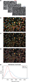
Fig. 1.
From microscope images to cellular phenotypes. (a) Fluorescent microscopy images from three channels of the same population of HeLa cells perturbed by siRluc. (b) A false colour image combining the actin (red), the tubulin (green) and the DNA (blue) channels. (c) Nuclei boundaries (yellow) were segmented with adaptive thresholding followed by connected set labeling. (d) Cell membranes (magenta) were determined by Voronoi segmentation. (e) Distribution of the cell sizes compared to a population of HeLa cells perturbed by siCLSPN. Cells treated with siCLSPN were significantly enlarged compared to those perturbed with siRluc (Wilcoxon rank sum test, P<10−15).
Similar articles
- InterMineR: an R package for InterMine databases.
Kyritsis KA, Wang B, Sullivan J, Lyne R, Micklem G. Kyritsis KA, et al. Bioinformatics. 2019 Sep 1;35(17):3206-3207. doi: 10.1093/bioinformatics/btz039. Bioinformatics. 2019. PMID: 30668641 Free PMC article. - cytoviewer: an R/Bioconductor package for interactive visualization and exploration of highly multiplexed imaging data.
Meyer L, Eling N, Bodenmiller B. Meyer L, et al. BMC Bioinformatics. 2024 Jan 3;25(1):9. doi: 10.1186/s12859-023-05546-z. BMC Bioinformatics. 2024. PMID: 38172724 Free PMC article. - oneChannelGUI: a graphical interface to Bioconductor tools, designed for life scientists who are not familiar with R language.
Sanges R, Cordero F, Calogero RA. Sanges R, et al. Bioinformatics. 2007 Dec 15;23(24):3406-8. doi: 10.1093/bioinformatics/btm469. Epub 2007 Sep 17. Bioinformatics. 2007. PMID: 17875544 - OsiriX: an open-source software for navigating in multidimensional DICOM images.
Rosset A, Spadola L, Ratib O. Rosset A, et al. J Digit Imaging. 2004 Sep;17(3):205-16. doi: 10.1007/s10278-004-1014-6. Epub 2004 Jun 29. J Digit Imaging. 2004. PMID: 15534753 Free PMC article. Review. - Visualization approaches for multidimensional biological image data.
Rueden CT, Eliceiri KW. Rueden CT, et al. Biotechniques. 2007 Jul;43(1 Suppl):31, 33-6. doi: 10.2144/000112511. Biotechniques. 2007. PMID: 17936940 Review.
Cited by
- Transcription cofactor GRIP1 differentially affects myeloid cell-driven neuroinflammation and response to IFN-β therapy.
Mimouna S, Rollins DA, Shibu G, Tharmalingam B, Deochand DK, Chen X, Oliver D, Chinenov Y, Rogatsky I. Mimouna S, et al. J Exp Med. 2021 Jan 4;218(1):e20192386. doi: 10.1084/jem.20192386. J Exp Med. 2021. PMID: 33045064 Free PMC article. - PUPAID: A R + ImageJ pipeline for thorough and semi-automated processing and analysis of multi-channel immunofluorescence data.
Régnier P, Montardi C, Maciejewski-Duval A, Marques C, Saadoun D. Régnier P, et al. PLoS One. 2024 Sep 19;19(9):e0308970. doi: 10.1371/journal.pone.0308970. eCollection 2024. PLoS One. 2024. PMID: 39298534 Free PMC article. - AdipoGauge software for analysis of biological microscopic images.
Yosofvand M, Liyanage S, Kalupahana NS, Scoggin S, Moustaid-Moussa N, Moussa H. Yosofvand M, et al. Adipocyte. 2020 Dec;9(1):360-373. doi: 10.1080/21623945.2020.1787583. Adipocyte. 2020. PMID: 32654628 Free PMC article. - Membrane-bound Heat Shock Protein mHsp70 Is Required for Migration and Invasion of Brain Tumors.
Shevtsov M, Bobkov D, Yudintceva N, Likhomanova R, Kim A, Fedorov E, Fedorov V, Mikhailova N, Oganesyan E, Shabelnikov S, Rozanov O, Garaev T, Aksenov N, Shatrova A, Ten A, Nechaeva A, Goncharova D, Ziganshin R, Lukacheva A, Sitovskaya D, Ulitin A, Pitkin E, Samochernykh K, Shlyakhto E, Combs SE. Shevtsov M, et al. Cancer Res Commun. 2024 Aug 1;4(8):2025-2044. doi: 10.1158/2767-9764.CRC-24-0094. Cancer Res Commun. 2024. PMID: 39015084 Free PMC article. - GBIQ: a non-arbitrary, non-biased method for quantification of fluorescent images.
Ninomiya Y, Zhao W, Saga Y. Ninomiya Y, et al. Sci Rep. 2016 May 23;6:26454. doi: 10.1038/srep26454. Sci Rep. 2016. PMID: 27211912 Free PMC article.
References
- Loo LH, et al. Image-based multivariate profiling of drug responses from single cells. Nat. Methods. 2007;4:445–453. - PubMed
Publication types
MeSH terms
LinkOut - more resources
Full Text Sources
Other Literature Sources
Molecular Biology Databases
