Systematic functional analysis reveals that a set of seven genes is involved in fine-tuning of the multiple functions mediated by type IV pili in Neisseria meningitidis - PubMed (original) (raw)
Systematic functional analysis reveals that a set of seven genes is involved in fine-tuning of the multiple functions mediated by type IV pili in Neisseria meningitidis
Daniel R Brown et al. Infect Immun. 2010 Jul.
Abstract
Type IV pili (Tfp), which mediate multiple phenotypes ranging from adhesion to motility, are one of the most widespread virulence factors in bacteria. However, the molecular mechanisms of Tfp biogenesis and associated functions remain poorly understood. One of the underlying reasons is that the roles played by the numerous genes involved in Tfp biology are unclear because corresponding mutants have been studied on a case-by-case basis, in different species, and using different assays, often generating heterogeneous results. Therefore, we have recently started a systematic functional analysis of the genes involved in Tfp biology in a well-characterized clinical isolate of the human pathogen Neisseria meningitidis. After previously studying 16 genes involved in Tfp biogenesis, here we report the characterization of 7 genes that are dispensable for piliation and potentially involved in Tfp biology. Using a battery of assays, we assessed piliation and each of the Tfp-linked functions in single mutants, double mutants in which filament retraction is abolished by a concurrent mutation in pilT, and strains overexpressing the corresponding proteins. This showed that each of the seven genes actually fine-tunes a Tfp-linked function(s), which brings us one step closer to a global view of Tfp biology in the meningococcus.
Figures
FIG. 1.
Immunoblot analysis of the minor pilins ComP, PilV, and PilX. (A) Immunoblot detection of ComP, PilV, and PilX (as a control) in whole-cell protein extracts in PilD+ and PilD− genetic backgrounds. Equal amounts of proteins were loaded in each lane. For ComP, which could not be detected in the WT strain, this was done in an overexpressing _comP_ind strain that contains a second copy of comP under the transcriptional control of an IPTG-inducible promoter. (B) Immunoblot detection of ComP, PilV, and PilX in “classical” pilus preparations from the WT strain and various mutants. Equal amounts of pili were loaded in each lane, as assessed by PilE immunodetection (data not shown). There was one major cross-reacting species in each immunoblot (indicated by an asterisk). As suggested by its size and shape, it is possible that this band corresponds to PilE, which is the overwhelmingly dominant protein in pilus preparations.
FIG. 2.
Qualitative (A and B) and quantitative (C) assays of piliation in N. meningitidis comP, pilT, pilT2, pilU, pilV, pilX, and pilZ mutants. The WT strain and a nonpiliated pilD mutant were included as positive and negative controls, respectively. (A and B) Tfp purified from the different strains using a “classical” shearing/ammonium sulfate precipitation method (A) or a shearing/immunoprecipitation method (B) were separated by SDS-PAGE and stained with Coomassie blue. Samples were prepared from equivalent numbers of CFU, and identical volumes were loaded in each lane. (C) Tfp were quantified by a whole-cell ELISA using a monoclonal antibody specific for strain 8013 fibers. Equivalent numbers of CFU were applied to the well of a microtiter plate, and Tfp were quantified by measuring the OD450. Results are expressed as CFU mutant/CFU WT strain giving an OD450 of 0.4 and are the means ± standard deviations from 4 to 12 independent experiments. Ratios for the WT strain (positive control) and the nonpiliated pilD mutant (negative control) have been set at 1 and 0, respectively. A ratio smaller than 1 indicates that the mutant is less piliated than the WT strain.
FIG. 3.
Quantification of the competence for DNA transformation in N. meningitidis comP, pilT, pilT2, pilU, pilV, pilX, and pilZ mutants. The WT strain and a nonpiliated pilD mutant were included as positive and negative controls, respectively. Equivalent numbers of recipient cells were transformed using 1 μg of chromosomal DNA purified from a Rifr strain, and Rifr transformants were counted. Results are expressed as percentages of recipient cells transformed and are the means ± standard deviations from four to six independent experiments.
FIG. 4.
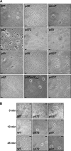
Aggregation as assessed by phase-contrast microscopy (scale bars, 10 μm). (A) Aggregates observed after 2 h in N. meningitidis comP, pilT, pilT2, pilU, pilV, pilX, and pilZ mutants. The WT strain and a nonpiliated pilD mutant were included as positive and negative controls, respectively. Aggregates restored in double pilXT and pilZT mutants are also shown. (B) Aggregation kinetics in pilT2 and pilV mutants. The WT strain was included as a control. Pictures were taken at 0, 10, and 45 min.
FIG. 5.
Quantification of adhesion to HUVEC. After a 30-min contact during which standard numbers of bacteria adhered to standard numbers of cells, nonadherent bacteria were removed by replacing the medium. After further incubation (with the medium being regularly replaced every hour), cells were washed and recovered by scraping, and adherent bacteria were counted. Results, expressed as CFU of adhering bacteria, were normalized for an inoculum of 107 CFU. (A) Adhesion or N. meningitidis comP, pilT, pilT2, pilU, pilV, pilX, and pilZ mutants to HUVEC after 270 min of infection. The WT strain and a nonpiliated pilD mutant were included as positive and negative controls, respectively. Restored adhesion in pilXT and pilZT double mutants is also shown. Results are the means ± standard deviations from 3 to 10 independent experiments. (B) Adhesion of the pilU mutant to HUVEC after 30 and 90 min of infection. The WT strain was included as a control. Results are the means ± standard deviations from five independent experiments.
FIG. 6.
Adhesion of a pilZ mutant to HUVEC as observed by phase-contrast microscopy (scale bars, 10 μm). Images were taken after 150 min of infection. The WT strain and a nonpiliated pilD mutant were included as positive and negative controls, respectively.
FIG. 7.
Immunoblot detection of PilV, PilX, and PilC1 in “classical” pilus preparations from a pilZ mutant. The WT strain and a pilE mutant were included as positive and negative controls, respectively. Equal amounts of pili were loaded in each lane, as assessed by PilE immunodetection (data not shown). As in Fig. 1, there was a major cross-reacting species (indicated by an asterisk) in the immunoblots in which minor pilins were detected, which might correspond to PilE.
FIG. 8.
Aggregation, competence, and/or adhesion in cross-complemented _pilX/comP_ind and _pilX/pilV_ind strains. (A) Aggregation in the _pilX/comP_ind and _pilX/pilV_ind mutants as observed by phase-contrast microscopy after 2 h of growth (scale bars, 10 μm). The _pilX/pilX_ind strain was included as a control. (B) Adhesion of the _pilX/comP_ind strain to HUVEC after 270 min of infection. The WT strain and the pilD and pilX mutants were included as controls. The results, expressed as CFU of adhering bacteria normalized for an inoculum of 107 CFU, are the means ± standard deviations from four to six independent experiments. (C) Quantification of the competence for DNA transformation in _pilX/comP_ind and _pilX/pilV_ind strains. The WT strain, the _pilX/pilX_ind strain, and the pilD and pilX mutants were included as controls. Results are expressed as percentages of recipient cells transformed and are the means ± standard deviations from three to six independent experiments.
Similar articles
- Pilus-mediated adhesion of Neisseria meningitidis is negatively controlled by the pilus-retraction machinery.
Yasukawa K, Martin P, Tinsley CR, Nassif X. Yasukawa K, et al. Mol Microbiol. 2006 Jan;59(2):579-89. doi: 10.1111/j.1365-2958.2005.04954.x. Mol Microbiol. 2006. PMID: 16390451 - Type IV pilus biogenesis in Neisseria meningitidis: PilW is involved in a step occurring after pilus assembly, essential for fibre stability and function.
Carbonnelle E, Hélaine S, Prouvensier L, Nassif X, Pelicic V. Carbonnelle E, et al. Mol Microbiol. 2005 Jan;55(1):54-64. doi: 10.1111/j.1365-2958.2004.04364.x. Mol Microbiol. 2005. PMID: 15612916 - Structure/function analysis of Neisseria meningitidis PilW, a conserved protein that plays multiple roles in type IV pilus biology.
Szeto TH, Dessen A, Pelicic V. Szeto TH, et al. Infect Immun. 2011 Aug;79(8):3028-35. doi: 10.1128/IAI.05313-11. Epub 2011 Jun 6. Infect Immun. 2011. PMID: 21646452 Free PMC article. - Interaction of pathogenic neisseriae with nonphagocytic cells.
Nassif X, So M. Nassif X, et al. Clin Microbiol Rev. 1995 Jul;8(3):376-88. doi: 10.1128/CMR.8.3.376. Clin Microbiol Rev. 1995. PMID: 7553571 Free PMC article. Review. - Type II secretion and type IV pili of Francisella.
Forsberg A, Guina T. Forsberg A, et al. Ann N Y Acad Sci. 2007 Jun;1105:187-201. doi: 10.1196/annals.1409.016. Epub 2007 Apr 13. Ann N Y Acad Sci. 2007. PMID: 17435117 Review.
Cited by
- Functional analysis of an unusual type IV pilus in the Gram-positive Streptococcus sanguinis.
Gurung I, Spielman I, Davies MR, Lala R, Gaustad P, Biais N, Pelicic V. Gurung I, et al. Mol Microbiol. 2016 Jan;99(2):380-92. doi: 10.1111/mmi.13237. Epub 2015 Oct 27. Mol Microbiol. 2016. PMID: 26435398 Free PMC article. - Type IV pilus retraction is required for Neisseria musculi colonization and persistence in a natural mouse model of infection.
Rhodes KA, Rendón MA, Ma MC, Agellon A, Johnson AC, So M. Rhodes KA, et al. mBio. 2024 Jan 16;15(1):e0279223. doi: 10.1128/mbio.02792-23. Epub 2023 Dec 12. mBio. 2024. PMID: 38084997 Free PMC article. - Neisseria genes required for persistence identified via in vivo screening of a transposon mutant library.
Rhodes KA, Ma MC, Rendón MA, So M. Rhodes KA, et al. PLoS Pathog. 2022 May 17;18(5):e1010497. doi: 10.1371/journal.ppat.1010497. eCollection 2022 May. PLoS Pathog. 2022. PMID: 35580146 Free PMC article. - Neisseria cinerea isolates can adhere to human epithelial cells by type IV pilus-independent mechanisms.
Wörmann ME, Horien CL, Johnson E, Liu G, Aho E, Tang CM, Exley RM. Wörmann ME, et al. Microbiology (Reading). 2016 Mar;162(3):487-502. doi: 10.1099/mic.0.000248. Epub 2016 Jan 26. Microbiology (Reading). 2016. PMID: 26813911 Free PMC article. - Construction of a complete set of Neisseria meningitidis mutants and its use for the phenotypic profiling of this human pathogen.
Muir A, Gurung I, Cehovin A, Bazin A, Vallenet D, Pelicic V. Muir A, et al. Nat Commun. 2020 Nov 2;11(1):5541. doi: 10.1038/s41467-020-19347-y. Nat Commun. 2020. PMID: 33139723 Free PMC article.
References
- Aas, F. E., C. Lovold, and M. Koomey. 2002. An inhibitor of DNA binding and uptake events dictates the proficiency of genetic transformation in Neisseria gonorrhoeae: mechanism of action and links to type IV pilus expression. Mol. Microbiol. 46:1441-1450. - PubMed
- Aas, F. E., M. Wolfgang, S. Frye, S. Dunham, C. Lovold, and M. Koomey. 2002. Competence for natural transformation in Neisseria gonorrhoeae: components of DNA binding and uptake linked to type IV pilus expression. Mol. Microbiol. 46:749-760. - PubMed
- Amikam, D., and M. Y. Galperin. 2006. PilZ domain is part of the bacterial c-di-GMP binding protein. Bioinformatics 22:3-6. - PubMed
Publication types
MeSH terms
Substances
LinkOut - more resources
Full Text Sources
Other Literature Sources


