Sec24D-dependent transport of extracellular matrix proteins is required for zebrafish skeletal morphogenesis - PubMed (original) (raw)
Sec24D-dependent transport of extracellular matrix proteins is required for zebrafish skeletal morphogenesis
Swapnalee Sarmah et al. PLoS One. 2010.
Abstract
Protein transport from endoplasmic reticulum (ER) to Golgi is primarily conducted by coated vesicular carriers such as COPII. Here, we describe zebrafish bulldog mutations that disrupt the function of the cargo adaptor Sec24D, an integral component of the COPII complex. We show that Sec24D is essential for secretion of cartilage matrix proteins, whereas the preceding development of craniofacial primordia and pre-chondrogenic condensations does not depend on this isoform. Bulldog chondrocytes fail to secrete type II collagen and matrilin to extracellular matrix (ECM), but membrane bound receptor beta1-Integrin and Cadherins appear to leave ER in Sec24D-independent fashion. Consequently, Sec24D-deficient cells accumulate proteins in the distended ER, although a subset of ER compartments and Golgi complexes as visualized by electron microscopy and NBD C(6)-ceramide staining appear functional. Consistent with the backlog of proteins in the ER, chondrocytes activate the ER stress response machinery and significantly upregulate BiP transcription. Failure of ECM secretion hinders chondroblast intercalations thus resulting in small and malformed cartilages and severe craniofacial dysmorphology. This defect is specific to Sec24D mutants since knockdown of Sec24C, a close paralog of Sec24D, does not result in craniofacial cartilage dysmorphology. However, craniofacial development in double Sec24C/Sec24D-deficient animals is arrested earlier than in bulldog/sec24d, suggesting that Sec24C can compensate for loss of Sec24D at initial stages of chondrogenesis, but Sec24D is indispensable for chondrocyte maturation. Our study presents the first developmental perspective on Sec24D function and establishes Sec24D as a strong candidate for cartilage maintenance diseases and craniofacial birth defects.
Conflict of interest statement
Competing Interests: The authors have declared that no competing interests exist.
Figures
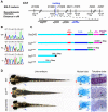
Figure 1. The bulldog mutations disrupt the sec24d gene and affect craniofacial development.
(A) The bulldog mutations map to chromosome 7 between markers Z13880 and Z13936. The number of recombinants and the corresponding distances in centiMorgans (cM) are indicated below. The genes in the critical region are listed. (B) Electropherograms of wild-type +/+, heterozygous +/– and bulm421 –/– genomic DNA shows the C→T transition (arrows) that results in an amber stop codon (TAG) in place of glutamine at position 811 (Q811X). (C) Schematic diagram of the Sec24D primary protein structure of wild-type and bulldog mutants. (D) bulm606 and sec24d morphant embryos (3.5 ng sec24d_-MO) have reduced head size (red arrows), shorter body length and pectoral fins (blue arrows). Alcian blue staining of the head skeleton in the right panels shows short Meckel's (red arrows), deformed ceratohyals (black arrows) and kinked pectoral fins (blue arrows) in bulm606 and s_ec24d morphant embryos compared to wild types. Toluidine blue stained coronal sections of craniofacial cartilage in bulm606 and sec24d morphant embryos demonstrate very low amount of ECM material (purple staining) and abnormal shape and packing density of chondrocytes as compared to wild types, all at 4 dpf. Abbreviation: ch, ceratohyal.
Figure 2. sec24d mRNA expression during embryogenesis and molecular analysis of the bulldog mutation.
(A) RT-PCR assay shows maternally deposited sec24d transcripts at 1 and 2 hpf and increasing levels thereafter. β-actin served as control. (B–H) Expression patterns of sec24d during zebrafish development using whole mount in situ hybridization staining of wild-type embryos. The cross section through the palatoquadrate (pq, arrowheads) at 3 dpf (I) shows robust riboprobe staining (h: hours, d: days). (J–O) Expression patterns of col2_α_1 (J,K) and sox9a (L,M) at 3 dpf. The expression is slightly upregulated in the posterior pharyngeal cartilages of bulm606 embryos. The Collagen-specific chaperone hsp47 (N,O) transcript is elevated in craniofacial skeleton (arrowheads), fins and the notochord. Arrowheads point to the jaw region in panels (G) through (O). (P) Relative sec24d sec24c, bip and sil1 mRNA levels were assessed by quantitative real-time PCR and adjusted against β-actin using total RNA samples from 4 dpf embryos. In bulm494, mRNA levels for bip and sil1 are induced 5-fold, whereas sec24d mRNA levels are reduced 0.7-fold. *, p<0.02; **, p<0.005. Abbreviation: b, brain, o, optic nerve, ot, otic capsule, r, retina. Anterior (D–O) is to the left.
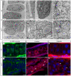
Figure 3. Sec24D-deficient cartilage contains sparse ECM and chondrocytes with distended ER.
(A–H) Electron micrographs show mature wild-type chondrocytes (cells 1–3 in A) are separated by ECM and contain dense ER membranes (A, E, arrows). In bulm494 mutants (B,F,H), cartilage matrix is sparse and large amounts of electron-dense material accumulate in the rough ER (asterisks, cells marked 1–6 in B). Wild-type chondrocytes (C) are devoid of cell-cell contacts, whereas the majority of bulm494 mutant cells (D) contain adherens junctions (arrowheads). bulm494 chondrocytes (F) contain both normal ER cisternae (arrows) and swollen, ribosome studded, ER (asterisks). _sec24d_-deficient cells (H) contain smaller and disorganized Golgi complexes (blue arrows) compared to wild-types (G). (I,J) Single-pass confocal images of C6-NBD ceramide stained chondrocytes of wild-type (I) and sec24d/bulm606 mutant (J) embryos at 4 dpf. (K,L) Adherens junctions are marked by β-catenin (red) in bulldog Meckel's cartilage (arrowheads), but are mostly absent in wild types (K). (M,N) Phalloidin (red) shows regular cortical distribution of polymerized actin in wild types (M), but uneven cellular distribution in bulm606 cartilage (arrowheads) (N). Nuclei are stained with TOPRO-3 (blue). Abbreviations: m, mitochondria; n, nucleus; ECM, extracellular matrix. Scale bars: 2 µM (A,B,E); 500 nm (F–H); 5 µM (I–N).
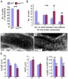
Figure 4. Analysis of chondrocyte shape and numbers in bulldog mutants.
(A) The number of cells in the ceratohyals at 5 dpf is not significantly different between wild-type and bulldog embryos (counted in single optical plane of Alcian blue stained preparations, six different animals each). (B) In contrast, the number of cells spanning the entire width of the ceratohyal at 5 dpf is notably higher in bulldog. (C,D) Single-pass confocal images of the Meckel's cartilage in live embryos marked with membrane tethered GFP tracer. Bulldog mutants (D) show multiple stacked chondrocytes as compared to a single spanning cell in wild-types (C). (E) The average chondrocyte width is comparable between wild-type and bulldog, whereas the length and the length-to-width ratio are significantly lower in the mutants. Cellular dimensions were counted in Meckel's (m) and ceratohyal (ch) cartilages in three different live embryos at 3 dpf. The number of cells used for measurements is indicated in the right graph (E). * denotes p<0.0001; **, _p_<0.0001; , p<0.003; ##, p<0.0001.
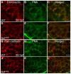
Figure 5. Analysis of mesenchymal condensations in cartilage primordia of bulldog mutants.
(A–F) Mesenchymal condensations, stained with Peanut Agglutinin (PNA, green) show similar distribution of Fibronectin (red) in wild-type (A–C) and bulldog hyosymplectic cartilages (D–F). (G–L) Cadherin expression in chondrocytes (marked by a Pan-cadherin antibody in red) is indistinguishable between wild-type and bulldog ceratohyals as shown by PNA staining. The right panels represent merged images of the left and middle panels. Scale bars are 5 µM.
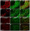
Figure 6. Sec24D-deficient chondrocytes do not transport ECM proteins.
(A–F) Collagen2α1 (red) and Wheat Germ Agglutinin (WGA, green) signals are concentrated at juxtanuclear compartments and in the extracellular space of wild-type chondrocytes (A–C). In bulldog, the signals are primarily localized in cytoplasmic vesicular compartments, although weak WGA signal is also detectable at the plasma membrane and the extracellular space (D–F). (G–L) Single pass confocal images of wild-type and bulm606 Meckel's cartilages labeled with the β1-integrin (red) recognizing antibody and counterstained with WGA (green). Merged channels show co-localization of the two labels in the cell boundaries in both wild-type and bulldog chondrocytes. The right panels represent merged images of the left and middle panels. Scale bars are 5 µM.
Figure 7. Sec24C expression during zebrafish development.
(A,B) sec24c is ubiquitously expressed at cleavage (8-cell, A) and gastrulation (shield, B) stages. (C–F) Sec24c expression at consecutive embryonic days is concentrated in the head and gut regions. Expression in the head overlaps with developing craniofacial structures.
Figure 8. Knockdown of sec24c, and combined loss of sec24d and sec24c phenotypes.
Analysis of wild types (A, E, I), _sec24c_-UTR-MO (B,F,J), bulldog (C, G, K) and double _sec24c_-UTR-MO+ _sec24d_-UTR-MO (D, H, L) injected embryos. (A–D) Images of live wild types (A), _sec24c_-UTR-MO, (2.0 ng injected, B), bulldog (C) and _sec24c_-UTR-MO+ _sec24d_-UTR-MO (2.0 ng each, D). The sec24c morphants have reduced body length (blue arrows), but the head appears normal. Double morphants for sec24d+sec24c are significantly shorter than bulldog larvae, almost completely lack fin-folds and have reduced jaw region (red arrow) and pronounced heart edema (D). (E–H) Alcian blue preparations of the head skeleton. The Alcian blue staining confirms normal craniofacial cartilages in sec24c morphants. (I–L) Histological sections counter-stained with toluidine blue at 4 dpf. Insets indicate the plane of the sections. (I,J) Histological, sagittal plastic sections stained with toluidine blue show normal jaw opening and surrounding cartilages in wild types (I) and sec24c morphants (J). Purple staining patterns for glycosaminoglycans of the ECM are also comparable. In contrast, the pharyngeal skeleton of sec24d+sec24c double mutants fails to stain with Alcian blue (H). Histological sections reveal patterned arches that are largely devoid of metachromatically stained cells (L). Abbreviations: cb, ceratobranchial; ch, ceratohyal; ep, ethmoid plate, m, Meckel's cartilage.
Figure 9. Migration of craniofacial primordia in double Sec24C and Sec24D morants.
(A–H) Images of transgenic sox10:mRFP embryos at 30 hpf (lateral view A–D) and 48 hpf (ventral view E–H) after injection with _sec24c_-UTR-MO, (2.0 ng), _sec24d_-UTR-MO (3.5 ng), and _sec24c_-UTR-MO+ _sec24d_-UTR-MO. Neural crest streams are numbered (1, 2, 3, 4, 5, 6 in A–D). Arrows point to migrating craniofacial primordia (E–H).
Similar articles
- The feelgood mutation in zebrafish dysregulates COPII-dependent secretion of select extracellular matrix proteins in skeletal morphogenesis.
Melville DB, Montero-Balaguer M, Levic DS, Bradley K, Smith JR, Hatzopoulos AK, Knapik EW. Melville DB, et al. Dis Model Mech. 2011 Nov;4(6):763-76. doi: 10.1242/dmm.007625. Epub 2011 Jul 4. Dis Model Mech. 2011. PMID: 21729877 Free PMC article. - Secretory COPII coat component Sec23a is essential for craniofacial chondrocyte maturation.
Lang MR, Lapierre LA, Frotscher M, Goldenring JR, Knapik EW. Lang MR, et al. Nat Genet. 2006 Oct;38(10):1198-203. doi: 10.1038/ng1880. Epub 2006 Sep 17. Nat Genet. 2006. PMID: 16980978 - Collagen has a unique SEC24 preference for efficient export from the endoplasmic reticulum.
Lu CL, Ortmeier S, Brudvig J, Moretti T, Cain J, Boyadjiev SA, Weimer JM, Kim J. Lu CL, et al. Traffic. 2022 Jan;23(1):81-93. doi: 10.1111/tra.12826. Epub 2021 Nov 22. Traffic. 2022. PMID: 34761479 Free PMC article. - Traffic jams in fish bones: ER-to-Golgi protein transport during zebrafish development.
Melville DB, Knapik EW. Melville DB, et al. Cell Adh Migr. 2011 Mar-Apr;5(2):114-8. doi: 10.4161/cam.5.2.14377. Epub 2011 Mar 1. Cell Adh Migr. 2011. PMID: 21178403 Free PMC article. Review. - ER-to-Golgi transport: form and formation of vesicular and tubular carriers.
Watson P, Stephens DJ. Watson P, et al. Biochim Biophys Acta. 2005 Jul 10;1744(3):304-15. doi: 10.1016/j.bbamcr.2005.03.003. Epub 2005 Mar 23. Biochim Biophys Acta. 2005. PMID: 15979504 Review.
Cited by
- Visualization of Procollagen IV Reveals ER-to-Golgi Transport by ERGIC-independent Carriers.
Matsui Y, Hirata Y, Wada I, Hosokawa N. Matsui Y, et al. Cell Struct Funct. 2020 Jul 23;45(2):107-119. doi: 10.1247/csf.20025. Epub 2020 Jun 18. Cell Struct Funct. 2020. PMID: 32554938 Free PMC article. - Transforming growth factor beta signaling and craniofacial development: modeling human diseases in zebrafish.
Fox SC, Waskiewicz AJ. Fox SC, et al. Front Cell Dev Biol. 2024 Feb 7;12:1338070. doi: 10.3389/fcell.2024.1338070. eCollection 2024. Front Cell Dev Biol. 2024. PMID: 38385025 Free PMC article. Review. - Evidence for nutrient-dependent regulation of the COPII coat by O-GlcNAcylation.
Bisnett BJ, Condon BM, Linhart NA, Lamb CH, Huynh DT, Bai J, Smith TJ, Hu J, Georgiou GR, Boyce M. Bisnett BJ, et al. Glycobiology. 2021 Sep 20;31(9):1102-1120. doi: 10.1093/glycob/cwab055. Glycobiology. 2021. PMID: 34142147 Free PMC article. - AP-4 loss in CRISPR-edited zebrafish affects early embryo development.
Pembridge OG, Wallace NS, Clements TP, Jackson LP. Pembridge OG, et al. Adv Biol Regul. 2023 Jan;87:100945. doi: 10.1016/j.jbior.2022.100945. Epub 2022 Dec 22. Adv Biol Regul. 2023. PMID: 36642642 Free PMC article. - KLHL12 can form large COPII structures in the absence of CUL3 neddylation.
Moretti T, Kim K, Tuladhar A, Kim J. Moretti T, et al. Mol Biol Cell. 2023 Mar 1;34(3):br4. doi: 10.1091/mbc.E22-08-0383. Epub 2023 Jan 18. Mol Biol Cell. 2023. PMID: 36652337 Free PMC article.
References
- Barlowe C, Orci L, Yeung T, Hosobuchi M, Hamamoto S, et al. COPII: a membrane coat formed by Sec proteins that drive vesicle budding from the endoplasmic reticulum. Cell. 1994;77:895–907. - PubMed
- Bonifacino JS, Glick BS. The mechanisms of vesicle budding and fusion. Cell. 2004;116:153–166. - PubMed
- Fromme JC, Orci L, Schekman R. Coordination of COPII vesicle trafficking by Sec23. Trends Cell Biol. 2008;18:330–336. - PubMed
- Boyadjiev SA, Fromme JC, Ben J, Chong SS, Nauta C, et al. Cranio-lenticulo-sutural dysplasia is caused by a SEC23A mutation leading to abnormal endoplasmic-reticulum-to-Golgi trafficking. Nat Genet. 2006;38:1192–1197. - PubMed
Publication types
MeSH terms
Substances
LinkOut - more resources
Full Text Sources
Molecular Biology Databases
