Evidence for the default network's role in spontaneous cognition - PubMed (original) (raw)
Evidence for the default network's role in spontaneous cognition
Jessica R Andrews-Hanna et al. J Neurophysiol. 2010 Jul.
Abstract
A set of brain regions known as the default network increases its activity when focus on the external world is relaxed. During such moments, participants change their focus of external attention and engage in spontaneous cognitive processes including remembering the past and imagining the future. However, the functional contributions of the default network to shifts in external attention versus internal mentation have been difficult to disentangle because the two processes are correlated under typical circumstances. To address this issue, the present study manipulated factors that promote spontaneous cognition separately from those that change the scope of external attention. Results revealed that the default network increased its activity when spontaneous cognition was maximized but not when participants increased their attention to unpredictable foveal or peripheral stimuli. To examine the nature of participants' spontaneous thoughts, a second experiment used self-report questionnaires to quantify spontaneous thoughts during extended fixation epochs. Thoughts about one's personal past and future comprised a major focus of spontaneous cognition with considerable variability. Activity correlations between the medial temporal lobe and distributed cortical regions within the default network predicted a small, but significant, portion of the observed variability. Collectively, these results suggest that during passive states, activity within the default network reflects spontaneous, internally directed cognitive processes.
Figures
Fig. 1.
Experimental paradigm to dissociate external attention from spontaneous cognition. The experimental paradigm contained 3 conditions that manipulated focus of attention through subject expectations. In the broad attention condition (top row), participants fixated and simultaneously pressed a button whenever a peripherally located flicker was detected. In the focal attention condition (middle row), participants detected centrally located flickers. In the passive condition (bottom row), participants remained fixated but did not expect flickers, thus increasing the tendency to engage in spontaneous cognition. Critically, embedded blocks within the broad and focal attention conditions contained no flickers. These analyzed blocks matched stimuli across all conditions (see blocks labeled analyzed time period). Thus only the expectations of the subject differed between conditions with subjects expecting peripheral events in the broad attention condition (top), foveal events in the focal attention condition (middle), or no events in the passive condition (bottom). The baseline condition for each critical fixation condition was abstract/concrete semantic classification of words.
Fig. 2.
Thought sampling and poststudy probes demonstrate differences in spontaneous cognition between conditions. To confirm that conditions varied with respect to spontaneous cognition, an independent sample of 30 participants completed the experimental paradigm in a mock magnetic resonance imaging (MRI) scanner that allowed spontaneous thought sampling during the paradigm itself and follow-up self-report questionnaires after the study was completed. A: significantly more task-unrelated thoughts were observed during the passive condition compared with that of either the broad or focal attention conditions. B: participants also reported mind-wandering significantly more during the passive condition in a postexperimental questionnaire (1 = spontaneous thoughts were never experienced to 7 = spontaneous thoughts were always experienced). These results confirm the passive condition was associated with an increased tendency to engage in spontaneous cognition. Error bars reflect SE. *P < 0.05; **P < 0.01.
Fig. 3.
Poststudy questionnaires confirm differences in spontaneous cognition during the functional magnetic resonance imaging (fMRI) study. The same surprise postexperimental questionnaire administered to the participants that completed the thought sampling study (Fig. 2) was also administered to the independent group of participants that completed the experimental task while in the MRI scanner. Following the MRI study, participants reported mind-wandering significantly more during the passive condition in a postscanning questionnaire. A nonsignificant trend was observed between the broad and focal conditions, with numerically more reports of mind-wandering during the broad condition. Error bars reflect SE. **P < 0.01, ***P < 0.001, †P = 0.07.
Fig. 4.
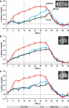
Hypothesis-driven analyses reveal increased activity within the default network during the passive condition. Significantly increased fMRI blood oxygenation level-dependent (BOLD) signal (an indirect measure of activity) was observed during the passive condition compared with either the broad or focal attention conditions. BOLD signal is measured as percentage signal change compared with baseline. This pattern was observed in A, a large region of interest comprising multiple regions within the default network; as well as in B, the anterior medial prefrontal cortex; and in C, the posterior cingulate cortex. The image insets (top left in each panel) show the regions plotted in either transverse (A) or sagittal (B and C) views. Error bars reflect SE. ***P < 0.001.
Fig. 5.
The time course of activity within the default network across conditions. The time course of the BOLD signal is plotted for the critical fixation epochs for each condition. BOLD signal is measured as percentage signal change compared with the abstract/concrete baseline task. The timing of each block is outlined below (C = cue). Dotted lines separate fixation blocks into 2 distinct 15 s epochs. In all regions, there was an increase in activity in the passive condition compared with either the broad or focal attention conditions. This difference emerged rapidly with the onset of the manipulation. What is also apparent is that the activity observed in the broad and focal conditions in Fig. 4 is largely accounted for by activity increases that occur late in the task epochs. This is most clear in A where the activity in the broad and focal conditions is absent in the first 15 s of the block. Error bars reflect SE.
Fig. 6.
Whole brain exploratory analyses demonstrate that the default network does not support broad external attention. Increased activity during broad attention compared with focal attention was observed in primary visual cortex, medial and lateral extrastriate cortex near the transverse occipital sulcus (TOS), and precuneus at or near area 7m (shown in red). In contrast, the focal attention condition elicited increased activity in the lateral occipital area (LO) and the lateral occipital sulcus (LOS) (shown in blue). Activity exceeding a threshold of P < 0.001 uncorrected is plotted on an inflated surface using Caret software (Van Essen 2005) as well as a flattened cortical map. Estimated areal boundaries are marked by a dotted line, whereas relevant sulci are highlighted in yellow.
Fig. 7.
Functional connectivity of the precuneus region at or near area 7m reveals it is not a component of the default network. An 8 mm sphere seed region centered at or near area 7m (16, −78, 56) from the broad > focal attention whole brain contrast in Fig. 6 was created (seed shown to the left). This region was used as a seed to examine low-frequency functional correlations during rest fixation runs in the same group of participants (similar to Kahn et al. 2008; Vincent et al. 2006, 2008). Warm colors represent voxels that exhibit positive correlations with the precuneus region at or near putative area 7m at a threshold of P < 0.0001 uncorrected, whereas cool colors represent voxels exhibiting negative correlations with the seed region. Note that a majority of the default network is negatively correlated with the precuneus, thus suggesting that precuneus area 7m and the default network belong to distinct brain systems (also see Buckner et al. 2008 for discussion).
Fig. 8.
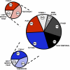
Spontaneous cognition is prominent during passive fixation. Postscanning questionnaires reveal that participants spend the majority of their time engaged in internal mentation during extended fixation blocks that prominently activate the default network. Suggesting an adaptive role, participants spend approximately half of the allotted time thinking about their past and future (particularly the recent past and near future).
Fig. 9.
Frequency of thoughts about the past and future predicts medial temporal lobe (MTL)-cortical functional correlations. Whole brain functional correlations with the MTL were extracted from 139 participants and regressed against their reported frequency of temporally oriented thoughts (past + future). A: the MTL seed is shown on coronal slices and includes a combined hippocampal formation (HF) and parahippocampal cortex (PHC) region of interest. B: a surface projection (Caret software; Van Essen 2005) shows voxels that exhibit a correlation of r > 0.22 (P < 0.01) for the brain–behavior relationships. In other words, individuals that report a greater frequency of temporally oriented thoughts exhibit greater functional correlation between the MTL and regions within the default network that are associated with an MTL subsystem (see Andrews-Hanna et al. 2010; Buckner et al. 2008). Note that the topography of the correlations overlaps those associated with the default network as traditionally defined. C: the same analysis is shown on transverse slices.
Fig. 10.
Exploratory analysis of medial temporal lobe functional correlations. For each participant, the mean _z_-transformed correlation coefficients between all pairs of regions comprising the medial temporal lobe and its correlated cortical regions (HF, PHC, Rsp, pIPL, vMPFC) is plotted along with the percentage of time participants reported thinking about the past and the future during extended fixation epochs. A significant (but modest) positive relationship is observed between the 2 variables (r = 0.18; P < 0.05), suggesting that participants who think more about the past and/or the future exhibit increased functional coupling within the “MTL subsystem” of the default network.
Similar articles
- Shaped by our thoughts--a new task to assess spontaneous cognition and its associated neural correlates in the default network.
O'Callaghan C, Shine JM, Lewis SJ, Andrews-Hanna JR, Irish M. O'Callaghan C, et al. Brain Cogn. 2015 Feb;93:1-10. doi: 10.1016/j.bandc.2014.11.001. Epub 2014 Nov 18. Brain Cogn. 2015. PMID: 25463243 - Default network activity, coupled with the frontoparietal control network, supports goal-directed cognition.
Spreng RN, Stevens WD, Chamberlain JP, Gilmore AW, Schacter DL. Spreng RN, et al. Neuroimage. 2010 Oct 15;53(1):303-17. doi: 10.1016/j.neuroimage.2010.06.016. Epub 2010 Jun 18. Neuroimage. 2010. PMID: 20600998 Free PMC article. - Attention Shifts Recruit the Monkey Default Mode Network.
Arsenault JT, Caspari N, Vandenberghe R, Vanduffel W. Arsenault JT, et al. J Neurosci. 2018 Jan 31;38(5):1202-1217. doi: 10.1523/JNEUROSCI.1111-17.2017. Epub 2017 Dec 20. J Neurosci. 2018. PMID: 29263238 Free PMC article. - A framework for understanding the relationship between externally and internally directed cognition.
Dixon ML, Fox KC, Christoff K. Dixon ML, et al. Neuropsychologia. 2014 Sep;62:321-30. doi: 10.1016/j.neuropsychologia.2014.05.024. Epub 2014 Jun 6. Neuropsychologia. 2014. PMID: 24912071 Review. - Cooperation between the default mode network and the frontal-parietal network in the production of an internal train of thought.
Smallwood J, Brown K, Baird B, Schooler JW. Smallwood J, et al. Brain Res. 2012 Jan 5;1428:60-70. doi: 10.1016/j.brainres.2011.03.072. Epub 2011 Apr 3. Brain Res. 2012. PMID: 21466793 Review.
Cited by
- Phenomenology of future-oriented mind-wandering episodes.
Stawarczyk D, Cassol H, D'Argembeau A. Stawarczyk D, et al. Front Psychol. 2013 Jul 16;4:425. doi: 10.3389/fpsyg.2013.00425. eCollection 2013. Front Psychol. 2013. PMID: 23882236 Free PMC article. - Self-views converge during enjoyable conversations.
Welker C, Wheatley T, Cason G, Gorman C, Meyer M. Welker C, et al. Proc Natl Acad Sci U S A. 2024 Oct 22;121(43):e2321652121. doi: 10.1073/pnas.2321652121. Epub 2024 Oct 14. Proc Natl Acad Sci U S A. 2024. PMID: 39401349 Free PMC article. - Mental time travel and default-mode network functional connectivity in the developing brain.
Østby Y, Walhovd KB, Tamnes CK, Grydeland H, Westlye LT, Fjell AM. Østby Y, et al. Proc Natl Acad Sci U S A. 2012 Oct 16;109(42):16800-4. doi: 10.1073/pnas.1210627109. Epub 2012 Oct 1. Proc Natl Acad Sci U S A. 2012. PMID: 23027942 Free PMC article. - Spatiotemporal complexity patterns of resting-state bioelectrical activity explain fluid intelligence: Sex matters.
Dreszer J, Grochowski M, Lewandowska M, Nikadon J, Gorgol J, Bałaj B, Finc K, Duch W, Kałamała P, Chuderski A, Piotrowski T. Dreszer J, et al. Hum Brain Mapp. 2020 Dec;41(17):4846-4865. doi: 10.1002/hbm.25162. Epub 2020 Aug 18. Hum Brain Mapp. 2020. PMID: 32808732 Free PMC article. - A functional architecture of the human brain: emerging insights from the science of emotion.
Lindquist KA, Barrett LF. Lindquist KA, et al. Trends Cogn Sci. 2012 Nov;16(11):533-40. doi: 10.1016/j.tics.2012.09.005. Epub 2012 Oct 2. Trends Cogn Sci. 2012. PMID: 23036719 Free PMC article. Review.
References
- Addis DR, Wong AT, Schacter DL. Age-related changes in the episodic simulation of future events. Psychol Sci 19: 33–41, 2008 - PubMed
- Andreasen NC, O'Leary DS, Cizadlo T, Arndt S, Rezai K, Watkins GL, Ponto LL, Hichwa RD. Remembering the past: two facets of episodic memory explored with positron emission tomography. Am J Psychiatry 152: 1576–1585, 1995 - PubMed
- Antrobus JS, Singer JL, Greenberg S. Studies in the stream of consciousness: experimental enhancement and suppression of spontaneous cognitive processes. Percept Mot Skills 23: 399–417, 1966
Publication types
MeSH terms
LinkOut - more resources
Full Text Sources
Miscellaneous


