Transforming growth factor-beta signaling curbs thymic negative selection promoting regulatory T cell development - PubMed (original) (raw)
Transforming growth factor-beta signaling curbs thymic negative selection promoting regulatory T cell development
Weiming Ouyang et al. Immunity. 2010.
Abstract
Thymus-derived naturally occurring regulatory T (nTreg) cells are necessary for immunological self-tolerance. nTreg cell development is instructed by the T cell receptor and can be induced by agonist antigens that trigger T cell-negative selection. How T cell deletion is regulated so that nTreg cells are generated is unclear. Here we showed that transforming growth factor-beta (TGF-beta) signaling protected nTreg cells and antigen-stimulated conventional T cells from apoptosis. Enhanced apoptosis of TGF-beta receptor-deficient nTreg cells was associated with high expression of proapoptotic proteins Bim, Bax, and Bak and low expression of the antiapoptotic protein Bcl-2. Ablation of Bim in mice corrected the Treg cell development and homeostasis defects. Our results suggest that nTreg cell commitment is independent of TGF-beta signaling. Instead, TGF-beta promotes nTreg cell survival by antagonizing T cell negative selection. These findings reveal a critical function for TGF-beta in control of autoreactive T cell fates with important implications for understanding T cell self-tolerance mechanisms.
Copyright 2010 Elsevier Inc. All rights reserved.
Conflict of interest statement
Competing Interest Statement
The authors declare that they have no competing financial interests.
Figures
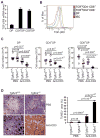
Figure 1. Enhanced Anti-CD3-induced T Cell Apoptosis in TGF-βRII-deficient Mice
(A, B) TGF-βRII expression in CD4+CD8+ double positive (DP), CD4+CD8− single positive (CD4+SP) and CD4−CD8+ (CD8+SP) thymocytes was determined by quantitative PCR (A) and Flow cytometric analysis (B). ISC stands for iso-type control. (C, D) Four-day-old wild-type (Tgfbr2+/+) and TGF-βRII-deficient (_Tgfbr2_−/−) mice were intraperitoneally injected with PBS or 20 μg/mouse of anti-CD3. Cell numbers of DP, CD4+SP, and CD8+SP thymocytes were determined 24 hr after the injection (C, n=6). Thymocyte apoptosis was examined by TUNEL staining 48 hr after the injection (D, left panel, original magnification, 20×). TUNEL staining-positive area of 4 sections per group was quantified by MetaMoph software (D, right panel). Data are representative of two (A) and three (B–D) independent experiments. The p values between the groups are shown. *depicts significant difference.
Figure 2. Exaggerated T cell Negative Selection in the absence of TGF-β Signaling
(A) Flow cytometric analysis of TCR-β expression in thymic OT-II T cells from 5-week-old Tgfbr2+/+ and _Tgfbr2_−/− OT-II mice in the absence or presence of RIP-mOva transgene (left panel). The numbers of thymic TCR-βhi OT-II T cells from 8 groups of mice are shown (right panel). (B) Flow cytometric analysis of CD69 and CD62L expression in thymic TCR-βhi OT-II T cells from 5-week-old Tgfbr2+/+ and _Tgfbr2_−/− OT-II mice in the absence or presence of RIP-mOva transgene (left panel). The numbers of thymic TCR-βhiCD69−CD62L+ OT-II T cells from 8 groups of mice are shown (right panel). (C) Survival of thymic OT-II T cells from Tgfbr2+/+ and _Tgfbr2_−/− mice. Thymic TCR-βhi OT-II T cells were purified by FACS sorting, and cultured for 12 hr. T cell viability before and after the cell culture was determined by annexin V staining (left panel). Percentages of viable T cells at 12 hr from 4 pairs of mice are presented (right panel). The p values between the groups are shown. *depicts significant difference.
Figure 3. Diabetes Development and T cell Activation in TGF-βRII-deficient OT-II RIP-mOva Mice
(A) The incidence of diabetes in Tgfbr2+/+ and _Tgfbr2_−/− OT-II RIP-mOva mice (n=11). (B) Hematoxylin and eosin staining of the pancreas of 8-week-old Tgfbr2+/+ and _Tgfbr2_−/− OT-II RIP-mOva mice (original magnification, 20×). These are representative results of four mice per group analyzed. (C) Flow cytometric analysis of CD44 and CD62L expression in OT-II T cells from non-pancreatic control lymph nodes (LN) and pancreatic lymph nodes (pLN) of 5-week-old Tgfbr2+/+ and _Tgfbr2_−/− OT-II mice in the absence or presence of RIP-mOva transgene. Data are representative of three independent experiments.
Figure 4. TGF-β Control of Thymic nTreg Survival
(A) Thymic nTregs from 5-week-old Tgfbr2+/+ and _Tgfbr2_−/− OT-II mice in the absence or presence RIP-mOva transgene. Foxp3 expression in TCR-βhi OT-II T cells (left panel), and the percentages (right top panel) and numbers (right bottom panel) of nTregs from Tgfbr2+/+ and _Tgfbr2_−/− OT-II RIP-mOva mice are shown (n=8). (B) Thymic nTregs in 3–5-day-old Tgfbr2+/+ and _Tgfbr2_−/− mice. Foxp3 expression in TCR-βhiCD4+ T cells (left panel), and nTreg numbers from Tgfbr2+/+ and _Tgfbr2_−/− mice (right panel) are shown (n=5). (C) Flow cytometric analysis of Ki67 expression in thymic nTregs from 3–5-day-old Tgfbr2+/+ and _Tgfbr2_−/− mice. These are representative results of four mice per group analyzed. (D) Thymic Foxp3+CD4+ single positive (SP) nTregs and Foxp3−CD4+ SP conventional T cells were purified by FACS sorting from 14–16-day-old Tgfbr2+/+ and _Tgfbr2_−/− mice, and cultured for 12 hr. T cell viability before and after the cell culture was determined by annexin V staining (left panel). Percentages of viable T cells at 12 hr from 5 pairs of mice are presented (right panel). The p values between the groups are shown. *depicts significant difference.
Figure 5. TGF-β Signaling Regulates Thymic nTreg Development via the Inhibition of Bim-dependent Apoptosis
(A) Flow cytometric analysis of caspase activation in 4-day-old thymic nTregs from Tgfbr2+/+ and _Tgfbr2_−/− mice. These are representative results of four mice per group analyzed. (B) Flow cytometric analysis of Bcl-2 expression in thymic nTregs from Tgfbr2+/+ and _Tgfbr2_−/− mice. ISC stands for the isotype control antibody. (C) The expression of Bim, Bak, and Bax in thymic nTregs from Tgfbr2+/+ and _Tgfbr2_−/− mice was determined by immunoblotting. β-actin was used as a protein-loading control. Band densities were quantified by ImageJ. The relative protein amounts are shown. Data are representative of three independent experiments. (D) Restoration of thymic nTreg development in _Tgfbr2_−/− mice by Bim deletion. Foxp3 expression in thymic TCR-βhiCD4+ T cells from 4-day-old Tgfbr2+/+Bcl2l11+/+, Tgfbr2_−/−_Bcl2l11+/+, Tgfbr2+/+_Bcl2l11_−/−, and _Tgfbr2_−/−_Bcl2l11_−/− mice (left panel), and nTreg numbers from 5 groups of 3–5-day-old mice are presented (right panel). The p values between the groups are shown. *depicts significant difference. (E) Flow cytometric analysis of caspase activation in thymic nTregs from Tgfbr2+/+Bcl2l11+/+, Tgfbr2_−/−_Bcl2l11+/+, Tgfbr2+/+_Bcl2l11_−/−, and _Tgfbr2_−/−_Bcl2l11_−/− mice. A representative of three independent experiments is shown.
Figure 6. Bim Ablation Restores Peripheral Tregs in TGF-βRII-deficient Mice
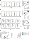
(A, B) Flow cytometric analysis of Foxp3 expression in splenic (A) and lymph node (LN) (B) CD4+ T cells from 16-day-old Tgfbr2+/+Bcl2l11+/+, Tgfbr2_−/−_Bcl2l11+/+, Tgfbr2+/+_Bcl2l11_−/−, and _Tgfbr2_−/−_Bcl2l11_−/− mice (left panels). Treg percentages from 8 groups of 14–16-day-old mice are shown (right panels). (C) Splenic and lymph node Tregs were purified from 16-day-old Tgfbr2+/+Bcl2l11+/+, Tgfbr2_−/−_Bcl2l11+/+, Tgfbr2+/+_Bcl2l11_−/− and _Tgfbr2_−/− _Bcl2l11_−/− mice by FACS sorting, and cultured for 12 hr. T cell viability before and after the cell culture was determined by annexin V staining (left panel). Percentages of viable T cells at 12 hr from 6 groups of 14–16-day-old mice are presented (right panel). The p values between the groups are shown. *depicts significant difference. (D) Suppressive function of Tgfbr2+/+Bcl2l11+/+, Tgfbr2_−/−_Bcl2l11+/+, Tgfbr2+/+_Bcl2l11_−/−, and _Tgfbr2_−/−_Bcl2l11_−/− Tregs. Splenic and lymph node Foxp3+CD4+ Tregs were isolated from Tgfbr2+/+Bcl2l11+/+, Tgfbr2_−/−_Bcl2l11+/+, Tgfbr2+/+_Bcl2l11_−/−, and _Tgfbr2_−/−_Bcl2l11_−/− mice. CD44loCD4+ T cells from Tgfbr2+/+Bcl2l11+/+ mice were labeled with CFSE, and used as responding T cells (Tresp). Cell division was assessed by CFSE dilution. The percentages of undivided Tresp cells (Y axis) and the ratios of cell numbers between Treg and Tresp cells (X axis) were plotted (left panel). A representative of CFSE dilution of Tresp cells cultured in the absence or presence of 1:2 ratio of Tregs to Tresp cells was shown (right panel). These are representative of three independent experiments.
Figure 7. Bim Ablation Partially Restores T Cell Tolerance in TGF-βRII-deficient Mice
(A) Survival of Tgfbr2_−/−_Bcl2l11+/+ (n=18) and _Tgfbr2_−/−_Bcl2l11_−/− (n=8) mice. (B) Flow cytometric analysis of CD44 and CD62L expression in splenic and lymph node (LN) CD4+ T cells from 14-day-old Tgfbr2+/+Bcl2l11+/+, Tgfbr2_−/−_Bcl2l11+/+, Tgfbr2+/+_Bcl2l11_−/−, and _Tgfbr2_−/−_Bcl2l11_−/− mice. These are representative results of four mice per group analyzed. (C) Splenic and LN CD4+ T cells from 14-day-old Tgfbr2+/+Bcl2l11+/+, Tgfbr2_−/−_Bcl2l11+/+, Tgfbr2+/+_Bcl2l11_−/−, and _Tgfbr2_−/−_Bcl2l11_−/− mice were stimulated with PMA and ionomycin for 4 hr, and analyzed for the expression of IFN-γ by intracellular staining. A representative of three independent experiments is shown.
Comment in
- TGF-Beta to the rescue.
Shevach EM. Shevach EM. Immunity. 2010 May 28;32(5):585-7. doi: 10.1016/j.immuni.2010.04.014. Immunity. 2010. PMID: 20510867 - Regulatory T cells: Nurtured by TGFbeta.
Bird L. Bird L. Nat Rev Immunol. 2010 Jul;10(7):466. doi: 10.1038/nri2812. Nat Rev Immunol. 2010. PMID: 20589991 No abstract available. - TGF-β promotes immune suppression by inhibiting Treg cell apoptosis.
Gigante M, Ranieri E. Gigante M, et al. Immunotherapy. 2010 Sep;2(5):608. Immunotherapy. 2010. PMID: 20919442 No abstract available.
Similar articles
- A regulatory role for TGF-β signaling in the establishment and function of the thymic medulla.
Hauri-Hohl M, Zuklys S, Holländer GA, Ziegler SF. Hauri-Hohl M, et al. Nat Immunol. 2014 Jun;15(6):554-61. doi: 10.1038/ni.2869. Epub 2014 Apr 13. Nat Immunol. 2014. PMID: 24728352 - Transforming Growth Factor-β Signaling in Regulatory T Cells Controls T Helper-17 Cells and Tissue-Specific Immune Responses.
Konkel JE, Zhang D, Zanvit P, Chia C, Zangarle-Murray T, Jin W, Wang S, Chen W. Konkel JE, et al. Immunity. 2017 Apr 18;46(4):660-674. doi: 10.1016/j.immuni.2017.03.015. Immunity. 2017. PMID: 28423340 - Effect of Regulatory T Cells on Promoting Apoptosis of T Lymphocyte and Its Regulatory Mechanism in Sepsis.
Luan YY, Yin CF, Qin QH, Dong N, Zhu XM, Sheng ZY, Zhang QH, Yao YM. Luan YY, et al. J Interferon Cytokine Res. 2015 Dec;35(12):969-80. doi: 10.1089/jir.2014.0235. Epub 2015 Aug 26. J Interferon Cytokine Res. 2015. PMID: 26309018 Free PMC article. - TGF-β1-induced regulatory T cells.
Hadaschik EN, Enk AH. Hadaschik EN, et al. Hum Immunol. 2015 Aug;76(8):561-4. doi: 10.1016/j.humimm.2015.06.015. Epub 2015 Jun 24. Hum Immunol. 2015. PMID: 26116540 Review. - TGF-β Control of Adaptive Immune Tolerance: A Break From Treg Cells.
Liu M, Li S, Li MO. Liu M, et al. Bioessays. 2018 Nov;40(11):e1800063. doi: 10.1002/bies.201800063. Epub 2018 Aug 29. Bioessays. 2018. PMID: 30159904 Free PMC article. Review.
Cited by
- A Genome-wide CRISPR Screen Reveals a Role for the Non-canonical Nucleosome-Remodeling BAF Complex in Foxp3 Expression and Regulatory T Cell Function.
Loo CS, Gatchalian J, Liang Y, Leblanc M, Xie M, Ho J, Venkatraghavan B, Hargreaves DC, Zheng Y. Loo CS, et al. Immunity. 2020 Jul 14;53(1):143-157.e8. doi: 10.1016/j.immuni.2020.06.011. Epub 2020 Jul 7. Immunity. 2020. PMID: 32640256 Free PMC article. - Immunomodulatory effects of transforming growth factor-β in the liver.
Schon HT, Weiskirchen R. Schon HT, et al. Hepatobiliary Surg Nutr. 2014 Dec;3(6):386-406. doi: 10.3978/j.issn.2304-3881.2014.11.06. Hepatobiliary Surg Nutr. 2014. PMID: 25568862 Free PMC article. Review. - The impact of aging on regulatory T-cells.
Fessler J, Ficjan A, Duftner C, Dejaco C. Fessler J, et al. Front Immunol. 2013 Aug 6;4:231. doi: 10.3389/fimmu.2013.00231. eCollection 2013. Front Immunol. 2013. PMID: 23964277 Free PMC article. - Selection of self-reactive T cells in the thymus.
Stritesky GL, Jameson SC, Hogquist KA. Stritesky GL, et al. Annu Rev Immunol. 2012;30:95-114. doi: 10.1146/annurev-immunol-020711-075035. Epub 2011 Dec 5. Annu Rev Immunol. 2012. PMID: 22149933 Free PMC article. Review. - Transforming Growth Factor-beta signaling in αβ thymocytes promotes negative selection.
McCarron MJ, Irla M, Sergé A, Soudja SM, Marie JC. McCarron MJ, et al. Nat Commun. 2019 Dec 19;10(1):5690. doi: 10.1038/s41467-019-13456-z. Nat Commun. 2019. PMID: 31857584 Free PMC article.
References
- Anderson MS, Venanzi ES, Chen Z, Berzins SP, Benoist C, Mathis D. The cellular mechanism of Aire control of T cell tolerance. Immunity. 2005;23:227–239. - PubMed
- Apostolou I, Sarukhan A, Klein L, von Boehmer H. Origin of regulatory T cells with known specificity for antigen. Nat Immunol. 2002;3:756–763. - PubMed
- Bouillet P, Purton JF, Godfrey DI, Zhang LC, Coultas L, Puthalakath H, Pellegrini M, Cory S, Adams JM, Strasser A. BH3-only Bcl-2 family member Bim is required for apoptosis of autoreactive thymocytes. Nature. 2002;415:922–926. - PubMed
- Bouneaud C, Kourilsky P, Bousso P. Impact of negative selection on the T cell repertoire reactive to a self-peptide: a large fraction of T cell clones escapes clonal deletion. Immunity. 2000;13:829–840. - PubMed
Publication types
MeSH terms
Substances
LinkOut - more resources
Full Text Sources
Other Literature Sources
Molecular Biology Databases
Research Materials


