Tyrosine residues in the cytoplasmic domains affect sorting and fusion activity of the Nipah virus glycoproteins in polarized epithelial cells - PubMed (original) (raw)
Tyrosine residues in the cytoplasmic domains affect sorting and fusion activity of the Nipah virus glycoproteins in polarized epithelial cells
Carolin Weise et al. J Virol. 2010 Aug.
Abstract
The highly pathogenic Nipah virus (NiV) is aerially transmitted and causes a systemic infection after entering the respiratory tract. Airway epithelia are thus important targets in primary infection. Furthermore, virus replication in the mucosal surfaces of the respiratory or urinary tract in later phases of infection is essential for virus shedding and transmission. So far, the mechanisms of NiV replication in epithelial cells are poorly elucidated. In the present study, we provide evidence that bipolar targeting of the two NiV surface glycoproteins G and F is of biological importance for fusion in polarized epithelia. We demonstrate that infection of polarized cells induces focus formation, with both glycoproteins located at lateral membranes of infected cells adjacent to uninfected cells. Supporting the idea of a direct spread of infection via lateral cell-to-cell fusion, we could identify basolateral targeting signals in the cytoplasmic domains of both NiV glycoproteins. Tyrosine 525 in the F protein is part of an endocytosis signal and is also responsible for basolateral sorting. Surprisingly, we identified a dityrosine motif at position 28/29 in the G protein, which mediates polarized targeting. A dileucine motif predicted to function as sorting signal is not involved. Mutation of the targeting signal in one of the NiV glycoproteins prevented the fusion of polarized cells, suggesting that basolateral or bipolar F and G expression facilitates the spread of NiV within epithelial cell monolayers, thereby contributing to efficient virus spread in mucosal surfaces in early and late phases of infection.
Figures
FIG. 1.
NiV infection of polarized MDCK cells. MDCK cells were grown on filter supports until full polarization was reached. Then cells either were left uninfected (mock) or were infected with NiV. At 18 h p.i. or 24 h p.i., cells were fixed with 4% PFA for 48 h. Subsequently, cells were stained with a NiV-specific guinea pig antiserum and Alexa Fluor 568-conjugated secondary antibodies. After permeabilization with methanol-acetone, cell junctions were visualized with a monoclonal antibody directed against E-cadherin and with Alexa Fluor 488-conjugated secondary antibodies. Magnification, ×630. Bar, 10 μm.
FIG. 2.
Distribution of F and G proteins in NiV-induced foci. MDCK cells were cultured on filter supports for 7 days and were then infected with NiV. At 18 h p.i., cells were fixed with 4% PFA for 48 h and were then incubated from the apical and basolateral sides with either F-specific (A) or G-specific (B) monoclonal antibodies and Alexa Fluor 568-conjugated secondary antibodies. (Top) Confocal horizontal (xy) sections through the apical part of the cell monolayer. Dashed lines indicate the lines along which vertical sections were recorded. (Bottom) Vertical (xz) sections through the foci. Arrows indicate lateral membranes. Bars, 10 μm.
FIG. 3.
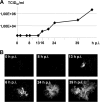
NiV release and syncytium formation at different time points p.i. MDCK cells were cultivated on filter supports until full polarization was reached. Then cells were infected with NiV at a multiplicity of infection of 10. (A) Virus titers in the supernatants were determined by the TCID50 method at 0, 2, 6, 8, 13, 16, 20, 24, 39, and 48 h p.i. (B) To visualize syncytium formation at the indicated time points p.i., cells were fixed, permeabilized with methanol-acetone, and inactivated with 4% PFA. Subsequently, cells were stained with a NiV-specific guinea pig antiserum and Alexa Fluor 568-conjugated secondary antibodies. Magnification, ×200.
FIG. 4.
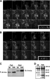
Surface distribution of wild-type F and G proteins in polarized MDCK cells. (A and B) At 7 days after seeding on filter supports, MDCK cells stably expressing either NiV Fwt (A) or NiV Gwt (B) were incubated with a NiV-specific antiserum from the apical and basolateral sides without prior fixation. Surface-bound antibodies were detected with Alexa Fluor 568-conjugated secondary antibodies. Confocal horizontal sections from top (upper left) to bottom (lower right) are shown. (Insets) Vertical sections. Bars, 20 μm. (C and D) Cell surface proteins were labeled with S-NHS biotin from either the apical (ap) or the basolateral (bas) side. (C) After cell lysis, F and G proteins were immunoprecipitated with NiV-specific antibodies. Precipitates were analyzed by SDS-PAGE under nonreducing (Fwt) or reducing (Gwt) conditions, transferred to nitrocellulose membranes, and probed with peroxidase-conjugated streptavidin. (D) To control surface-selective biotinylation after apical and basolateral labeling, 0.5% of the total-cell lysates was directly subjected to SDS-PAGE, blotting, and streptavidin detection. #, proteins selectively expressed on the apical surface; *, proteins found predominantly after basolateral biotinylation.
FIG. 5.
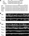
Surface distribution of mutant F and G proteins. (A) Amino acid sequences of the cytoplasmic domains of wild-type and mutant F and G proteins. Numbers above the sequences indicate amino acid positions. Boldface letters indicate exchanged amino acid residues. Vertical lines indicate the beginning of the predicted transmembrane domains. (B and C) Surface distribution of mutant NiV glycoproteins. MDCK cells stably expressing either wild-type or mutant NiV F (B) or G (C) proteins were immunostained as described in the legend to Fig. 4. Vertical sections through the cell monolayers are shown. Bar, 10 μm.
FIG. 6.
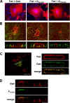
Fusion activity and surface distribution of wild-type and mutant NiV F and G proteins upon coexpression. (A) Syncytium formation in nonpolarized MDCK cells. Subconfluent MDCK cells stably expressing NiV Fwt were transfected with plasmids encoding either Gwt protein (Fwt + Gwt) or a mutant NiV G protein (Fwt + GL41/42A or Fwt + GY28/29A). At 24 h p.t., syncytia were visualized by staining with a G-specific monoclonal antibody without prior fixation, and the primary antibody was detected with Alexa Fluor 568-conjugated secondary antibodies. Cell nuclei were counterstained with DAPI. Magnification, ×200. (B) Syncytium formation in polarized MDCK cells. MDCK cells stably expressing NiV F protein were grown on filter supports. At 5 days postseeding, cells were transfected with plasmids encoding either wild-type or mutant NiV G. G-positive cells were stained at 48 h p.t. as described above. After permeabilization, cell junctions were visualized with an E-cadherin-specific monoclonal antibody and Alexa Fluor 488-conjugated secondary antibodies. Confocal horizontal sections (top) and side views (bottom) are shown. (C) Distribution of wild-type F and G proteins in polarized MDCK cells upon coexpression. MDCK cells stably expressing Fwt were grown on filter supports until full polarization was reached. Then the cells were transfected with a plasmid encoding Gwt. At 48 h p.t., stably expressed Fwt was surface stained from both sides with F-specific monoclonal antibodies and Alexa Fluor 568-conjugated secondary antibodies. The transiently expressed Gwt was then detected with G-specific antibodies and Alexa Fluor 488-conjugated secondary antibodies. (Left) Confocal xy section through the apical part of the monolayer; (right) vertical section recorded along the line indicated by the blue line in the right panel. (D) Distribution of wild-type G protein upon coexpression with FY525A. Polarized Gwt-expressing MDCK cells were transfected with mutant FY525A. At 48 h p.t., Gwt was surface stained from both sides with G-specific monoclonal antibodies and Alexa Fluor 568-conjugated secondary antibodies. The transiently expressed FY525A was then detected with F-specific antibodies and Alexa Fluor 488-conjugated secondary antibodies. Confocal vertical sections are shown. Bars, 10 μm.
Similar articles
- Structure-guided mutagenesis of Henipavirus receptor-binding proteins reveals molecular determinants of receptor usage and antibody-binding epitopes.
Oguntuyo KY, Haas GD, Azarm KD, Stevens CS, Brambilla L, Kowdle SS, Avanzato VA, Pryce R, Freiberg AN, Bowden TA, Lee B. Oguntuyo KY, et al. J Virol. 2024 Mar 19;98(3):e0183823. doi: 10.1128/jvi.01838-23. Epub 2024 Mar 1. J Virol. 2024. PMID: 38426726 Free PMC article. - Depressing time: Waiting, melancholia, and the psychoanalytic practice of care.
Salisbury L, Baraitser L. Salisbury L, et al. In: Kirtsoglou E, Simpson B, editors. The Time of Anthropology: Studies of Contemporary Chronopolitics. Abingdon: Routledge; 2020. Chapter 5. In: Kirtsoglou E, Simpson B, editors. The Time of Anthropology: Studies of Contemporary Chronopolitics. Abingdon: Routledge; 2020. Chapter 5. PMID: 36137063 Free Books & Documents. Review. - A monoclonal antibody targeting the Nipah virus fusion glycoprotein apex imparts protection from disease.
Avanzato VA, Bushmaker T, Oguntuyo KY, Yinda CK, Duyvesteyn HME, Stass R, Meade-White K, Rosenke R, Thomas T, van Doremalen N, Saturday G, Doores KJ, Lee B, Bowden TA, Munster VJ. Avanzato VA, et al. J Virol. 2024 Oct 22;98(10):e0063824. doi: 10.1128/jvi.00638-24. Epub 2024 Sep 6. J Virol. 2024. PMID: 39240113 - Dynamic Field Theory of Executive Function: Identifying Early Neurocognitive Markers.
McCraw A, Sullivan J, Lowery K, Eddings R, Heim HR, Buss AT. McCraw A, et al. Monogr Soc Res Child Dev. 2024 Dec;89(3):7-109. doi: 10.1111/mono.12478. Monogr Soc Res Child Dev. 2024. PMID: 39628288 Free PMC article. - Treatments for intractable constipation in childhood.
Gordon M, Grafton-Clarke C, Rajindrajith S, Benninga MA, Sinopoulou V, Akobeng AK. Gordon M, et al. Cochrane Database Syst Rev. 2024 Jun 19;6(6):CD014580. doi: 10.1002/14651858.CD014580.pub2. Cochrane Database Syst Rev. 2024. PMID: 38895907 Review.
Cited by
- Nipah virus fusion protein: Importance of the cytoplasmic tail for endosomal trafficking and bioactivity.
Weis M, Maisner A. Weis M, et al. Eur J Cell Biol. 2015 Jul-Sep;94(7-9):316-22. doi: 10.1016/j.ejcb.2015.05.005. Epub 2015 May 30. Eur J Cell Biol. 2015. PMID: 26059400 Free PMC article. - A functional henipavirus envelope glycoprotein pseudotyped lentivirus assay system.
Khetawat D, Broder CC. Khetawat D, et al. Virol J. 2010 Nov 12;7:312. doi: 10.1186/1743-422X-7-312. Virol J. 2010. PMID: 21073718 Free PMC article. - Comparison of the pathogenicity of Nipah virus isolates from Bangladesh and Malaysia in the Syrian hamster.
DeBuysscher BL, de Wit E, Munster VJ, Scott D, Feldmann H, Prescott J. DeBuysscher BL, et al. PLoS Negl Trop Dis. 2013;7(1):e2024. doi: 10.1371/journal.pntd.0002024. Epub 2013 Jan 17. PLoS Negl Trop Dis. 2013. PMID: 23342177 Free PMC article. - Morbillivirus and henipavirus attachment protein cytoplasmic domains differently affect protein expression, fusion support and particle assembly.
Sawatsky B, Bente DA, Czub M, von Messling V. Sawatsky B, et al. J Gen Virol. 2016 May;97(5):1066-1076. doi: 10.1099/jgv.0.000415. Epub 2016 Jan 26. J Gen Virol. 2016. PMID: 26813519 Free PMC article. - Nipah virus entry and egress from polarized epithelial cells.
Lamp B, Dietzel E, Kolesnikova L, Sauerhering L, Erbar S, Weingartl H, Maisner A. Lamp B, et al. J Virol. 2013 Mar;87(6):3143-54. doi: 10.1128/JVI.02696-12. Epub 2013 Jan 2. J Virol. 2013. PMID: 23283941 Free PMC article.
References
- Bello, V., J. W. Goding, V. Greengrass, A. Sali, V. Dubljevic, C. Lenoir, G. Trugnan, and M. Maurice. 2001. Characterization of a di-leucine-based signal in the cytoplasmic tail of the nucleotide-pyrophosphatase NPP1 that mediates basolateral targeting but not endocytosis. Mol. Biol. Cell 12:3004-3015. - PMC - PubMed
- Benedicto, I., F. Molina-Jimenez, O. Barreiro, A. Maldonado-Rodriguez, J. Prieto, R. Moreno-Otero, R. Aldabe, M. Lopez-Cabrera, and P. L. Majano. 2008. Hepatitis C virus envelope components alter localization of hepatocyte tight junction-associated proteins and promote occludin retention in the endoplasmic reticulum. Hepatology 48:1044-1053. - PubMed
- Bonaparte, M. I., A. S. Dimitrov, K. N. Bossart, G. Crameri, B. A. Mungall, K. A. Bishop, V. Choudhry, D. S. Dimitrov, L. F. Wang, B. T. Eaton, and C. C. Broder. 2005. Ephrin-B2 ligand is a functional receptor for Hendra virus and Nipah virus. Proc. Natl. Acad. Sci. U. S. A. 102:10652-10657. - PMC - PubMed
- Bonifacino, J. S., and L. M. Traub. 2003. Signals for sorting of transmembrane proteins to endosomes and lysosomes. Annu. Rev. Biochem. 72:395-447. - PubMed
Publication types
MeSH terms
Substances
LinkOut - more resources
Full Text Sources
Other Literature Sources


