FoXS: a web server for rapid computation and fitting of SAXS profiles - PubMed (original) (raw)
. 2010 Jul;38(Web Server issue):W540-4.
doi: 10.1093/nar/gkq461. Epub 2010 May 27.
Affiliations
- PMID: 20507903
- PMCID: PMC2896111
- DOI: 10.1093/nar/gkq461
FoXS: a web server for rapid computation and fitting of SAXS profiles
Dina Schneidman-Duhovny et al. Nucleic Acids Res. 2010 Jul.
Abstract
Small angle X-ray scattering (SAXS) is an increasingly common technique for low-resolution structural characterization of molecules in solution. SAXS experiment determines the scattering intensity of a molecule as a function of spatial frequency, termed SAXS profile. SAXS profiles can contribute to many applications, such as comparing a conformation in solution with the corresponding X-ray structure, modeling a flexible or multi-modular protein, and assembling a macromolecular complex from its subunits. These applications require rapid computation of a SAXS profile from a molecular structure. FoXS (Fast X-Ray Scattering) is a rapid method for computing a SAXS profile of a given structure and for matching of the computed and experimental profiles. Here, we describe the interface and capabilities of the FoXS web server (http://salilab.org/foxs).
Figures
Figure 1.
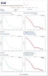
Snapshot of a FoXS output page. Computed profiles of two PDB files are compared to the experimental SAXS profile of malic enzyme (data from
, PF1026). The first structure (pdb_model) includes a model of the unfolded His tag region (35 residues), while the second structure (2dvm) does not. The server was run with the default parameters and the hydration layer modeling was disabled. Plots on the left display the theoretical profiles and plots on the right display their fit to the experimental profile. The top two plots are for the structure with the modeled unfolded region (pdb_model), the middle two plots are for the original PDB file (2dvm), the bottom left plot overlay the profiles for the two input structures, and the bottom right plot shows their fit to the experimental profile. The structure with the modeled unfolded region shows a better fit to the experimental profile with the value of χ = 2.88, compared to χ = 6.33 for the original crystallographic structure. The user can follow the links to download the computed profiles and their fittings.
Similar articles
- FoXS, FoXSDock and MultiFoXS: Single-state and multi-state structural modeling of proteins and their complexes based on SAXS profiles.
Schneidman-Duhovny D, Hammel M, Tainer JA, Sali A. Schneidman-Duhovny D, et al. Nucleic Acids Res. 2016 Jul 8;44(W1):W424-9. doi: 10.1093/nar/gkw389. Epub 2016 May 5. Nucleic Acids Res. 2016. PMID: 27151198 Free PMC article. - Accurate SAXS profile computation and its assessment by contrast variation experiments.
Schneidman-Duhovny D, Hammel M, Tainer JA, Sali A. Schneidman-Duhovny D, et al. Biophys J. 2013 Aug 20;105(4):962-74. doi: 10.1016/j.bpj.2013.07.020. Biophys J. 2013. PMID: 23972848 Free PMC article. - ModBase, a database of annotated comparative protein structure models and associated resources.
Pieper U, Webb BM, Dong GQ, Schneidman-Duhovny D, Fan H, Kim SJ, Khuri N, Spill YG, Weinkam P, Hammel M, Tainer JA, Nilges M, Sali A. Pieper U, et al. Nucleic Acids Res. 2014 Jan;42(Database issue):D336-46. doi: 10.1093/nar/gkt1144. Epub 2013 Nov 23. Nucleic Acids Res. 2014. PMID: 24271400 Free PMC article. - Hybrid Methods for Modeling Protein Structures Using Molecular Dynamics Simulations and Small-Angle X-Ray Scattering Data.
Ekimoto T, Ikeguchi M. Ekimoto T, et al. Adv Exp Med Biol. 2018;1105:237-258. doi: 10.1007/978-981-13-2200-6_15. Adv Exp Med Biol. 2018. PMID: 30617833 Review. - Structural Modeling Using Solution Small-Angle X-ray Scattering (SAXS).
Gräwert TW, Svergun DI. Gräwert TW, et al. J Mol Biol. 2020 Apr 17;432(9):3078-3092. doi: 10.1016/j.jmb.2020.01.030. Epub 2020 Feb 6. J Mol Biol. 2020. PMID: 32035901 Review.
Cited by
- PqqD is a novel peptide chaperone that forms a ternary complex with the radical S-adenosylmethionine protein PqqE in the pyrroloquinoline quinone biosynthetic pathway.
Latham JA, Iavarone AT, Barr I, Juthani PV, Klinman JP. Latham JA, et al. J Biol Chem. 2015 May 15;290(20):12908-18. doi: 10.1074/jbc.M115.646521. Epub 2015 Mar 27. J Biol Chem. 2015. PMID: 25817994 Free PMC article. - Quantitative selection of sample structures in small-angle scattering using Bayesian methods.
Hayashi Y, Katakami S, Kuwamoto S, Nagata K, Mizumaki M, Okada M. Hayashi Y, et al. J Appl Crystallogr. 2024 Jun 18;57(Pt 4):955-965. doi: 10.1107/S1600576724004138. eCollection 2024 Aug 1. J Appl Crystallogr. 2024. PMID: 39108817 Free PMC article. - Comprehensive phylogenetic analysis of the ribonucleotide reductase family reveals an ancestral clade.
Burnim AA, Spence MA, Xu D, Jackson CJ, Ando N. Burnim AA, et al. Elife. 2022 Sep 1;11:e79790. doi: 10.7554/eLife.79790. Elife. 2022. PMID: 36047668 Free PMC article. - Crystal Structure of PKG I:cGMP Complex Reveals a cGMP-Mediated Dimeric Interface that Facilitates cGMP-Induced Activation.
Kim JJ, Lorenz R, Arold ST, Reger AS, Sankaran B, Casteel DE, Herberg FW, Kim C. Kim JJ, et al. Structure. 2016 May 3;24(5):710-720. doi: 10.1016/j.str.2016.03.009. Epub 2016 Apr 7. Structure. 2016. PMID: 27066748 Free PMC article. - Novel bacterial clade reveals origin of form I Rubisco.
Banda DM, Pereira JH, Liu AK, Orr DJ, Hammel M, He C, Parry MAJ, Carmo-Silva E, Adams PD, Banfield JF, Shih PM. Banda DM, et al. Nat Plants. 2020 Sep;6(9):1158-1166. doi: 10.1038/s41477-020-00762-4. Epub 2020 Aug 31. Nat Plants. 2020. PMID: 32868887
References
- Koch MH, Vachette P, Svergun DI. Small-angle scattering: a view on the properties, structures and structural changes of biological macromolecules in solution. Q. Rev. Biophys. 2003;36:147–227. - PubMed
- Petoukhov MV, Svergun DI. Analysis of X-ray and neutron scattering from biomacromolecular solutions. Curr. Opin. Struct. Biol. 2007;17:562–571. - PubMed
- Putnam CD, Hammel M, Hura GL, Tainer JA. X-ray solution scattering (SAXS) combined with crystallography and computation: defining accurate macromolecular structures, conformations and assemblies in solution. Q. Rev. Biophys. 2007;40:191–285. - PubMed
Publication types
MeSH terms
LinkOut - more resources
Full Text Sources
Other Literature Sources
