Metadata matters: access to image data in the real world - PubMed (original) (raw)
. 2010 May 31;189(5):777-82.
doi: 10.1083/jcb.201004104.
Curtis T Rueden, Chris Allan, Jean-Marie Burel, Will Moore, Andrew Patterson, Brian Loranger, Josh Moore, Carlos Neves, Donald Macdonald, Aleksandra Tarkowska, Caitlin Sticco, Emma Hill, Mike Rossner, Kevin W Eliceiri, Jason R Swedlow
Affiliations
- PMID: 20513764
- PMCID: PMC2878938
- DOI: 10.1083/jcb.201004104
Metadata matters: access to image data in the real world
Melissa Linkert et al. J Cell Biol. 2010.
Abstract
Data sharing is important in the biological sciences to prevent duplication of effort, to promote scientific integrity, and to facilitate and disseminate scientific discovery. Sharing requires centralized repositories, and submission to and utility of these resources require common data formats. This is particularly challenging for multidimensional microscopy image data, which are acquired from a variety of platforms with a myriad of proprietary file formats (PFFs). In this paper, we describe an open standard format that we have developed for microscopy image data. We call on the community to use open image data standards and to insist that all imaging platforms support these file formats. This will build the foundation for an open image data repository.
Figures
Figure 1.
Example data in the JCB DataViewer. An example of original image data associated with this paper, viewed in the JCB DataViewer. The image shows the following: a 3D stack of a fixed HeLa cell stained with DAPI (blue), anti-INCENP (red), and anti-tubulin (green), recorded using a wide-field microscope; a time-lapse video of a C. elegans embryo expressing GFP-tubulin, recorded using a multiphoton microscope; a transmission electron microscope (TEM) image of bacteriophages visualized using negative stain; a 3D stack of a fixed HeLa cell stained with anti-tubulin, recorded using an OMX 3D structured illumination microscope; a TEM image of Rb bound to DNA; and a 5D image of GFP-coilin and YFP-histone H2B in a HeLa cell, recorded by wide-field microscopy (Platani et al., 2000). An example view of metadata is included at the bottom left. Note that available metadata differ substantially between the different images, depending on the metadata that are stored in the original files. These images and their associated metadata are available at
http://jcb-dataviewer.rupress.org/jcb/browse/2859/
.
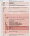
Figure 2.
Recommendations for OME Compliant image metadata. The Image and Instrument Elements from the OME Data Model, with attributes and hierarchies shown in diagrammatic form. The Image Element contains core metadata that can be used for display and processing of the associated binary image data. Currently, an OME Compliant image completes all of the metadata in the Image Element. By the end of 2010, we aim to include the Instrument Element in the OME Compliant specification. The Bio-Formats library provides support for writing OME-XML either as a stand-alone file or within the header of an OME-TIFF file. The full XML Schema version of the OME Data Model is available at
http://ome-xml.org/browser/Schemas/OME/2010-04/ome.xsd
. Updates to the OME Data Model are announced on the project’s roadmap site (
).
Similar articles
- A global view of standards for open image data formats and repositories.
Swedlow JR, Kankaanpää P, Sarkans U, Goscinski W, Galloway G, Malacrida L, Sullivan RP, Härtel S, Brown CM, Wood C, Keppler A, Paina F, Loos B, Zullino S, Longo DL, Aime S, Onami S. Swedlow JR, et al. Nat Methods. 2021 Dec;18(12):1440-1446. doi: 10.1038/s41592-021-01113-7. Nat Methods. 2021. PMID: 33948027 No abstract available. - Towards a repository for standardized medical image and signal case data annotated with ground truth.
Deserno TM, Welter P, Horsch A. Deserno TM, et al. J Digit Imaging. 2012 Apr;25(2):213-26. doi: 10.1007/s10278-011-9428-4. J Digit Imaging. 2012. PMID: 22075810 Free PMC article. - Formats of image data files that can be used for routine digital light micrography. Part one.
Entwistle A. Entwistle A. Biotech Histochem. 2003 Apr;78(2):77-89. doi: 10.1080/10520290310001593810. Biotech Histochem. 2003. PMID: 14533844 - Common file formats.
Leonard SA, Littlejohn TG, Baxevanis AD. Leonard SA, et al. Curr Protoc Bioinformatics. 2007 Jan;Appendix 1:Appendix 1B. doi: 10.1002/0471250953.bia01bs16. Curr Protoc Bioinformatics. 2007. PMID: 18428774 Review. - Visualization approaches for multidimensional biological image data.
Rueden CT, Eliceiri KW. Rueden CT, et al. Biotechniques. 2007 Jul;43(1 Suppl):31, 33-6. doi: 10.2144/000112511. Biotechniques. 2007. PMID: 17936940 Review.
Cited by
- Modality specific roles for metabotropic GABAergic signaling and calcium induced calcium release mechanisms in regulating cold nociception.
Patel AA, Sakurai A, Himmel NJ, Cox DN. Patel AA, et al. Front Mol Neurosci. 2022 Sep 9;15:942548. doi: 10.3389/fnmol.2022.942548. eCollection 2022. Front Mol Neurosci. 2022. PMID: 36157080 Free PMC article. - DeepLIIF: An Online Platform for Quantification of Clinical Pathology Slides.
Ghahremani P, Marino J, Dodds R, Nadeem S. Ghahremani P, et al. Proc IEEE Comput Soc Conf Comput Vis Pattern Recognit. 2022;2022:21399-21405. Proc IEEE Comput Soc Conf Comput Vis Pattern Recognit. 2022. PMID: 36159229 Free PMC article. - Imaging methods are vastly underreported in biomedical research.
Marqués G, Pengo T, Sanders MA. Marqués G, et al. Elife. 2020 Aug 11;9:e55133. doi: 10.7554/eLife.55133. Elife. 2020. PMID: 32780019 Free PMC article. - BioImageXD: an open, general-purpose and high-throughput image-processing platform.
Kankaanpää P, Paavolainen L, Tiitta S, Karjalainen M, Päivärinne J, Nieminen J, Marjomäki V, Heino J, White DJ. Kankaanpää P, et al. Nat Methods. 2012 Jun 28;9(7):683-9. doi: 10.1038/nmeth.2047. Nat Methods. 2012. PMID: 22743773 - Combined Porogen Leaching and Emulsion Templating to produce Bone Tissue Engineering Scaffolds.
Owen R, Sherborne C, Evans R, Reilly GC, Claeyssens F. Owen R, et al. Int J Bioprint. 2020 Apr 30;6(2):265. doi: 10.18063/ijb.v6i2.265. eCollection 2020. Int J Bioprint. 2020. PMID: 32782992 Free PMC article.
References
- Committee on Science, Engineering, and Public Policy (US), and Committee on Ensuring the Utility and Integrity of Research Data in a Digital Age 2009. Ensuring the Integrity, Accessibility, and Stewardship of Research Data in the Digital Age. National Academies Press, Washington, DC: 162 pp - PubMed
- Goldberg I.G., Allan C., Burel J.-M., Creager D., Falconi A., Hochheiser H.S., Johnston J., Mellen J., Sorger P.K., Swedlow J.R. 2005. The Open Microscopy Environment (OME) data model and XML file: open tools for informatics and quantitative analysis in biological imaging. Genome Biol. 6:R47 10.1186/gb-2005-6-5-r47 - DOI - PMC - PubMed
Publication types
MeSH terms
Grants and funding
- 085982/WT_/Wellcome Trust/United Kingdom
- BB/G022585/BB_/Biotechnology and Biological Sciences Research Council/United Kingdom
- BB/D00151X/1/BB_/Biotechnology and Biological Sciences Research Council/United Kingdom
- WT_/Wellcome Trust/United Kingdom
- BB/G022585/1/BB_/Biotechnology and Biological Sciences Research Council/United Kingdom
LinkOut - more resources
Full Text Sources
Other Literature Sources
Miscellaneous

