The bacteriology of pouchitis: a molecular phylogenetic analysis using 16S rRNA gene cloning and sequencing - PubMed (original) (raw)
Comparative Study
The bacteriology of pouchitis: a molecular phylogenetic analysis using 16S rRNA gene cloning and sequencing
Simon D McLaughlin et al. Ann Surg. 2010 Jul.
Abstract
Objective: To identify, compare, and contrast the microbiota in patients with and without pouchitis after restorative proctocolectomy (RPC) for ulcerative colitis (UC) and familial adenomatous polyposis (FAP).
Summary background data: Pouchitis is the most common complication following RPC. An abnormal host-microbial interaction has been implicated. We investigated the pouch microbiota in patients with and without pouchitis undergoing restorative proctocolectomy for UC and FAP.
Methods: Mucosal pouch biopsies, taken from 16 UC (pouchitis 8) and 8 FAP (pouchitis 3) patients were analyzed to the species (or phylotype) level by cloning and sequencing of 3184 full-length bacterial 16S rRNA genes.
Results: There was a significant increase in Proteobacteria (P = 0.019) and a significant decrease in Bacteroidetes (P = 0.001) and Faecalibacterium prausnitzii (P = 0.029) in the total UC compared with the total FAP cohort, but only limited differences were found between the UC nonpouchitis and pouchitis groups and the FAP pouchitis and nonpouchitis groups. Bacterial diversity in the FAP nonpouchitis group was significantly greater than in UC nonpouchitis (P = 0.019) and significantly greater in UC nonpouchitis compared with UC pouchitis (P = 0.009). No individual species or phylotype specifically associated with either UC or FAP pouchitis was found.
Conclusions: UC pouch patients have a different, less diverse, gut microbiota than FAP patients. A further reduction in bacterial diversity but no significant dysbiosis occurs in those with pouchitis. The study suggests that a dysbiosis occurs in the ileal pouch of UC RPC patients which predisposes to, but may not directly cause, pouchitis.
Figures
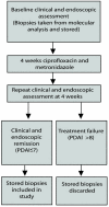
Figure 1
Selection of patient samples from pouchitis group
Figure 2
Box plot comparing the percentage of sequences identified from the four predominant bacterial phyla in UC patient samples compared with FAP patient samples
Figure 3
Box-plot comparing the percentage of sequences identified from the four predominant bacterial phyla in UC pouchitis patient samples compared with UC non-pouchitis patient samples.
Figure 4
Median percentage of clones identified from each bacterial family in samples from UC pouchitis, UC non-pouchitis, FAP pouchitis and FAP non-pouchitis patients. Significant differences at the 5% level are asterisked (full details given in text).
Figure 5
Boxplot comparing the Shannon Diversity index in samples from UC pouchitis patients compared to UC non-pouchitis patient samples.
Figure 6
Box plot comparing the Shannon Diversity index in the total UC cohort and total FAP cohort
Comment in
- Ileal pouch microbial diversity.
Rowan F, Docherty NG, Murphy M, Murphy TB, Coffey JC, O'Connell PR. Rowan F, et al. Ann Surg. 2011 Oct;254(4):669; author reply 669-70. doi: 10.1097/SLA.0b013e3182306578. Ann Surg. 2011. PMID: 21892071 No abstract available.
Similar articles
- Distinct microbiome in pouchitis compared to healthy pouches in ulcerative colitis and familial adenomatous polyposis.
Zella GC, Hait EJ, Glavan T, Gevers D, Ward DV, Kitts CL, Korzenik JR. Zella GC, et al. Inflamm Bowel Dis. 2011 May;17(5):1092-100. doi: 10.1002/ibd.21460. Epub 2010 Sep 15. Inflamm Bowel Dis. 2011. PMID: 20845425 - Bacterial community diversity in cultures derived from healthy and inflamed ileal pouches after restorative proctocolectomy.
Johnson MW, Rogers GB, Bruce KD, Lilley AK, von Herbay A, Forbes A, Ciclitira PJ, Nicholls RJ. Johnson MW, et al. Inflamm Bowel Dis. 2009 Dec;15(12):1803-11. doi: 10.1002/ibd.21022. Epub 2009 Jul 27. Inflamm Bowel Dis. 2009. PMID: 19637361 - Relationship between pouch microbiota and pouchitis following restorative proctocolectomy for ulcerative colitis.
Angriman I, Scarpa M, Castagliuolo I. Angriman I, et al. World J Gastroenterol. 2014 Aug 7;20(29):9665-74. doi: 10.3748/wjg.v20.i29.9665. World J Gastroenterol. 2014. PMID: 25110406 Free PMC article. Review. - Pouch Inflammation Is Associated With a Decrease in Specific Bacterial Taxa.
Reshef L, Kovacs A, Ofer A, Yahav L, Maharshak N, Keren N, Konikoff FM, Tulchinsky H, Gophna U, Dotan I. Reshef L, et al. Gastroenterology. 2015 Sep;149(3):718-27. doi: 10.1053/j.gastro.2015.05.041. Epub 2015 May 27. Gastroenterology. 2015. PMID: 26026389 - Prevalence of pouchitis in both ulcerative colitis and familial adenomatous polyposis: A systematic review and meta-analysis.
Sriranganathan D, Kilic Y, Nabil Quraishi M, Segal JP. Sriranganathan D, et al. Colorectal Dis. 2022 Jan;24(1):27-39. doi: 10.1111/codi.15995. Epub 2021 Dec 3. Colorectal Dis. 2022. PMID: 34800326 Review.
Cited by
- Inflammatory pouch disease: The spectrum of pouchitis.
Zezos P, Saibil F. Zezos P, et al. World J Gastroenterol. 2015 Aug 7;21(29):8739-52. doi: 10.3748/wjg.v21.i29.8739. World J Gastroenterol. 2015. PMID: 26269664 Free PMC article. Review. - Exploring the influence of the gut microbiota and probiotics on health: a symposium report.
Thomas LV, Ockhuizen T, Suzuki K. Thomas LV, et al. Br J Nutr. 2014 Jul;112 Suppl 1(Suppl 1):S1-18. doi: 10.1017/S0007114514001275. Br J Nutr. 2014. PMID: 24953670 Free PMC article. - The Structure and Function of the Human Small Intestinal Microbiota: Current Understanding and Future Directions.
Kastl AJ Jr, Terry NA, Wu GD, Albenberg LG. Kastl AJ Jr, et al. Cell Mol Gastroenterol Hepatol. 2020;9(1):33-45. doi: 10.1016/j.jcmgh.2019.07.006. Epub 2019 Jul 22. Cell Mol Gastroenterol Hepatol. 2020. PMID: 31344510 Free PMC article. Review. - Probiotics for the treatment of inflammatory bowel disease.
Veerappan GR, Betteridge J, Young PE. Veerappan GR, et al. Curr Gastroenterol Rep. 2012 Aug;14(4):324-33. doi: 10.1007/s11894-012-0265-5. Curr Gastroenterol Rep. 2012. PMID: 22581276 Review. - Fecal Microbiota Transplantation in the Treatment of Chronic Pouchitis: A Systematic Review.
Cold F, Kousgaard SJ, Halkjaer SI, Petersen AM, Nielsen HL, Thorlacius-Ussing O, Hansen LH. Cold F, et al. Microorganisms. 2020 Sep 18;8(9):1433. doi: 10.3390/microorganisms8091433. Microorganisms. 2020. PMID: 32962069 Free PMC article. Review.
References
- Sartor RB. Microbial influences in inflammatory bowel diseases. Gastroenterology. 2008;134:577–94. - PubMed
- Schultz M, Sartor RB. Probiotics and inflammatory bowel diseases. Am J Gastroenterol. 2000;95(1 Suppl):S19–S21. - PubMed
- Shen B. Diagnosis and treatment of patients with pouchitis. Drugs. 2003;63:453–61. - PubMed
- McLaughlin SD, Clark SK, Bell AJ, et al. An open study of antibiotics for the treatment of pre-pouch ileitis following restorative proctocolectomy with ileal pouch-anal anastomosis. Aliment Pharmacol Ther. 2008 - PubMed
- Gionchetti P, Rizzello F, Venturi A, et al. Oral bacteriotherapy as maintenance treatment in patients with chronic pouchitis: a double-blind, placebo-controlled trial. Gastroenterology. 2000;119:305–9. - PubMed
Publication types
MeSH terms
Substances
LinkOut - more resources
Full Text Sources
Other Literature Sources
Miscellaneous


