Fibroblast growth factor 21 regulates energy metabolism by activating the AMPK-SIRT1-PGC-1alpha pathway - PubMed (original) (raw)
Fibroblast growth factor 21 regulates energy metabolism by activating the AMPK-SIRT1-PGC-1alpha pathway
Mary D L Chau et al. Proc Natl Acad Sci U S A. 2010.
Abstract
Fibroblast growth factor 21 (FGF21) has been identified as a potent metabolic regulator. Administration of recombinant FGF21 protein to rodents and rhesus monkeys with diet-induced or genetic obesity and diabetes exerts strong antihyperglycemic and triglyceride-lowering effects and reduction of body weight. Despite the importance of FGF21 in the regulation of glucose, lipid, and energy homeostasis, the mechanisms by which FGF21 functions as a metabolic regulator remain largely unknown. Here we demonstrate that FGF21 regulates energy homeostasis in adipocytes through activation of AMP-activated protein kinase (AMPK) and sirtuin 1 (SIRT1), resulting in enhanced mitochondrial oxidative function. AMPK phosphorylation levels were increased by FGF21 treatment in adipocytes as well as in white adipose tissue from ob/ob mice. FGF21 treatment increased cellular NAD(+) levels, leading to activation of SIRT1 and deacetylation of its downstream targets, peroxisome proliferator-activated receptor-gamma coactivator-1alpha (PGC-1alpha) and histone 3. Activation of AMPK and SIRT1 by FGF21 in adipocytes enhanced mitochondrial oxidative capacity as demonstrated by increases in oxygen consumption, citrate synthase activity, and induction of key metabolic genes. The effects of FGF21 on mitochondrial function require serine/threonine kinase 11 (STK11/LKB1), which activates AMPK. Inhibition of AMPK, SIRT1, and PGC-1alpha activities attenuated the effects of FGF21 on oxygen consumption and gene expression, indicating that FGF21 regulates mitochondrial activity and enhances oxidative capacity through an AMPK-SIRT1-PGC1alpha-dependent mechanism in adipocytes.
Conflict of interest statement
The authors declare no conflict of interest.
Figures
Fig. 1.
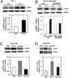
FGF21 increases AMPK activity. (A and B) Western blot and quantification of (A) p-AMPK in 3T3-L1 cells treated for 3 d with FGF21 (4.0 μg/mL) and (B) human adipocytes. Data are average of three independent experiments. (C and D) Western blot and quantification of (C) p-AMPK and (D) p-ACC in WAT from vehicle-treated (white bar), FGF21-treated (gray bar), and paired-fed (black bar) mice. n = 8 animals/group. *P < 0.05 (Student's t test). **P < 0.01.
Fig. 2.
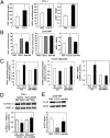
FGF21 increases cellular NAD+ and decreases H3 acetylation. (A) NAD+/NADH levels in 3T3-L1 adipocytes. 3T3-L1 adipocytes were treated for 3 d with FGF21 (4.0 μg/mL; black bars) or PBS (white bars). (B) NAD+/NADH levels in WAT from vehicle-treated (white bar), FGF21-treated (gray bar), and paired-fed (black bar) mice. n = 8 animals/group. (C) NAD+/NADH levels in human adipocytes transduced with adenovirus expressing control vector or DN-AMPK. Transduced adipocytes were treated for 3 d with FGF21 (4.0 μg/mL; black bars) or PBS (white bars). (D) Western blot and quantification of acetylated PGC-1α (Ac-PGC-1α) in 3T3-L1 adipocytes. 3T3-L1 adipocytes were transduced with adenovirus expressing SIRT1-shRNA and Flag–PGC-1α. Flag–PGC-1α was immunoprecipitated and blotted with pan acetylated lysine antibody. t-PGC-1α, total PGC-1α. (E) Western blot and quantification of acetylated H3 (Ac-H3) in WAT from vehicle-treated (white bar), FGF21-treated (gray bar), and paired-fed (black bar) mice. All data are averages of three independent experiments. n = 8 animals/group. *P < 0.05 (Student's t test).
Fig. 3.
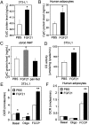
FGF21 increases mitochondrial protein expression and function. CytC protein levels in (A) FGF21-treated 3T3-L1 cells and (B) human adipocytes. White bars: PBS treatment; black bars: FGF21 treatment. (C) CytC protein levels in WAT from vehicle-treated (white bar), FGF21-treated (gray bar), and paired-fed (black bar) mice. n = 8 animals/group. (D) Citrate synthase (CS) activity in 3T3-L1 adipocytes treated with FGF21. Oxygen consumption rate (OCR) in (E) 3T3-L1 cells and (F) human adipocytes treated with PBS (white bars) or FGF21 (black bars). Data are averages of three independent experiments. *P < 0.05; **P < 0.01 (Student's t test).
Fig. 4.
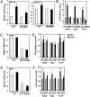
FGF21 effects in adipocytes require AMPK activity. (A) Gene expression in human adipocytes infected with adenovirus expressing DN-AMPK or GFP (Ctrl) and treated with PBS (white bars) or FGF21 (black bars). (B) Oxygen consumption in human adipocytes infected with adenovirus expressing DN-AMPK or GFP (Ctrl) and treated with PBS (white bars) or FGF21 (black bars). (C) Gene expression in human adipocytes infected with lentivirus expressing shRNA against LKB1 or control shRNA (Ctrl) and treated with PBS (white bars) or FGF21 (black bars). (D) Oxygen consumption in 3T3-L1 adipocytes infected with lentivirus expressing shRNA against LKB1 or control shRNA (Ctrl) and treated with PBS (white bars) or FGF21 (black bars). (E) Gene expression in human adipocytes infected with adenovirus expressing shRNA against SIRT1 or control shRNA (Ctrl) and treated with PBS (white bars) or FGF21 (black bars). (F) Oxygen consumption in 3T3-L1 adipocytes infected with adenovirus expressing shRNA against SIRT1 or control shRNA (Ctrl) and treated with PBS (white bars) or FGF21 (black bars). Data are averages of three independent experiments. All gene-expression data are quantitative RT-PCRs, normalized to the housekeeping gene, TATA box binding protein. *P < 0.05; **P < 0.01 (Student's t test). ***P < 0.001.
Fig. 5.
FGF21 effects in adipocytes require PGC-1α. (A) PGC-1α expression in 3T3-L1 adipocytes infected with shRNA-cytomegalovirus (CMV) (Ctrl) or shRNA–PGC-1α adenovirus and treated with PBS (white bars) or FGF21 (black bars). Data shown are from quantitative RT-PCRs, normalized to the housekeeping gene, TATA box binding protein. (B) Oxygen consumption in 3T3-L1 adipocytes infected with shRNA-CMV (Ctrl) or shRNA–PGC-1α adenovirus and treated with PBS (white bars) or FGF21 (black bars). Data are averages of three independent experiments. *P < 0.05; **P < 0.01 (Student's t test). (C) Schematic diagram showing FGF21 activation of AMPK via LKB1, which indirectly activates SIRT1 by increasing the cellular NAD+/NADH ratio. The pathway converges on regulation of PGC-1α activity and, ultimately, on mitochondrial oxidative function.
Similar articles
- The protective effect of FGF21 on diabetes-induced male germ cell apoptosis is associated with up-regulated testicular AKT and AMPK/Sirt1/PGC-1α signaling.
Jiang X, Chen J, Zhang C, Zhang Z, Tan Y, Feng W, Skibba M, Xin Y, Cai L. Jiang X, et al. Endocrinology. 2015 Mar;156(3):1156-70. doi: 10.1210/en.2014-1619. Epub 2015 Jan 5. Endocrinology. 2015. PMID: 25560828 Free PMC article. - The diabetes medication canagliflozin promotes mitochondrial remodelling of adipocyte via the AMPK-Sirt1-Pgc-1α signalling pathway.
Yang X, Liu Q, Li Y, Tang Q, Wu T, Chen L, Pu S, Zhao Y, Zhang G, Huang C, Zhang J, Zhang Z, Huang Y, Zou M, Shi X, Jiang W, Wang R, He J. Yang X, et al. Adipocyte. 2020 Dec;9(1):484-494. doi: 10.1080/21623945.2020.1807850. Adipocyte. 2020. PMID: 32835596 Free PMC article. - Skeletal muscle increases FGF21 expression in mitochondrial disorders to compensate for energy metabolic insufficiency by activating the mTOR-YY1-PGC1α pathway.
Ji K, Zheng J, Lv J, Xu J, Ji X, Luo YB, Li W, Zhao Y, Yan C. Ji K, et al. Free Radic Biol Med. 2015 Jul;84:161-170. doi: 10.1016/j.freeradbiomed.2015.03.020. Epub 2015 Apr 3. Free Radic Biol Med. 2015. PMID: 25843656 - Antagonistic crosstalk between NF-κB and SIRT1 in the regulation of inflammation and metabolic disorders.
Kauppinen A, Suuronen T, Ojala J, Kaarniranta K, Salminen A. Kauppinen A, et al. Cell Signal. 2013 Oct;25(10):1939-48. doi: 10.1016/j.cellsig.2013.06.007. Epub 2013 Jun 11. Cell Signal. 2013. PMID: 23770291 Review. - PGC-1alpha, SIRT1 and AMPK, an energy sensing network that controls energy expenditure.
Cantó C, Auwerx J. Cantó C, et al. Curr Opin Lipidol. 2009 Apr;20(2):98-105. doi: 10.1097/MOL.0b013e328328d0a4. Curr Opin Lipidol. 2009. PMID: 19276888 Free PMC article. Review.
Cited by
- From fasting to fat reshaping: exploring the molecular pathways of intermittent fasting-induced adipose tissue remodeling.
Vo N, Zhang Q, Sung HK. Vo N, et al. J Pharm Pharm Sci. 2024 Jul 22;27:13062. doi: 10.3389/jpps.2024.13062. eCollection 2024. J Pharm Pharm Sci. 2024. PMID: 39104461 Free PMC article. Review. - The protective effect of FGF21 on diabetes-induced male germ cell apoptosis is associated with up-regulated testicular AKT and AMPK/Sirt1/PGC-1α signaling.
Jiang X, Chen J, Zhang C, Zhang Z, Tan Y, Feng W, Skibba M, Xin Y, Cai L. Jiang X, et al. Endocrinology. 2015 Mar;156(3):1156-70. doi: 10.1210/en.2014-1619. Epub 2015 Jan 5. Endocrinology. 2015. PMID: 25560828 Free PMC article. - Betaine prevented high-fat diet-induced NAFLD by regulating the FGF10/AMPK signaling pathway in ApoE-/- mice.
Chen W, Zhang X, Xu M, Jiang L, Zhou M, Liu W, Chen Z, Wang Y, Zou Q, Wang L. Chen W, et al. Eur J Nutr. 2021 Apr;60(3):1655-1668. doi: 10.1007/s00394-020-02362-6. Epub 2020 Aug 17. Eur J Nutr. 2021. PMID: 32808060 - Emerging role of AMP-activated protein kinase in endocrine control of metabolism in the liver.
Hasenour CM, Berglund ED, Wasserman DH. Hasenour CM, et al. Mol Cell Endocrinol. 2013 Feb 25;366(2):152-62. doi: 10.1016/j.mce.2012.06.018. Epub 2012 Jul 14. Mol Cell Endocrinol. 2013. PMID: 22796337 Free PMC article. Review. - The Effect of Endotoxin-Induced Inflammation on the Activity of the Somatotropic Axis in Sheep.
Wójcik M, Zieba DA, Bochenek J, Krawczyńska A, Barszcz M, Gajewska A, Antushevich H, Herman AP. Wójcik M, et al. Int J Inflam. 2024 Aug 7;2024:1057299. doi: 10.1155/2024/1057299. eCollection 2024. Int J Inflam. 2024. PMID: 39149693 Free PMC article.
References
- Hardie DG. AMP-activated/SNF1 protein kinases: Conserved guardians of cellular energy. Nat Rev Mol Cell Biol. 2007;8:774–785. - PubMed
- Fryer LGD, Parbu-Patel A, Carling D. The anti-diabetic drugs rosiglitazone and metformin stimulate AMP-activated protein kinase through distinct signaling pathways. J Biol Chem. 2002;277:25226–25232. - PubMed
MeSH terms
Substances
LinkOut - more resources
Full Text Sources
Other Literature Sources
Molecular Biology Databases
Miscellaneous


