Effects of estrogen and aging on the synaptic distribution of phosphorylated Akt-immunoreactivity in the CA1 region of the female rat hippocampus - PubMed (original) (raw)
Comparative Study
Effects of estrogen and aging on the synaptic distribution of phosphorylated Akt-immunoreactivity in the CA1 region of the female rat hippocampus
Murat Yildirim et al. Brain Res. 2011.
Abstract
The estrogen 17β-estradiol (E) increases the axospinous synaptic density and plasticity in the hippocampal CA1 region of young female rats but fails to do so in aged female rats. This E stimulus on synaptic plasticity is associated with the phosphorylation-dependent activation of Akt kinase. Our previous findings demonstrated that increased estrogen levels subsequently increase phosphorylated Akt (pAkt)-immunoreactivity (-IR) within the dendritic shafts and spines of pyramidal neurons in young female rats. Therefore, because Akt can promote cell survival and growth, we tested the hypothesis that the less plastic synapses of aged female rats would contain less E-stimulated pAkt-IR. Here, young (3-4 months) and aged (22-23 months) female rats were ovariectomized 7 days prior to a 48-h administration of either vehicle or E. The pAkt-IR synaptic distribution was then analyzed using post-embedding electron microscopy. In both young and aged rats, pAkt-IR was found in dendritic spines and terminals, and pAkt-IR was particularly abundant at the post-synaptic density. Quantitative analyses revealed that the percentage of pAkt-labeled synapses was significantly greater in young rats compared to aged rats. Nonetheless, E treatment significantly increased pAkt-IR in pre- and post-synaptic profiles of both young and aged rats, although the stimulus in young rats was notably more widespread. These data support the evidence that hormone-activated signaling associated with cell growth and survival is diminished in the aged brain. However, the observation that E can still increase pAkt-IR in aged synapses presents this signaling component as a candidate target for hormone replacement therapies.
Copyright © 2010. Published by Elsevier B.V.
Figures
Fig. 1
Representative electron micrographs show the distribution of pAkt-IR (black particles) in the CA1 stratum radiatum of young OVX + estrogen (E) (A), young OVX + vehicle (B), aged OVX + E (C) and aged OVX + vehicle (D) rats. t, terminal; sp, dendritic spine. Bar, 100 nm.
Fig. 2
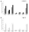
The overall percentage of pAkt immunoreactive axospinous synapses is significantly decreased in aged OVX rats compared to young OVX rats and significantly increased following E-replacement in both young and aged OVX rats. *p < 0.02.
Fig. 3
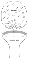
Schematic drawing showing the eight bin divisions of a pre- and post-synaptic profile.
Fig 4
Both age and estrogen altered the subcellular distribution of pAkt immunogold particles in pre- and post-synaptic profiles in the CA1 stratum radiatum region. A. In young OVX rats, E administration significantly increased the number of pAkt immunogold particles in all synaptic bins except 6 and 7 (although there was a tends in bin 7, p = 0.05). B. In aged OVX rats, E administration significantly increased the number of pAkt immunogold particles only in bins 1, 2 and 6. Moreover, E administration to aged OVX animals significantly decreased the number of pAkt immunogold particles in bin 4. *p < 0.05
Fig. 5
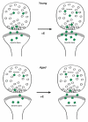
Schematic diagram showing the effects of age and estrogen replacement on the distribution of pAkt in synaptic complexes of the female rat stratum radiatum CA1 region. Synaptic complexes from young OVX rats have more pAkt-IR, especially in the post-synaptic region compared to aged OVX rats. Following E treatment, pAkt-IR is increased in synaptic complexes in both young and aged OVX rats, although the stimulus in young rats is more widespread.
Similar articles
- Estrogen and aging affect the synaptic distribution of estrogen receptor β-immunoreactivity in the CA1 region of female rat hippocampus.
Waters EM, Yildirim M, Janssen WG, Lou WY, McEwen BS, Morrison JH, Milner TA. Waters EM, et al. Brain Res. 2011 Mar 16;1379:86-97. doi: 10.1016/j.brainres.2010.09.069. Epub 2010 Sep 25. Brain Res. 2011. PMID: 20875808 Free PMC article. - Estrogen and aging affect synaptic distribution of phosphorylated LIM kinase (pLIMK) in CA1 region of female rat hippocampus.
Yildirim M, Janssen WG, Tabori NE, Adams MM, Yuen GS, Akama KT, McEwen BS, Milner TA, Morrison JH. Yildirim M, et al. Neuroscience. 2008 Mar 18;152(2):360-70. doi: 10.1016/j.neuroscience.2008.01.004. Epub 2008 Jan 12. Neuroscience. 2008. PMID: 18294775 Free PMC article. - Estrogen levels regulate the subcellular distribution of phosphorylated Akt in hippocampal CA1 dendrites.
Znamensky V, Akama KT, McEwen BS, Milner TA. Znamensky V, et al. J Neurosci. 2003 Mar 15;23(6):2340-7. doi: 10.1523/JNEUROSCI.23-06-02340.2003. J Neurosci. 2003. PMID: 12657693 Free PMC article. - Interactive effects of age and estrogen on cortical neurons: implications for cognitive aging.
Bailey ME, Wang AC, Hao J, Janssen WG, Hara Y, Dumitriu D, Hof PR, Morrison JH. Bailey ME, et al. Neuroscience. 2011 Sep 15;191:148-58. doi: 10.1016/j.neuroscience.2011.05.045. Epub 2011 Jun 2. Neuroscience. 2011. PMID: 21664255 Free PMC article. Review. - Estradiol and the relationship between dendritic spines, NR2B containing NMDA receptors, and the magnitude of long-term potentiation at hippocampal CA3-CA1 synapses.
Smith CC, Vedder LC, McMahon LL. Smith CC, et al. Psychoneuroendocrinology. 2009 Dec;34 Suppl 1:S130-42. doi: 10.1016/j.psyneuen.2009.06.003. Psychoneuroendocrinology. 2009. PMID: 19596521 Free PMC article. Review.
Cited by
- Sex Differences in Neuroplasticity- and Stress-Related Gene Expression and Protein Levels in the Rat Hippocampus Following Oxycodone Conditioned Place Preference.
Randesi M, Contoreggi NH, Zhou Y, Rubin BR, Bellamy JR, Yu F, Gray JD, McEwen BS, Milner TA, Kreek MJ. Randesi M, et al. Neuroscience. 2019 Jul 1;410:274-292. doi: 10.1016/j.neuroscience.2019.04.047. Epub 2019 May 7. Neuroscience. 2019. PMID: 31071414 Free PMC article. - Estrogen receptors in the central nervous system and their implication for dopamine-dependent cognition in females.
Almey A, Milner TA, Brake WG. Almey A, et al. Horm Behav. 2015 Aug;74:125-38. doi: 10.1016/j.yhbeh.2015.06.010. Epub 2015 Jun 27. Horm Behav. 2015. PMID: 26122294 Free PMC article. Review. - Inositol pyrophosphates mediate the effects of aging on bone marrow mesenchymal stem cells by inhibiting Akt signaling.
Zhang Z, Zhao C, Liu B, Liang D, Qin X, Li X, Zhang R, Li C, Wang H, Sun D, Cao F. Zhang Z, et al. Stem Cell Res Ther. 2014 Mar 26;5(2):33. doi: 10.1186/scrt431. Stem Cell Res Ther. 2014. PMID: 24670364 Free PMC article. - Sex differences in stress-related psychiatric disorders: neurobiological perspectives.
Bangasser DA, Valentino RJ. Bangasser DA, et al. Front Neuroendocrinol. 2014 Aug;35(3):303-19. doi: 10.1016/j.yfrne.2014.03.008. Epub 2014 Apr 12. Front Neuroendocrinol. 2014. PMID: 24726661 Free PMC article. Review. - Intriguing roles of hippocampus-synthesized 17β-estradiol in the modulation of hippocampal synaptic plasticity.
Bian C, Zhu H, Zhao Y, Cai W, Zhang J. Bian C, et al. J Mol Neurosci. 2014;54(2):271-81. doi: 10.1007/s12031-014-0285-8. Epub 2014 Apr 15. J Mol Neurosci. 2014. PMID: 24729128 Review.
References
- Adams MM, Morrison JH, Gore AC. N-methyl-D-aspartate receptor mRNA levels change during reproductive senescence in the hippocampus of female rats. Exp Neurol. 2001a;170:171–9. - PubMed
- Adams MM, Oung T, Morrison JH, Gore AC. Length of postovariectomy interval and age, but not estrogen replacement, regulate N-methyl-D-aspartate receptor mRNA levels in the hippocampus of female rats. Exp Neurol. 2001b;170:345–56. - PubMed
- Adams MM, Morrison JH. Estrogen and the aging hippocampal synapse. Cereb Cortex. 2003;13:1271–5. - PubMed
Publication types
MeSH terms
Substances
Grants and funding
- NS07080/NS/NINDS NIH HHS/United States
- DA08259/DA/NIDA NIH HHS/United States
- R01 NS007080/NS/NINDS NIH HHS/United States
- R01 DA008259/DA/NIDA NIH HHS/United States
- P01-AG16765/AG/NIA NIH HHS/United States
- P01 AG016765/AG/NIA NIH HHS/United States
LinkOut - more resources
Full Text Sources
Medical
Miscellaneous


