microRNA miR-144 modulates oxidative stress tolerance and associates with anemia severity in sickle cell disease - PubMed (original) (raw)
microRNA miR-144 modulates oxidative stress tolerance and associates with anemia severity in sickle cell disease
Carolyn Sangokoya et al. Blood. 2010.
Abstract
Although individuals with homozygous sickle cell disease (HbSS) share the same genetic mutation, the severity and manifestations of this disease are extremely heterogeneous. We have previously shown that the microRNA expression in normal and HbSS erythrocytes exhibit dramatic differences. In this study, we identify a subset of HbSS patients with higher erythrocytic miR-144 expression and more severe anemia. HbSS erythrocytes are known to have reduced tolerance for oxidative stress, yet the basis for this phenotype remains unknown. This study reveals that miR-144 directly regulates nuclear factor-erythroid 2-related factor 2, a central regulator of cellular response to oxidative stress, and modulates the oxidative stress response in K562 cell line and primary erythroid progenitor cells. We further demonstrate that increased miR-144 is associated with reduced NRF2 levels in HbSS reticulocytes and with decreased glutathione regeneration and attenuated antioxidant capacity in HbSS erythrocytes, thereby providing a possible mechanism for the reduced oxidative stress tolerance and increased anemia severity seen in HbSS patients. Taken together, our findings suggest that erythroid microRNAs can serve as genetic modifiers of HbS-related anemia and can provide novel insights into the clinical heterogeneity and pathobiology of sickle cell disease.
Figures
Figure 1
Erythrocyte microRNA expression identifies SCD subtypes. (A) The separation of all HbSS patients into 2 groups (HbSS group I, brown, and HbSS group II, red) based on unsupervised analysis of the erythrocyte microRNA expression pattern. (B) The mean expression values of the 83 reticulocyte microRNAs were not significantly different between the 2 HbSS groups (P = .87). (C) The average reticulocyte percentages for group I were significantly higher than group II HbSS patients (P = .001). (D) HbSS subtype classification: training errors are shown as a function of the threshold parameter δ. The value δ = 4.34 is chosen and yields a subset of 6 selected genes and leads to zero error rate. (E) The heat map showing the expression values for the 6 top selected genes in the HbSS groups I and II.
Figure 2
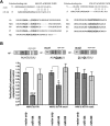
NRF2 is a direct regulatory target of miR-144. (A) Sequence alignment and evolutionary conservation between miR-144 and its 2 putative binding sites in the 3′UTR of NRF2 mRNA from several indicated species, with sequences recognized by miR-144 seed sequence shown in bold. (B) NRF2 3′UTR luciferase reporters (upper panel) were cotransfected with empty vector, miR-144, or miR-320 expression constructs (detailed in supplemental Figure 3). The overexpression of miR-144 significantly repressed the luciferase activity of a NRF2 3′UTR reporter construct. While mutation of the first miR-144 binding site (mut1) only modestly decreased the miR-144–mediated repression, mutation of the second site (mut 2) abolished this effect. MiR-320 does not contain a NRF2 3′UTR binding site and did not significantly alter the luciferase activity of any of the 3 reporters compared with empty vector control. *Significantly different by Student t test: *P ≤ .05, ***P ≤ .0001.
Figure 3
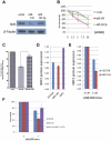
MiR-144 modulates cellular response to oxidative stress. (A) Western blot analysis of NRF2 protein levels after the overexpression of miR-144 and miR-142-5p in K562 cells and H2O2-induced oxidative stress. (B) MiR-144 overexpression leads to significantly increased sensitivity to oxidative stress at indicated concentrations of H2O2 as measured by MTS assay. Each curve represents the indicated sample relative to its unstressed (baseline) control. (C) MiR-144–mediated sensitivity to H2O2 oxidative stress is partially rescued by NRF2 overexpression. (D) ELISA analysis of baseline NRF2 protein levels in primary erythroid cells after miR-144 overexpression compared with vector control. NRF2 overexpression is shown as a positive control. (E) ELISA analysis of NRF2 protein levels demonstrates decreased NRF2 levels after H2O2-induced oxidative stress in the miR-144 transfected primary erythroid cells compared with vector control. (F) MiR-144–mediated sensitivity to H2O2 oxidative stress is fully rescued by NRF2 overexpression without its 3′UTR. *Significantly different by Student t test: *P ≤ .05.
Figure 4
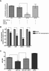
MiR-144 reduces ARE-driven gene expression and cellular GSH levels. (A) MiR-144, but not miR-320, repressed reporter activity of an antioxidant response element (ARE) luciferase construct in K562 cells. (B) MiR-144 overexpression decreased gene expression of indicated ARE-driven genes and (C) led to significant reduction in cellular GSH after H2O2-induced oxidative stress. This GSH reduction is partially rescued by exogenous NRF2 expression. *Significantly different by Student t test: *P ≤ .05, **P ≤ .001, ***P ≤ .0001.
Figure 5
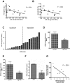
MiR-144 expression levels correlate with antioxidant protein expression in HbSS mature erythrocytes. (A) Hemoglobin and (B) hematocrit levels correlated inversely with miR-144 expression in mature erythrocytes. (C) Eighteen HbSS samples were grouped into 2 groups based on miR-144 expression. (D) GCLC and (E) GCLM intracellular protein expression in mature erythrocytes were compared and shown as mean fluorescence intensity (MFI) for samples from each group (n = 5 in panel D, n = 4 in panel E). (F) HbSS erythrocytes (n = 8) expressed significantly decreased SOD activity compared with normal HbAA erythrocytes (n = 4). (G) MiR-144 expression in HbSS erythrocytes had a negative correlation with superoxide dismutase activity. Each HbSS sample miR-144 expression (fold relative to HbAA) is shown with corresponding SOD activity.
Figure 6
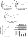
Correlation of miR-144 and NRF2 protein level in HbSS reticulocytes and during in vitro differentiation. (A) Hemoglobin and (B) hematocrit levels were inversely correlated with miR-144 expression in an independent cohort of reticulocyte samples from 25 HbSS patients. (C) NRF2 protein levels measured by NRF2 ELISA assay in HbSS reticulocytes (n = 15). (D) The induction of miR-144 expression during indicated days of erythroid maturation of HbSS CD34+ progenitors. (E) NRF2 protein levels (upper panel) with densitometric analysis (lower panel) during the corresponding days of erythroid maturation in HbSS progenitors were determined by Western blot and normalized to glyceraldehyde 3-phosphate dehydrogenase. Glycophorin A expression is shown as an additional control for erythroid maturation.
Figure 7
Proposed model for biologic consequences of miR-144 overexpression in SCD. We propose that miR-144 is a genetic modifier of oxidative stress tolerance in HbSS erythrocytes and can contribute to the varying clinical severity of SCD.
Comment in
- Micro-mismanaging sickle cell stress.
Wojchowski DM. Wojchowski DM. Blood. 2010 Nov 18;116(20):4040-2. doi: 10.1182/blood-2010-09-304386. Blood. 2010. PMID: 21088142 No abstract available.
Similar articles
- MIR-144-mediated NRF2 gene silencing inhibits fetal hemoglobin expression in sickle cell disease.
Li B, Zhu X, Ward CM, Starlard-Davenport A, Takezaki M, Berry A, Ward A, Wilder C, Neunert C, Kutlar A, Pace BS. Li B, et al. Exp Hematol. 2019 Feb;70:85-96.e5. doi: 10.1016/j.exphem.2018.11.002. Epub 2018 Nov 6. Exp Hematol. 2019. PMID: 30412705 Free PMC article. - Proteasome inhibition induces both antioxidant and hb f responses in sickle cell disease via the nrf2 pathway.
Pullarkat V, Meng Z, Tahara SM, Johnson CS, Kalra VK. Pullarkat V, et al. Hemoglobin. 2014;38(3):188-95. doi: 10.3109/03630269.2014.898651. Epub 2014 Mar 26. Hemoglobin. 2014. PMID: 24670032 Clinical Trial. - miR-144 regulates oxidative stress tolerance of thalassemic erythroid cell via targeting NRF2.
Srinoun K, Sathirapongsasuti N, Paiboonsukwong K, Sretrirutchai S, Wongchanchailert M, Fucharoen S. Srinoun K, et al. Ann Hematol. 2019 Sep;98(9):2045-2052. doi: 10.1007/s00277-019-03737-4. Epub 2019 Jun 26. Ann Hematol. 2019. PMID: 31243572 - Crosstalk between NRF2 and Dicer through metastasis regulating MicroRNAs; mir-34a, mir-200 family and mir-103/107 family.
Nabih HK. Nabih HK. Arch Biochem Biophys. 2020 Jun 15;686:108326. doi: 10.1016/j.abb.2020.108326. Epub 2020 Mar 3. Arch Biochem Biophys. 2020. PMID: 32142889 Review. - Regulation of the Nrf2 antioxidant pathway by microRNAs: New players in micromanaging redox homeostasis.
Cheng X, Ku CH, Siow RC. Cheng X, et al. Free Radic Biol Med. 2013 Sep;64:4-11. doi: 10.1016/j.freeradbiomed.2013.07.025. Epub 2013 Jul 21. Free Radic Biol Med. 2013. PMID: 23880293 Review.
Cited by
- The Role of NRF2 in Trinucleotide Repeat Expansion Disorders.
Chang KH, Chen CM. Chang KH, et al. Antioxidants (Basel). 2024 May 26;13(6):649. doi: 10.3390/antiox13060649. Antioxidants (Basel). 2024. PMID: 38929088 Free PMC article. Review. - MicroRNAs: potential mediators and biomarkers of diabetic complications.
Kato M, Castro NE, Natarajan R. Kato M, et al. Free Radic Biol Med. 2013 Sep;64:85-94. doi: 10.1016/j.freeradbiomed.2013.06.009. Epub 2013 Jun 12. Free Radic Biol Med. 2013. PMID: 23770198 Free PMC article. Review. - Common patterns and disease-related signatures in tuberculosis and sarcoidosis.
Maertzdorf J, Weiner J 3rd, Mollenkopf HJ; TBornotTB Network; Bauer T, Prasse A, Müller-Quernheim J, Kaufmann SH. Maertzdorf J, et al. Proc Natl Acad Sci U S A. 2012 May 15;109(20):7853-8. doi: 10.1073/pnas.1121072109. Epub 2012 Apr 30. Proc Natl Acad Sci U S A. 2012. PMID: 22547807 Free PMC article. - Targeting Genetic Modifiers of HBG Gene Expression in Sickle Cell Disease: The miRNA Option.
Starlard-Davenport A, Gu Q, Pace BS. Starlard-Davenport A, et al. Mol Diagn Ther. 2022 Sep;26(5):497-509. doi: 10.1007/s40291-022-00589-z. Epub 2022 May 12. Mol Diagn Ther. 2022. PMID: 35553407 Free PMC article. Review. - MicroRNAs miR-451a and Let-7i-5p Profiles in Circulating Exosomes Vary among Individuals with Different Sickle Hemoglobin Genotypes and Malaria.
Oxendine Harp K, Bashi A, Botchway F, Dei-Adomakoh Y, Iqbal SA, Wilson MD, Adjei AA, Stiles JK, Driss A. Oxendine Harp K, et al. J Clin Med. 2022 Jan 19;11(3):500. doi: 10.3390/jcm11030500. J Clin Med. 2022. PMID: 35159951 Free PMC article.
References
- Platt OS, Brambilla DJ, Rosse WF, et al. Mortality in sickle cell disease. Life expectancy and risk factors for early death. N Engl J Med. 1994;330(23):1639–1644. - PubMed
- Gill FM, Sleeper LA, Weiner SJ, et al. Clinical events in the first decade in a cohort of infants with sickle cell disease. Cooperative Study of Sickle Cell Disease [see comments]. Blood. 1995;86(2):776–783. - PubMed
- Castro O, Brambilla DJ, Thorington B, et al. The acute chest syndrome in sickle cell disease: incidence and risk factors. The Cooperative Study of Sickle Cell Disease. Blood. 1994;84(2):643–649. - PubMed
- Vichinsky EP, Styles LA, Colangelo LH, et al. Acute chest syndrome in sickle cell disease: clinical presentation and course. Blood. 1997;89(5):1787–1792. - PubMed
- Kensler TW, Wakabayashi N, Biswal S. Cell survival responses to environmental stresses via the Keap1-Nrf2-ARE pathway. Annu Rev Pharmacol Toxicol. 2007;47:89–116. - PubMed
Publication types
MeSH terms
Substances
Grants and funding
- R01 HL079915/HL/NHLBI NIH HHS/United States
- R21 DK080994/DK/NIDDK NIH HHS/United States
- R01HL079915/HL/NHLBI NIH HHS/United States
- R21DK080994/DK/NIDDK NIH HHS/United States
LinkOut - more resources
Full Text Sources
Other Literature Sources
Medical

