Glutamate receptor ion channels: structure, regulation, and function - PubMed (original) (raw)
Review
Glutamate receptor ion channels: structure, regulation, and function
Stephen F Traynelis et al. Pharmacol Rev. 2010 Sep.
Erratum in
- Pharmacol Rev. 2014 Oct;66(4):1141
Abstract
The mammalian ionotropic glutamate receptor family encodes 18 gene products that coassemble to form ligand-gated ion channels containing an agonist recognition site, a transmembrane ion permeation pathway, and gating elements that couple agonist-induced conformational changes to the opening or closing of the permeation pore. Glutamate receptors mediate fast excitatory synaptic transmission in the central nervous system and are localized on neuronal and non-neuronal cells. These receptors regulate a broad spectrum of processes in the brain, spinal cord, retina, and peripheral nervous system. Glutamate receptors are postulated to play important roles in numerous neurological diseases and have attracted intense scrutiny. The description of glutamate receptor structure, including its transmembrane elements, reveals a complex assembly of multiple semiautonomous extracellular domains linked to a pore-forming element with striking resemblance to an inverted potassium channel. In this review we discuss International Union of Basic and Clinical Pharmacology glutamate receptor nomenclature, structure, assembly, accessory subunits, interacting proteins, gene expression and translation, post-translational modifications, agonist and antagonist pharmacology, allosteric modulation, mechanisms of gating and permeation, roles in normal physiological function, as well as the potential therapeutic use of pharmacological agents acting at glutamate receptors.
Figures
Fig. 1.
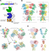
Structure and domain organization of glutamate receptors. A, linear representation of the subunit polypeptide chain and schematic illustration of the subunit topology. Glutamate receptor subunits have a modular structure composed of two large extracellular domains [the ATD (green) and the LBD (blue)]; a TMD (orange) that forms part of the ion channel pore; and an intracellular CTD. The LBD is defined by two segments of amino acids termed S1 and S2. The TMD contains three membrane-spanning helices (M1, M3, and M4) and a membrane re-entrant loop (M2). The isolated S1 and S2 segments have been constructed by deleting the ATD along with the TMD and joining S1 and S2 with a hydrophilic linker (dotted line). SP, signal peptide. B, crystal structure at 3.6 Å of the membrane-spanning tetrameric GluA2 AMPA receptor (PDB code
3KG2
). C, subunit interfaces between the ATD, LBD, and TMD of the four subunits in the membrane-spanning tetrameric GluA2 AMPA receptor. The subunits are viewed from top down the 2-fold axis of symmetry. The ATDs and LBDs have a 2-fold axis of symmetry, whereas the TMDs have 4-fold axis of symmetry. D, the symmetry mismatch between the TMDs and the extracellular domains (ATDs and LBDs) as well as the subunit crossover (or domain swapping) from the LBD to the ATD give rise to two distinct types of subunits in the homotetrameric GluA2 receptor with two distinct conformations. The subunits are referred to as the A/C and B/D subunits. [Adapted from Sobolevsky AI, Rosconi MP, and Gouaux E (2009) X-ray structure, symmetry and mechanism of an AMPA-subtype glutamate receptor. Nature **462:**745–756. Copyright © 2009 Nature Publishing Group. Used with permission.]
Fig. 2.
Alignments of agonist-binding residues of glutamate receptor subunits. Residue numbering is according to the total protein including the signal peptide (initiating methionine is 1). For reference, the predicted size of the signal peptide (SP) is included in parenthesis at the end of the alignment. Amino acid numbering in AMPA and kainate receptor subunits has historically been for the mature protein without the signal peptide, whereas amino acid numbering of NMDA and GluD receptor subunits has started with the initiating methionine as 1. Fully conserved residues are yellow, conserved residues are blue, and similar residues are green. # denotes residues capable of forming hydrogen bonds or electrostatic interactions with the agonist; + denotes residues capable of forming van der Waals contacts with the agonist.
Fig. 3.
Conformational changes in the functioning AMPA receptor. Ribbon diagrams of the crystal structures of the GluA2 LBD dimer in conformations that correspond to the resting state (apo form; PDB code
1FT0
), active state (glutamate-bound; PDB code
1FTJ
) and desensitized state (glutamate-bound; PDB code
2I3V
). In these structures, the LBD exists in a bilobed clamshell-like arrangement with the agonist-binding pocket located deep within the cleft between the two lobes referred to as D1 and D2. Binding of glutamate induces a transition of D2 that leads to separation of the linker segments that replace the TMDs in the full-length subunits (represented here by cylinders). The NTD and CTD are omitted for clarity. Distances between the linkers that face the TMD and distances between a glycine residue (Gly739) at the top of the dimer are taken from Armstrong et al. (2006). Upon glutamate binding and domain closure, separation of the linkers can result in reorientation of the transmembrane helices and opening of the ion channel. The active, nondesensitized receptor conformation is unstable, and stability can be restored either by reopening of the ABD or by rearrangement at the dimer interface. Rearrangement at the dimer interface results in desensitization by repositioning the transmembrane helices such that the ion channel is closed.
Fig. 4.
Schematic diagram of the proximal promoter regulatory regions of glutamate receptors. The proximal promoter regions of the GluN1, GluN2A, GluN2B, and GluN2C NMDA receptors, the GluA1 and GluA2 AMPA receptors, and the GluK5 kainate receptor are shown. Promoters are shown as thin lines and introns as thin lines with hashmarks. The 5′-untranslated exon sequences are represented by open bars; blackened bars designate the protein coding domains. Glutamate receptor regulatory elements are identified; those requiring further confirmation are in parentheses. The promoter regions are not drawn to scale.
Fig. 5.
Post-translational modifications of AMPA and kainate receptor C-terminal domains. Multiple forms of post-translational modifications (including palmitoylation, phosphorylation, and SUMOylation) that influence receptor trafficking, channel activity, and interactions with other proteins are shown. The C-terminal domains of GluA1–4 and GluK1–5 given in the center column. The left column contains the receptor subunit with the UniProt-SwissProt human accession number. The length of the subunit, including the signal peptide, is given in the column at right, with residue numbering beginning with the initiating methionine. The beginning of the CTD is defined by hydrophobicity analyses. Modified residues are in red, with the enzyme (if known) indicated by a symbol above the residue. When no enzyme is given, the modification has been identified through fragmentation and mass spectrometry (Munton et al., 2007; Ballif et al., 2008; Trinidad et al., 2008).
Fig. 6.
Post-translational modifications of GluN1 and GluN2A NMDA receptor C-terminal domains. The GluN1 and GluN2A NMDA receptor subunits undergo the indicated post-translational modification. The left column is the NMDA receptor subunit with the UniProt-SwissProt human accession number. The C-terminal domains of the GluN1 and GluN2A subunits are listed in the center column. The length of the receptor subunit is given in the right column, the numbering beginning with the initiating methionine. The beginning of the CTD is defined by hydrophobicity analyses. Modified residues are in red, with the enzyme (when known) indicated by a symbol above.
Fig. 7.
Post-translational modifications of the GluN2B-D NMDA and GluD1–2 δ receptor C-terminal domains. The post-translational modifications of the NMDA and δ receptors are shown in red. The enzymes mediating the modifications are identified (when known) by a symbol above. When no enzyme is designated, the modification has been identified by fragmentation and mass spectrometry. The left column is the receptor subunit and the UniProt-SwissProt human accession number. The C-terminal domains are shown in the center column, and the length of the subunit, beginning with the initiating methionine, is in the right column. The beginning of the CTD is defined by hydrophobicity analyses.
Fig. 8.
Agonist binding pockets of glutamate receptors. A, binding of glutamate (yellow) in the agonist binding pocket of GluA2 (PDB code
1FTJ
). Only side chains of interacting residues are shown. Not all residues are labeled. B, binding of glutamate in GluK2 (PDB code
1S7Y
). Compared with the glutamate-bound ligand binding pocket of GluA2, there is a loss of a direct hydrogen bond to the α-amino group of glutamate at position Ala487 in GluK2, which is the site equivalent to Thr480 in GluA2. An additional water molecule forms a hydrogen bond to the α-amino group of glutamate in GluK2. C, binding of glutamate in GluN2A (PDB code
2A5T
). Compared with glutamate bound in GluA2, the salt bridge between Asp731 and the positively charged α-amino group of glutamate is absent. Instead, the α-amino group of glutamate forms water-mediated hydrogen bonds to Glu413 and Tyr761. D, binding of glycine in GluN1 (PDB code
2A5T
). Specificity of GluN1 for glycine can be explained by the hydrophobic environment created by Val689 and the steric barrier formed by Trp731. E, binding of glycine in GluN3A (PDB code
2RC7
). Trp731 of GluN1 is replaced by M844, allowing room for a water molecule in the pocket. F, binding of
d
-serine in GluD2 (PDB code
2V3U
).
Fig. 9.
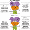
Binding sites for the agonists, antagonists, and modulators described in sections V and VI are shown for the glutamate receptor. The receptor targets of ligands selective for one or several subunits are listed in parenthesis. AMPA and kainate indicates that the ligand selectively targets GluA or GluK receptor subunits, respectively. The ATDs, LBDs, TMDs, and linkers are shown in purple, orange, green, and gray, respectively. [Adapted from Sobolevsky AI, Rosconi MP, and Gouaux E (2009) X-ray structure, symmetry and mechanism of an AMPA-subtype glutamate receptor. Nature **462:**745–756. Copyright © 2009 Nature Publishing Group. Used with permission.]
Fig. 10.
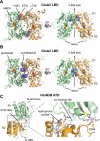
Allosteric regulation of glutamate receptors. A, the structure of the dimer formed between LBDs of the L483Y mutated GluA2 (PDB code
1LB8
) is shown from the top (left) and perpendicular (right) to the 2-fold axis. Mutation of residue 483 (blue) located on D1 from Leu to Tyr attenuates desensitization and stabilizes the dimer interface by interactions with Leu748 and Lys752 on the opposing protomer. B, the LBD dimer interface contains two binding sites for the positive AMPA receptor modulator cyclothiazide (blue) that inhibits receptor desensitization (PDB code
1LBC
). Cyclothiazide stabilizes the dimer interface by forming additional intersubunit interactions in the dimer interface. C, structure of the GluN2B ATD with bound Zn2+ (PDB code
3JPY
). The cleft formed by the upper R1 and the lower R2 lobes can be divided into three pockets: the hydrophobic pocket (gray carbon atoms), the ion binding site with Na+ and Cl−, and the hydrophilic pocket with the Zn2+ binding site. The hydrophobic pocket is thought to bind ifenprodil and its analogs.
Fig. 11.
Desensitization of recombinant AMPA, kainate, and NMDA receptors expressed in the absence of accessory proteins, which can alter response time course (section II). AMPA and kainate receptors activated by
l
-glutamate undergo pronounced and rapid desensitization that occurs within milliseconds after activation and results in steady-state currents less than 5% of the peak response. A and B, voltage-clamp recordings are shown from outside-out patches excised from human embryonic kidney 293 cells expressing recombinant rat GluA1 AMPA receptors (A) or recombinant rat GluK2 kainate receptors (B). Receptors are activated by saturating glutamate (10 mM) for 75 ms. C, voltage-clamp recordings from excised outside-out patches for GluN1/GluN2A-C and a whole-cell voltage-clamp recording of GluN1/GluN2D are given in which the receptors are activated for 1 s by saturating
l
-glutamate and glycine. The degree and time course of desensitization is subunit-dependent. GluN2A-containing receptors desensitize rapidly, GluN2B-containg receptors show slower desensitization, and GluN2C- and GluN2D-containing receptors undergo little to no desensitization. All traces are shown with the peak amplitude normalized to 1. Bottom, steady-state single-channel recordings of GluA1, GluK2, GluN1/GluN2A and GluN1/GluN2D are shown beneath appropriate panels, and illustrate qualitative differences in unitary currents exhibited by AMPA, kainate, and NMDA receptors. Unpublished data for GluK2, GluN2A, GluN2C, and GluN2D, from S. M. Dravid, K. M. Vance, and S. F. Traynelis. Data for GluA1 single-channel recordings were from Banke et al. (2000), GluK2 single channel recordings were from Zhang et al. (2009c), and GluN2B macroscopic current recordings were from Banke et al. (2005).
Fig. 12.
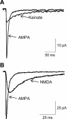
Contribution of glutamate receptor subtypes to synaptic activity. A, a recording of spontaneous mEPSCs in the presence of 100 μM (R)-2-amino-5-phosphonopentanoate (
d
-APV), 100 μM bicuculline, and 1 μM tetrodotoxin from a CA3 pyramidal cell shows the contribution of AMPA and kainate receptors to synaptic activity. Fast mEPSCs mediated by synaptic AMPA receptors and slower mEPSCs mediated by synaptic kainate receptors were both present before application of 100 μM benzenamine (GYKI-52466), but only the slower kainate receptor-mediated mEPSC persisted in the presence of GYKI-52466. CNQX blocks both fast AMPA-mediated and slow kainate receptor-mediated mEPSCs. B, AMPA and NMDA receptor-mediated EPSCs at the pyramidal to multipolar interneuron synapse in the visual cortex. The contributions of AMPA and NMDA receptors to synaptic time course are contrasted using evoked responses from a synaptically connected pair of neurons (pyramidal-to-interneuron). In the presence of the AMPA receptor antagonist CNQX and the absence of Mg2+, the NMDA receptor activity is isolated, highlighting the slow rise time and deactivation time course. There is no significant kainate receptor component at this synapse. Data in A is from Mott et al., 2008; unpublished data in B is from L. P. Wollmuth.
Fig. 13.
A, the structure of GluA2 with two subunits (A and C) transparent. Red dashed line indicates interface between LBD and TMD. B, expanded view of the LBD-TMD regions of subunits B and D. The structure of the water-soluble GluA2 LBD (S1S2) crystallized in complex with glutamate has been superimposed, using the D1 domain, on the corresponding region of GluA2cryst and is shown in green. Helical regions of the ion channel as well as parts of LBD that are proposed to move upon activation are shown as cylinders. Purple and green spheres indicate positions of the α-carbons for the residues Lys393 and Pro632. Stick models of ZK200775 and glutamate are shown in purple and green, respectively. Red arrows indicate proposed movement during receptor activation. C, the transmembrane domain architecture is shown for subunit A parallel to the channel pore as a ribbon structure (left). The transmembrane domains for all four subunits are shown viewed from the intracellular side down the axis of the pore (center), and as a surface representation for subunits B, C, and D with subunit A membrane-associated helices shown as green cylinders (right). [Adapted from Sobolevsky AI, Rosconi MP, and Gouaux E (2009) X-ray structure, symmetry and mechanism of an AMPA-subtype glutamate receptor. Nature **462:**745–756. Copyright © 2009 Nature Publishing Group. Used with permission.]
Fig. 14.
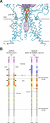
A, surface representation of a closed ion conduction pathway and the pore diameter as a function of distance along the central axis of the channel (red < 1.4 Å < green < 2.8 Å < purple). The residues most proximal to each other that form the activation gate in the closed state are located at the top of the ion channel pore. B, AMPA receptor subunit, GluA2 (left). Subunits A/C and B/D are indicated (see section II). Sequence of the M3 segment (α-helical portion highlighted in gray), the M3–S2 linker, and the S2 lobe (highlighted in magenta). Positions highlighted in yellow are conserved across all mammalian glutamate receptor subunits, including the most highly conserved sequence SYTANLAAF. Also indicated is the border for the M3 segment defined from hydropathy plots (M3 hydrophobic border). Black triangles indicate positions that are located in proximity to each other in the structure of the closed state (red representation in A) and presumably reflect the activation gate. Positions below the dashed line (zδ = 0) show voltage-dependent reactivity to cysteine-reactive reagents, whereas those above do not (Sobolevsky et al., 2003). The Lurcher (Lc) position is highlighted (Zuo et al., 1997; Kohda et al., 2000). Mutations of positions highlighted red have been identified to increase leak current or potentiate glutamate-activated current when modified by cysteine-reactive reagents (Sobolevsky et al., 2003), suggesting that they alter gating. The approximate location of the QRN site is indicated, because this region is disordered in the crystal structure (see sections II.F and VIII.A) (Sobolevsky et al., 2009). NMDA receptors subunits GluN1 and GluN2A (right). Arrangement is the same as in A except that triangles refer to positions that when mutated to cysteine formed cross-linked dimers (black triangle) or did not form dimers (white triangle). Based on these results, the GluN1 subunits are presumed to adopt the A/C conformation and the GluN2 subunits to adopt the B/D conformation (Sobolevsky et al., 2009). Mutations of positions highlighted red either alter leak currents or potentiate glutamate-activated currents when modified by cysteine-reactive reagents (Beck et al., 1999; Jones et al., 2002; Sobolevsky et al., 2002ab, 2007; Yuan et al., 2005), show increases in leak current with single amino acid substitutions (Yuan et al., 2005; Chang and Kuo, 2008), alter channel block (Kashiwagi et al., 2002), and/or alter proton sensitivity (Low et al., 2003). The DRPEER motif in the GluN1 subunit that affects Ca2+ permeability (Watanabe et al., 2002) is highlighted blue, as are corresponding negative charges in the GluN2A subunit that do not affect Ca2+ permeability. Data in A are from Sobolevsky et al. (2009).
Similar articles
- Functional analysis of Caenorhabditis elegans glutamate receptor subunits by domain transplantation.
Strutz-Seebohm N, Werner M, Madsen DM, Seebohm G, Zheng Y, Walker CS, Maricq AV, Hollmann M. Strutz-Seebohm N, et al. J Biol Chem. 2003 Nov 7;278(45):44691-701. doi: 10.1074/jbc.M305497200. Epub 2003 Aug 20. J Biol Chem. 2003. PMID: 12930835 - Structure and gating of the glutamate receptor ion channel.
Wollmuth LP, Sobolevsky AI. Wollmuth LP, et al. Trends Neurosci. 2004 Jun;27(6):321-8. doi: 10.1016/j.tins.2004.04.005. Trends Neurosci. 2004. PMID: 15165736 Review. - Regulation of ligand-gated ion channels by protein phosphorylation.
Swope SL, Moss SJ, Raymond LA, Huganir RL. Swope SL, et al. Adv Second Messenger Phosphoprotein Res. 1999;33:49-78. doi: 10.1016/s1040-7952(99)80005-6. Adv Second Messenger Phosphoprotein Res. 1999. PMID: 10218114 Review. - Mu Receptors.
Herman TF, Cascella M, Muzio MR. Herman TF, et al. 2024 Jun 8. In: StatPearls [Internet]. Treasure Island (FL): StatPearls Publishing; 2025 Jan–. 2024 Jun 8. In: StatPearls [Internet]. Treasure Island (FL): StatPearls Publishing; 2025 Jan–. PMID: 31855381 Free Books & Documents. - Arabidopsis thaliana glutamate receptor ion channel function demonstrated by ion pore transplantation.
Tapken D, Hollmann M. Tapken D, et al. J Mol Biol. 2008 Oct 31;383(1):36-48. doi: 10.1016/j.jmb.2008.06.076. Epub 2008 Jul 3. J Mol Biol. 2008. PMID: 18625242
Cited by
- NMDA Receptors: Distribution, Role, and Insights into Neuropsychiatric Disorders.
Beaurain M, Salabert AS, Payoux P, Gras E, Talmont F. Beaurain M, et al. Pharmaceuticals (Basel). 2024 Sep 25;17(10):1265. doi: 10.3390/ph17101265. Pharmaceuticals (Basel). 2024. PMID: 39458906 Free PMC article. Review. - PCDH7 interacts with GluN1 and regulates dendritic spine morphology and synaptic function.
Wang Y, Kerrisk Campbell M, Tom I, Foreman O, Hanson JE, Sheng M. Wang Y, et al. Sci Rep. 2020 Jul 2;10(1):10951. doi: 10.1038/s41598-020-67831-8. Sci Rep. 2020. PMID: 32616769 Free PMC article. - A eukaryotic specific transmembrane segment is required for tetramerization in AMPA receptors.
Salussolia CL, Gan Q, Kazi R, Singh P, Allopenna J, Furukawa H, Wollmuth LP. Salussolia CL, et al. J Neurosci. 2013 Jun 5;33(23):9840-5. doi: 10.1523/JNEUROSCI.2626-12.2013. J Neurosci. 2013. PMID: 23739980 Free PMC article. - The role of glutamate and the immune system in organophosphate-induced CNS damage.
Eisenkraft A, Falk A, Finkelstein A. Eisenkraft A, et al. Neurotox Res. 2013 Aug;24(2):265-79. doi: 10.1007/s12640-013-9388-1. Epub 2013 Mar 27. Neurotox Res. 2013. PMID: 23532600 Review. - Ethanol effects on N-methyl-D-aspartate receptors in the bed nucleus of the stria terminalis.
Wills TA, Winder DG. Wills TA, et al. Cold Spring Harb Perspect Med. 2013 Apr 1;3(4):a012161. doi: 10.1101/cshperspect.a012161. Cold Spring Harb Perspect Med. 2013. PMID: 23426579 Free PMC article. Review.
References
- aan het Rot M, Collins KA, Murrough JW, Perez AM, Reich DL, Charney DS, Mathew SJ. (2010) Safety and efficacy of repeated-dose intravenous ketamine for treatment-resistant depression. Biol Psychiatry 67:139–145 - PubMed
- Aarsland D, Ballard C, Walker Z, Bostrom F, Alves G, Kossakowski K, Leroi I, Pozo-Rodriguez F, Minthon L, Londos E. (2009) Memantine in patients with Parkinson's disease dementia or dementia with Lewy bodies: a double-blind, placebo-controlled, multicentre trial. Lancet Neurol 8:613–618 - PubMed
- Abbott LF, Regehr WG. (2004) Synaptic computation. Nature 431:796–803 - PubMed
- Abe T, Matsumura S, Katano T, Mabuchi T, Takagi K, Xu L, Yamamoto A, Hattori K, Yagi T, Watanabe M, et al. (2005) Fyn kinase-mediated phosphorylation of NMDA receptor NR2B subunit at Tyr1472 is essential for maintenance of neuropathic pain. Eur J Neurosci 22:1445–1454 - PubMed
- Abele R, Keinanen K, Madden DR. (2000) Agonist-induced isomerization in a glutamate receptor ligand-binding domain. A kinetic and mutagenetic analysis. J Biol Chem 275:21355–21363 - PubMed
Publication types
MeSH terms
Substances
Grants and funding
- T32-DA01504006/DA/NIDA NIH HHS/United States
- R37 NS036654/NS/NINDS NIH HHS/United States
- NS068464/NS/NINDS NIH HHS/United States
- R01 NS036654/NS/NINDS NIH HHS/United States
- R01 NS068464/NS/NINDS NIH HHS/United States
- NS036604/NS/NINDS NIH HHS/United States
- R01 NS065371/NS/NINDS NIH HHS/United States
- R01 MH066892/MH/NIMH NIH HHS/United States
- NS036654/NS/NINDS NIH HHS/United States
- MH066892/MH/NIMH NIH HHS/United States
- T32 GM008602/GM/NIGMS NIH HHS/United States
- T32-GM008602/GM/NIGMS NIH HHS/United States
- EY01697905/EY/NEI NIH HHS/United States
- T32 ES012870/ES/NIEHS NIH HHS/United States
- R01 NS036604/NS/NINDS NIH HHS/United States
- T32-ES012870/ES/NIEHS NIH HHS/United States
- R01 EY016979/EY/NEI NIH HHS/United States
- NS065371/NS/NINDS NIH HHS/United States
LinkOut - more resources
Full Text Sources
Other Literature Sources
Molecular Biology Databases
