An interaction between kynurenine and the aryl hydrocarbon receptor can generate regulatory T cells - PubMed (original) (raw)
An interaction between kynurenine and the aryl hydrocarbon receptor can generate regulatory T cells
Joshua D Mezrich et al. J Immunol. 2010.
Abstract
The aryl hydrocarbon receptor (AHR) has been known to cause immunosuppression after binding dioxin. It has recently been discovered that the receptor may be central to T cell differentiation into FoxP3(+) regulatory T cells (Tregs) versus Th17 cells. In this paper, we demonstrate that kynurenine, the first breakdown product in the IDO-dependent tryptophan degradation pathway, activates the AHR. We furthermore show that this activation leads to AHR-dependent Treg generation. We additionally investigate the dependence of TGF-beta on the AHR for optimal Treg generation, which may be secondary to the upregulation of this receptor that is seen in T cells postexposure to TGF-beta. These results shed light on the relationship of IDO to the generation of Tregs, in addition to highlighting the central importance of the AHR in T cell differentiation. All tissues and cells were derived from mice.
Figures
Figure 1. AHR activation in mouse BMDCs leads to IDO
BMDCs were generated from the bone marrow of C57BL/6J wild type (WT) and AHR null mice as described in Material and Methods. Cells were harvested on day 6 as immature BMDCs or on day 7 as mature BMDCs following addition on day 6 of LPS at a dose of 50ng/ml, a concentration which itself does not cause IDO expression, confirmed in supplemental figure 1. BMDCs were cultured in the presence or absence of TCDD (10nM) added on day 0 of culture. mRNA was isolated from immature or mature BMDCs and assayed for the expression of Cyp1a1 (left panel), a marker of AHR activation, and IDO1 (right panel). Data was normalized to WT control. Post ANOVA testing comparisons are to cultures without TCDD; ***, p < 0.001. Cyp1a1 mRNA was undetectable in all AHR null PCR reactions. Each graph is representative of three independent experiments.
FIGURE 2. Kynurenine, but not other tryptophan breakdown products, directly activates the AHR
Mouse Hepa1 cells that were transfected with luciferase reporter gene fused to the dioxin-responsive elements (DRE) were seeded at 0.6×10⁶ cells per well. Cells were then exposed to tryptophan breakdown products along the kynurenine pathway (50 µM, except as indicated) for 4 hours except as indicated. Luciferase activity was measured on a luminometer. A, Data was converted as a percent of response to TCDD (10nM) to determine the dose response curve (left) and time-course (right) of luciferase activity. B, mRNA was isolated from Hepa1 cells following 2 and 5 hour exposure to TCDD (10nM) and kynurenine (50 µM). qPCR analysis was performed to determine expression levels of Cyp1a1 (upper) and Cyp1b1 to confirm activation of the AHR. Post ANOVA testing comparisons are to control; ***, p < 0.001; **, p < 0.01. C, Time-course of luciferase activity following exposure of Hepa1 cells to FICZ (200nM) or kynurenine (50 µM). All figures represent 1 of 3 independent experiments.
Figure 3. Presence of the AHR is necessary in T cells for optimal generation of FoxP3+ Tregs in Treg-polarizing conditions with and without cell-cell contact
A, Naïve CD4 T cells (CD4+CD62L+ T cells) were generated by magnetic bead separation. 5×10⁵ cells/well were cultured for 5 days with anti CD3/CD28 beads in the presence of no or 2ng/ml of TGF-β. qPCR was used to test the generation of FoxP3. B, Naïve CD4 T cells (CD4+CD62L+ T cells) were generated by magnetic bead separation. 5×10⁵ cells/well were cultured for 5 days with anti CD3/CD28 beads in the presence of titrating doses of TGF-β. Flow cytometry was used to analyze for CD25 and intracellular FoxP3. C, Graphical representation of a similar experiment as B, utilizing titrating doses of TGF-β and titrating numbers of T cells per well. D, Similar to C, except some naïve cells were exposed to a soluble AHR antagonist. E, Naïve CD4+ CD25− T cells were isolated from B6 WT and AHR-null mice and co-cultured with pDCs isolated from BALB/c mice using the Miltenyi mouse pDC isolation kit at a ratio of 20 to 1. CpG was added at the start of culture in some experiments. On day 5, cells were harvested and subjected to flow cytometric analysis. Percentages are the fraction of gated live CD4+ cells that were FoxP3/CD25 double positive. The figures are representative of 3 independent experiments.
Figure 4. Kynurenine induces generation of FoxP3+ Tregs in an AHR-dependent manner
A, Naïve CD4+ CD25− T cells were isolated from B6 WT (top) and AHR-null (bottom) mice. Cells at varying cell densities (50 – 200 × 103 cell per well) were cultured in F10 + 5% FCS in wells coated with anti-CD3/ anti-CD28 antibodies for 5 days. Human TGF-β, kynurenine, TCDD and FICZ were added at the start of the culture. mRNA was isolated from harvested cells and qPCR was performed to determine FoxP3 expression levels. Data is relative to WT or AHR-null cells cultured without TGF-β. The WT experiments were conducted 11 times, and are presented as the mean values. Null experiments were conducted 3 times, with mean values presented. Post ANOVA testing comparisons are against the WT or AHR-null control; *, p < 0.05; ***, p < 0.001. B, Naïve T cells were separated and cultured with antibody stimulation as in A, with and without kynurenine (50µM) in the culture. mRNA was isolated after 5 days and qPCR was performed for Cyp1a1, Cyp1b1, and TGF-β. Post ANOVA testing comparisons are against the vehicle control; ***, p < 0.001. C, (top) Naïve CD4+ CD25− T cells were cultured in the presence of immunomagnetic microbeads coated with anti-CD3/ anti-CD28 antibodies with (transparent peak) and without (gray peak) kynurenine (50 µM). Results were measured by flow cytometry with intracellular FoxP3 staining after 5 days of culture. These results are representative of the protein induction obtained in 4 of 6 separate biological assays. C, (bottom) Varying concentrations of kynurenine +/− AHR antagonist were added at the start of culture. Cells were then harvested and subjected to flow cytometry. Percentages are the fraction of gated Live CD4+ cells that are FoxP3 positive. Post ANOVA testing comparisons are against the vehicle control; ***, p < 0.001. D, Naïve CD4+ CD25− T cells were isolated from B6 WT and AHR-null mice and co-cultured with pDCs isolated from BALB/c mice using the Miltenyi mouse pDC isolation kit at a ratio of 20 to 1. CpG, FICZ and kynurenine were added at the start of culture at the concentrations indicated. On day 5, cells were harvested and subjected to flow cytometric analysis. Percentages are the fraction of gated live CD4+ cells that were FoxP3/CD25 double positive. E, The experiments in D were repeated at the same ratio of pDCs to naïve CD4+ T cells (1 to 20), and results are expressed graphically for WT and Null cells. These figures represent 3 independent experiments. Post ANOVA testing comparisons are against the untreated control; *, p < 0.05; **, p < 0.01.
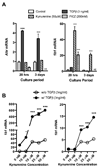
Figure 5. TGF-β upregulates AHR expression, potentiating activation of the DRE by kynurenine
A, (left) Total RNA was extracted from naïve T cells (separated by magnetic beads), either fresh or after 20 hours or 3 days of culture, and qPCR was performed for AHR expression. Culture conditions included antibody stimulation, FICZ 200nM, kynurenine 50uM, or TGF-β 3ng/ml. A, (right) Cyp1b1 mRNA expression was also examined by qPCR at 20 hours and 3 days after the same culture conditions. The FICZ sample was not tested at 3 days (nt). B, To assess whether AHR upregulation secondary to TGF-β would potentiate binding of ligands to the AHR, Cyp1a1 and Cyp1b1 expression after kynurenine exposure in culture for 3 days with and without TGF-β. The response is strongly enhanced after TGF-β exposure, shifting the curve up significantly, implying that TGF-β does potentiate the binding of kynurenine to the AHR when this ligand is present in the culture. Post ANOVA testing comparisons are against the vehicle control; *, p < 0.05; ***, p < 0.001.
Figure 6. FICZ but not Kynurenine enhances TH17 cell differentiation in vitro
Naïve CD4+ CD25− T cells were isolated from B6 WT spleens and cultured in the presence of immunomagnetic microbeads coated with anti-CD3/ anti-CD28 antibodies, TGF-β (4 ng/ml) and IL-6 (20ng/ml) for 5 days. FICZ (200 nM) or kynurenine (50 µM) were added at the start of culture. Cells were then stimulated for 6 hours in the presence of PMA, Ionomycin and Brefeldin A at which time they were harvested and surfaced stained for CD4. This was followed by intracellular staining for IL-17. Numbers are the percent of live CD4 T cells expressing IL-17. Solid thick line is experimental histogram and shaded histogram is control (anti-CD3/CD28, TGF-β and IL-6 alone). Number above gate is the percent IL-17 positive of experimental histogram. Percent IL-17 positive of control histogram is 15.4. This figure is representative of 2 independent experiments.
Similar articles
- Aryl hydrocarbon receptor negatively regulates dendritic cell immunogenicity via a kynurenine-dependent mechanism.
Nguyen NT, Kimura A, Nakahama T, Chinen I, Masuda K, Nohara K, Fujii-Kuriyama Y, Kishimoto T. Nguyen NT, et al. Proc Natl Acad Sci U S A. 2010 Nov 16;107(46):19961-6. doi: 10.1073/pnas.1014465107. Epub 2010 Nov 1. Proc Natl Acad Sci U S A. 2010. PMID: 21041655 Free PMC article. - Blockade of IDO-Kynurenine-AhR Axis Ameliorated Colitis-Associated Colon Cancer via Inhibiting Immune Tolerance.
Zhang X, Liu X, Zhou W, Du Q, Yang M, Ding Y, Hu R. Zhang X, et al. Cell Mol Gastroenterol Hepatol. 2021;12(4):1179-1199. doi: 10.1016/j.jcmgh.2021.05.018. Epub 2021 Jun 1. Cell Mol Gastroenterol Hepatol. 2021. PMID: 34087454 Free PMC article. - IDO upregulates regulatory T cells via tryptophan catabolite and suppresses encephalitogenic T cell responses in experimental autoimmune encephalomyelitis.
Yan Y, Zhang GX, Gran B, Fallarino F, Yu S, Li H, Cullimore ML, Rostami A, Xu H. Yan Y, et al. J Immunol. 2010 Nov 15;185(10):5953-61. doi: 10.4049/jimmunol.1001628. Epub 2010 Oct 13. J Immunol. 2010. PMID: 20944000 Free PMC article. - Indoleamine 2,3-dioxygenase: from catalyst to signaling function.
Fallarino F, Grohmann U, Puccetti P. Fallarino F, et al. Eur J Immunol. 2012 Aug;42(8):1932-7. doi: 10.1002/eji.201242572. Eur J Immunol. 2012. PMID: 22865044 Review. - Targeting the IDO1/TDO2-KYN-AhR Pathway for Cancer Immunotherapy - Challenges and Opportunities.
Cheong JE, Sun L. Cheong JE, et al. Trends Pharmacol Sci. 2018 Mar;39(3):307-325. doi: 10.1016/j.tips.2017.11.007. Epub 2017 Dec 15. Trends Pharmacol Sci. 2018. PMID: 29254698 Review.
Cited by
- Activation of the aryl hydrocarbon receptor in inflammatory bowel disease: insights from gut microbiota.
Hou JJ, Ma AH, Qin YH. Hou JJ, et al. Front Cell Infect Microbiol. 2023 Oct 24;13:1279172. doi: 10.3389/fcimb.2023.1279172. eCollection 2023. Front Cell Infect Microbiol. 2023. PMID: 37942478 Free PMC article. Review. - The causative role and therapeutic potential of the kynurenine pathway in neurodegenerative disease.
Amaral M, Outeiro TF, Scrutton NS, Giorgini F. Amaral M, et al. J Mol Med (Berl). 2013 Jun;91(6):705-13. doi: 10.1007/s00109-013-1046-9. Epub 2013 May 1. J Mol Med (Berl). 2013. PMID: 23636512 Review. - Gut-Brain Axis in the Early Postnatal Years of Life: A Developmental Perspective.
Jena A, Montoya CA, Mullaney JA, Dilger RN, Young W, McNabb WC, Roy NC. Jena A, et al. Front Integr Neurosci. 2020 Aug 5;14:44. doi: 10.3389/fnint.2020.00044. eCollection 2020. Front Integr Neurosci. 2020. PMID: 32848651 Free PMC article. Review. - Indole and Tryptophan Metabolism: Endogenous and Dietary Routes to Ah Receptor Activation.
Hubbard TD, Murray IA, Perdew GH. Hubbard TD, et al. Drug Metab Dispos. 2015 Oct;43(10):1522-35. doi: 10.1124/dmd.115.064246. Epub 2015 Jun 3. Drug Metab Dispos. 2015. PMID: 26041783 Free PMC article. Review. - T cell metabolic fitness in antitumor immunity.
Siska PJ, Rathmell JC. Siska PJ, et al. Trends Immunol. 2015 Apr;36(4):257-64. doi: 10.1016/j.it.2015.02.007. Epub 2015 Mar 12. Trends Immunol. 2015. PMID: 25773310 Free PMC article. Review.
References
- Gershon RK. T cell control of antibody production. Contemp Top Immunobiol. 1974;3:1–40. - PubMed
- Sakaguchi S, Sakaguchi N, Asano M, Itoh M, Toda M. Immunologic self-tolerance maintained by activated T cells expressing IL-2 receptor alpha-chains (CD25). Breakdown of a single mechanism of self-tolerance causes various autoimmune diseases. J Immunol. 1995;155:1151–1164. - PubMed
- Hori S, Nomura T, Sakaguchi S. Control of regulatory T cell development by the transcription factor Foxp3. Science. 2003;299:1057–1061. - PubMed
- Gavin MA, Clarke SR, Negrou E, Gallegos A, Rudensky A. Homeostasis and anergy of CD4(+)CD25(+) suppressor T cells in vivo. Nat Immunol. 2002;3:33–41. - PubMed
Publication types
MeSH terms
Substances
Grants and funding
- R37 ES005703/ES/NIEHS NIH HHS/United States
- UL1 RR025011/RR/NCRR NIH HHS/United States
- T32ES007015-32/ES/NIEHS NIH HHS/United States
- T32 ES007015/ES/NIEHS NIH HHS/United States
- UL1 RR025011-01/RR/NCRR NIH HHS/United States
- R01AI066219/AI/NIAID NIH HHS/United States
- P30CA014520/CA/NCI NIH HHS/United States
- R37ES00570/ES/NIEHS NIH HHS/United States
- R01 ES013566/ES/NIEHS NIH HHS/United States
- 1UL1RR025011/RR/NCRR NIH HHS/United States
LinkOut - more resources
Full Text Sources
Other Literature Sources
Molecular Biology Databases
Research Materials
Miscellaneous


