TDP-43 proteinopathy and motor neuron disease in chronic traumatic encephalopathy - PubMed (original) (raw)
doi: 10.1097/NEN.0b013e3181ee7d85.
Brandon E Gavett, Robert A Stern, Christopher J Nowinski, Robert C Cantu, Neil W Kowall, Daniel P Perl, E Tessa Hedley-Whyte, Bruce Price, Chris Sullivan, Peter Morin, Hyo-Soon Lee, Caroline A Kubilus, Daniel H Daneshvar, Megan Wulff, Andrew E Budson
Affiliations
- PMID: 20720505
- PMCID: PMC2951281
- DOI: 10.1097/NEN.0b013e3181ee7d85
TDP-43 proteinopathy and motor neuron disease in chronic traumatic encephalopathy
Ann C McKee et al. J Neuropathol Exp Neurol. 2010 Sep.
Abstract
Epidemiological evidence suggests that the incidence of amyotrophic lateral sclerosis is increased in association with head injury. Repetitive head injury is also associated with the development of chronic traumatic encephalopathy (CTE), a tauopathy characterized by neurofibrillary tangles throughout the brain in the relative absence of β-amyloid deposits. We examined 12 cases of CTE and, in 10, found a widespread TAR DNA-binding protein of approximately 43kd (TDP-43) proteinopathy affecting the frontal and temporal cortices, medial temporal lobe, basal ganglia, diencephalon, and brainstem. Three athletes with CTE also developed a progressive motor neuron disease with profound weakness, atrophy, spasticity, and fasciculations several years before death. In these 3 cases, there were abundant TDP-43-positive inclusions and neurites in the spinal cord in addition to tau neurofibrillary changes, motor neuron loss, and corticospinal tract degeneration. The TDP-43 proteinopathy associated with CTE is similar to that found in frontotemporal lobar degeneration with TDP-43 inclusions, in that widespread regions of the brain are affected. Akin to frontotemporal lobar degeneration with TDP-43 inclusions, in some individuals with CTE, the TDP-43 proteinopathy extends to involve the spinal cord and is associated with motor neuron disease. This is the first pathological evidence that repetitive head trauma experienced in collision sports might be associated with the development of a motor neuron disease.
Figures
FIGURE 1
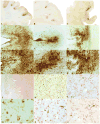
Cases of chronic traumatic encephalopathy (CTE) with widespread TAR DNA-binding protein of approximately 43 kd (TDP-43) immunoreactivity and motor neuron disease (CTE + MND). (A–C) Tau-immunoreactive neurofibrillary degeneration in the frontal cortex of the 3 cases of CTE + MND (whole-mount 50-μm sections immunostained for CP13, original magnification: 1×). (D–I) Tau-positive neurofibrillary tangles, glial tangles, and neuropil neurites are particularly dense at the depth of the cortical sulci, CP13 immunostain, original magnification: (D–F) 50×; (G–I) 100×. (J) TDP-43 immunostaining reveals abundant TDP-positive pathology in all cortical layers in the frontal cortex of Case 1, original magnification: 50×. (K) Numerous TDP-43–positive ring-shaped neurites (RNs) and ring-shaped glial inclusions (RGIs) in the frontal cortex of Case 2, original magnification: 200×. (L) Double immunostaining shows that most TDP-43–positive RNs and RGIs (red) are not colocalized with tau-positive neurites (PHF-1 brown, arrows), original magnification: 400×. (M) TDP-43–positive filamentous neuronal inclusions, original magnification: 600×. (N) Double immunostaining shows a tau-positive pretangle (PHF-1 brown, arrow) that is not associated with TDP-43 immunoreactivity (red), original magnification: 400×. (O) Double immunostaining shows a tau-positive tangle (PHF-1, arrow) that is not associated with TDP-43 immunoreactivity (red), original magnification: 400×.
FIGURE 2
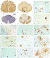
Spinal cord pathology in chronic traumatic encephalopathy (CTE) with TAR DNA-binding protein of approximately 43 kd (TDP-43) proteinopathy, tauopathy, and motor neuron disease (CTE + MND). (A, B) Whole-mount 50-μm sections of lumbar and thoracic spinal cord immunostained with antibody AT8 showing abundant tau immunostaining in the ventral horns. (C) Tau-positive astrocytic tangles in the ventral horn of Case 1 (AT8 immunostain, original magnification: 100×). (D) Whole-mount 10-μm section through high thoracic spinal cord showing marked myelin and axonal loss in the lateral and ventral corticospinal tracts. Atrophic ventral roots are not visible. Luxol fast blue hematoxylin and eosin stain, original magnification: 1×. (E) Whole-mount 50-μm section of high thoracic cord showing intense immunoreactivity for activated microglia and macrophages in lateral and corticospinal tracts (LN-3 immunostain, original magnification: 1×). (F) Tau-positive neurofibrillary tangles in ventral horn of Case 3, AT8 immunostain, original magnification: 600×. (G) TDP-43 immunoreactivity in ventral horn, original magnification: 50×. (H) TDP-43 immunoreactive filamentous neuronal inclusions (FNIs), ring-shaped glial inclusions (RGIs), and ring-shaped neurites (RNs) in the ventral horns of the lumbar spinal cord in Case 3, original magnification: 200×. (I) Tau-positive astrocytes and their processes surrounding degenerating anterior horn cells in the thoracic spinal cord (AT8 immunostain, original magnification: 350×). (J) TDP-43–positive FNIs, RGIs, and RNs in the ventral horns of the lumbar spinal cord in Case 2, original magnification: 200×. (K) TDP-43–positive FNI in the anterior horn, original magnification: 400×.(L) Double immunostained sections show tau-positive astrocytes (brown) and their processes surrounding anterior horn neurons containing TDP-43–positive filamentous inclusions (red), PHF-1 and TDP-43 immunostains, original magnification: 400×. (M) TDP-43–positive FNIs, RGIs, and RNs in the lumbar ventral horns in Case 1, original magnification: 200×. (N) TDP-43–positive FNIs in the anterior horn, original magnification: 400×. (O) Double immunostained sections showing tau-positive astrocytes (brown) and their processes surrounding anterior horn neurons containing TDP-43–positive FNIs (red), PHF-1 and TDP-43 immunostains, original magnification: 600×.
FIGURE 3
TAR DNA-binding protein of approximately 43 kd (TDP-43) immunoreactivity in chronic traumatic encephalopathy (CTE). TDP-43 immunoreactivity is found as glial cytoplasmic inclusions (GCIs) and neuropil neurites in multiple brainstem nuclei including hypoglossal nucleus (A), oculomotor nucleus (B), substantia nigra (C) (original magnification: 200×). TDP-43 immunoreactivity in the medial temporal lobe structures consists primarily of dotlike neurites. (D) Hippocampus, CA1 (original magnification: 200×). TDP-43–positive dystrophic neurites and GCIs are also found in white matter. (E) Subcortical frontal white matter (original magnification: 200×). No ubiquitinated or TDP-43–positive inclusions are found in the dentate gyrus of the hippocampus. (F) Dentate gyrus of the hippocampus (original magnification: 400×).
FIGURE 4
Limited colocalization of tau and TAR DNA-binding protein of approximately 43 kd (TDP-43). (A, B) Laser scanning confocal microscopy was carried out on sections from the frontal cortex of Case 1 (A) and Case 2 (B). Sections were double immunostained for TDP-43 (green) and PHF-1 (red). Areas of colocalization appear yellow. In (A), there are abundant tau-positive neurites around a small blood vessel; white arrowhead indicates a small cluster of tau-positive neurites that seem to be colocalized with TDP-43. (B) Many TDP-43–positive neurites (green) and a few tau-positive neurites (red). Most neurites do not seem to be colocalized, but arrowheads point to occasional colocalized intraneuronal inclusions and neurites. White bars = 200 μm.
Comment in
- Correspondence regarding: TDP-43 proteinopathy and motor neuron disease in chronic traumatic encephalopathy. J Neuropathol Exp Neurol 2010:69;918-29.
Bedlack RS, Genge A, Amato AA, Shaibani A, Jackson CE, Kissel JT, Wall C, King WM, Cupler E, Lou JS, Ensrud E, Tan E, Goldstein JM, Katz J, Dimachkie MM, Barohn RJ, Mozaffar T. Bedlack RS, et al. J Neuropathol Exp Neurol. 2011 Jan;70(1):96-7; author reply 98-100. doi: 10.1097/NEN.0b013e318204782b. J Neuropathol Exp Neurol. 2011. PMID: 21173608 No abstract available. - Correspondence regarding: TDP-43 proteinopathy and motor neuron disease in chronic traumatic encephalopathy. J Neuropathol Exp Neurol 2010:69;918-29.
Armon C, Miller RG. Armon C, et al. J Neuropathol Exp Neurol. 2011 Jan;70(1):97-8; author reply 98-100. doi: 10.1097/01.JNEN.0000392910.86750.32. J Neuropathol Exp Neurol. 2011. PMID: 21173609 No abstract available.
Similar articles
- Globular Glial Mixed Four Repeat Tau and TDP-43 Proteinopathy with Motor Neuron Disease and Frontotemporal Dementia.
Takeuchi R, Toyoshima Y, Tada M, Tanaka H, Shimizu H, Shiga A, Miura T, Aoki K, Aikawa A, Ishizawa S, Ikeuchi T, Nishizawa M, Kakita A, Takahashi H. Takeuchi R, et al. Brain Pathol. 2016 Jan;26(1):82-94. doi: 10.1111/bpa.12262. Epub 2015 May 19. Brain Pathol. 2016. PMID: 25787090 Free PMC article. - Acute and chronically increased immunoreactivity to phosphorylation-independent but not pathological TDP-43 after a single traumatic brain injury in humans.
Johnson VE, Stewart W, Trojanowski JQ, Smith DH. Johnson VE, et al. Acta Neuropathol. 2011 Dec;122(6):715-26. doi: 10.1007/s00401-011-0909-9. Epub 2011 Nov 19. Acta Neuropathol. 2011. PMID: 22101322 Free PMC article. - The spectrum of disease in chronic traumatic encephalopathy.
McKee AC, Stern RA, Nowinski CJ, Stein TD, Alvarez VE, Daneshvar DH, Lee HS, Wojtowicz SM, Hall G, Baugh CM, Riley DO, Kubilus CA, Cormier KA, Jacobs MA, Martin BR, Abraham CR, Ikezu T, Reichard RR, Wolozin BL, Budson AE, Goldstein LE, Kowall NW, Cantu RC. McKee AC, et al. Brain. 2013 Jan;136(Pt 1):43-64. doi: 10.1093/brain/aws307. Epub 2012 Dec 2. Brain. 2013. PMID: 23208308 Free PMC article. - Chronic traumatic encephalopathy in athletes: progressive tauopathy after repetitive head injury.
McKee AC, Cantu RC, Nowinski CJ, Hedley-Whyte ET, Gavett BE, Budson AE, Santini VE, Lee HS, Kubilus CA, Stern RA. McKee AC, et al. J Neuropathol Exp Neurol. 2009 Jul;68(7):709-35. doi: 10.1097/NEN.0b013e3181a9d503. J Neuropathol Exp Neurol. 2009. PMID: 19535999 Free PMC article. Review. - Chronic traumatic encephalopathy: a potential late effect of sport-related concussive and subconcussive head trauma.
Gavett BE, Stern RA, McKee AC. Gavett BE, et al. Clin Sports Med. 2011 Jan;30(1):179-88, xi. doi: 10.1016/j.csm.2010.09.007. Clin Sports Med. 2011. PMID: 21074091 Free PMC article. Review.
Cited by
- Current pathways for epidemiological research in amyotrophic lateral sclerosis.
Factor-Litvak P, Al-Chalabi A, Ascherio A, Bradley W, Chío A, Garruto R, Hardiman O, Kamel F, Kasarskis E, McKee A, Nakano I, Nelson LM, Eisen A. Factor-Litvak P, et al. Amyotroph Lateral Scler Frontotemporal Degener. 2013 May;14 Suppl 1(Suppl 1):33-43. doi: 10.3109/21678421.2013.778565. Amyotroph Lateral Scler Frontotemporal Degener. 2013. PMID: 23678878 Free PMC article. Review. - Clinical features of repetitive traumatic brain injury and chronic traumatic encephalopathy.
Montenigro PH, Bernick C, Cantu RC. Montenigro PH, et al. Brain Pathol. 2015 May;25(3):304-17. doi: 10.1111/bpa.12250. Brain Pathol. 2015. PMID: 25904046 Free PMC article. Review. - Alzheimer's disease and chronic traumatic encephalopathy: Distinct but possibly overlapping disease entities.
Turner RC, Lucke-Wold BP, Robson MJ, Lee JM, Bailes JE. Turner RC, et al. Brain Inj. 2016;30(11):1279-1292. doi: 10.1080/02699052.2016.1193631. Epub 2016 Aug 11. Brain Inj. 2016. PMID: 27715315 Free PMC article. Review. - Chronic traumatic encephalopathy (CTE): criteria for neuropathological diagnosis and relationship to repetitive head impacts.
McKee AC, Stein TD, Huber BR, Crary JF, Bieniek K, Dickson D, Alvarez VE, Cherry JD, Farrell K, Butler M, Uretsky M, Abdolmohammadi B, Alosco ML, Tripodis Y, Mez J, Daneshvar DH. McKee AC, et al. Acta Neuropathol. 2023 Apr;145(4):371-394. doi: 10.1007/s00401-023-02540-w. Epub 2023 Feb 10. Acta Neuropathol. 2023. PMID: 36759368 Free PMC article. Review. - Metal content and kinetic properties of yeast RNA lariat debranching enzyme Dbr1.
Clark NE, Katolik A, Taggart AJ, Buerer L, Holloway SP, Miller N, Phillips JD, Farrell CP, Damha MJ, Fairbrother WG. Clark NE, et al. RNA. 2022 Jul;28(7):927-936. doi: 10.1261/rna.079159.122. Epub 2022 Apr 22. RNA. 2022. PMID: 35459748 Free PMC article.
References
- Bruijn LI, Miller TM, Cleveland DW. Unraveling the mechanisms involved in motor neuron degeneration in ALS. Annu Rev Neurosci. 2004;27:723–49. - PubMed
- Mulder DW, Kurland LT, Offord KP, et al. Familial adult motor neuron disease: Amyotrophic lateral sclerosis. Neurology. 1986;36:511–17. - PubMed
- Okamoto K, Murakami N, Kusaka H, et al. Ubiquitin-positive intraneuronal inclusions in the extramotor cortices of presenile dementia patients with motor neuron disease. J Neurol. 1992;239:426–30. - PubMed
- Wightman G, Anderson VE, Martin J, et al. Hippocampal and neocortical ubiquitin-immunoreactive inclusions in amyotrophic lateral sclerosis with dementia. Neurosci Lett. 1992;139:269–74. - PubMed
Publication types
MeSH terms
Substances
Grants and funding
- P30 AG013846/AG/NIA NIH HHS/United States
- P01 AG002219-179002/AG/NIA NIH HHS/United States
- P01 AG002219/AG/NIA NIH HHS/United States
- P30 AG13846/AG/NIA NIH HHS/United States
- P30 AG013846-029002/AG/NIA NIH HHS/United States
- P50AG05138/AG/NIA NIH HHS/United States
- P50 AG005138-149002/AG/NIA NIH HHS/United States
- P50AG05134/AG/NIA NIH HHS/United States
- P01AG02219/AG/NIA NIH HHS/United States
- P50 AG005134/AG/NIA NIH HHS/United States
- P50 AG005134-149001/AG/NIA NIH HHS/United States
- P50 AG005138/AG/NIA NIH HHS/United States
LinkOut - more resources
Full Text Sources
Other Literature Sources
Miscellaneous


