Cellularity and adipogenic profile of the abdominal subcutaneous adipose tissue from obese adolescents: association with insulin resistance and hepatic steatosis - PubMed (original) (raw)
. 2010 Sep;59(9):2288-96.
doi: 10.2337/db10-0113.
Markus Eszlinger, Deepak Narayan, Teresa Liu, Merlijn Bazuine, Anna M G Cali, Ebe D'Adamo, Melissa Shaw, Bridget Pierpont, Gerald I Shulman, Samuel W Cushman, Arthur Sherman, Sonia Caprio
Affiliations
- PMID: 20805387
- PMCID: PMC2927952
- DOI: 10.2337/db10-0113
Cellularity and adipogenic profile of the abdominal subcutaneous adipose tissue from obese adolescents: association with insulin resistance and hepatic steatosis
Romy Kursawe et al. Diabetes. 2010 Sep.
Abstract
Objective: We explored whether the distribution of adipose cell size, the estimated total number of adipose cells, and the expression of adipogenic genes in subcutaneous adipose tissue are linked to the phenotype of high visceral and low subcutaneous fat depots in obese adolescents.
Research design and methods: A total of 38 adolescents with similar degrees of obesity agreed to have a subcutaneous periumbilical adipose tissue biopsy, in addition to metabolic (oral glucose tolerance test and hyperinsulinemic euglycemic clamp) and imaging studies (MRI, DEXA, (1)H-NMR). Subcutaneous periumbilical adipose cell-size distribution and the estimated total number of subcutaneous adipose cells were obtained from tissue biopsy samples fixed in osmium tetroxide and analyzed by Beckman Coulter Multisizer. The adipogenic capacity was measured by Affymetrix GeneChip and quantitative RT-PCR.
Results: Subjects were divided into two groups: high versus low ratio of visceral to visceral + subcutaneous fat (VAT/[VAT+SAT]). The cell-size distribution curves were significantly different between the high and low VAT/(VAT+SAT) groups, even after adjusting for age, sex, and ethnicity (MANOVA P = 0.035). Surprisingly, the fraction of large adipocytes was significantly lower (P < 0.01) in the group with high VAT/(VAT+SAT), along with the estimated total number of large adipose cells (P < 0.05), while the mean diameter was increased (P < 0.01). From the microarray analyses emerged a lower expression of lipogenesis/adipogenesis markers (sterol regulatory element binding protein-1, acetyl-CoA carboxylase, fatty acid synthase) in the group with high VAT/(VAT+SAT), which was confirmed by RT-PCR.
Conclusions: A reduced lipo-/adipogenic capacity, fraction, and estimated number of large subcutaneous adipocytes may contribute to the abnormal distribution of abdominal fat and hepatic steatosis, as well as to insulin resistance in obese adolescents.
Figures
FIG. 1.
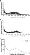
Multisizer adipose cell profiles of 20 subjects with a low VAT/(VAT+SAT) ratio (A) and 18 subjects with a high VAT/(VAT+SAT) ratio (B), plotting cell diameter using linear bins against relative frequency in percent C: Cell-size profiles of the adipose cell size using the mean parameters from the curve-fitting formula for subjects with a low VAT/(VAT+SAT) ratio (dashed line) and subjects with a high VAT/(VAT+SAT) ratio (solid line) (P = 0.035 using MANCOVA).
FIG. 2.
Differences in cell-size parameters between the groups with the low (white bar) and the high (black bar) VAT/(VAT+SAT) ratio. (A) nadir, (B) peak diameter (cp), (C) fraction of large cells (fraclarge), (D) number of large cells (means ± SD).
FIG. 3.
Diagram of the insulin signaling pathway from PathVisio. Red colored boxes indicate significantly increased expression, whereas green colored boxes indicate a significantly decreased expression in the high versus low VAT/(VAT+SAT) group. Besides the gene boxes, the P value is given.
FIG. 4.
Box-plots for the expression of SREBF1, FASN, ACACA, LPIN1, ADIPOQ, PPARγ2, and LPL, normalized to the expression of 18S rRNA and based on the expression of a human control adipose tissue (2ΔΔCt). The white boxes represent the means and SD for the group with the low VAT/(VAT+SAT) ratio, and the black boxes represent the means and SD for the group with the high VAT/(VAT+SAT) ratio. The Mann-Whitney test between the two groups was significant at the <0.05 level (*) or at the <0.005 level (**).
FIG. 5.
Schematic representation of the proposed interactions among size of the subcutaneous fat depot, adipogenesis/lipogenesis, and cell size in the subcutaneous adipose tissue, plasma lipid level, hepatic steatosis, and insulin resistance. (A high-quality digital representation of this figure is available in the online issue.)
Comment in
- Obesity and insulin resistance: an ongoing saga.
Kim SH, Reaven G. Kim SH, et al. Diabetes. 2010 Sep;59(9):2105-6. doi: 10.2337/db10-0766. Diabetes. 2010. PMID: 20805385 Free PMC article. No abstract available.
Similar articles
- A Role of the Inflammasome in the Low Storage Capacity of the Abdominal Subcutaneous Adipose Tissue in Obese Adolescents.
Kursawe R, Dixit VD, Scherer PE, Santoro N, Narayan D, Gordillo R, Giannini C, Lopez X, Pierpont B, Nouws J, Shulman GI, Caprio S. Kursawe R, et al. Diabetes. 2016 Mar;65(3):610-8. doi: 10.2337/db15-1478. Epub 2015 Dec 30. Diabetes. 2016. PMID: 26718495 Free PMC article. - Increased fat accumulation in liver may link insulin resistance with subcutaneous abdominal adipocyte enlargement, visceral adiposity, and hypoadiponectinemia in obese individuals.
Koska J, Stefan N, Permana PA, Weyer C, Sonoda M, Bogardus C, Smith SR, Joanisse DR, Funahashi T, Krakoff J, Bunt JC. Koska J, et al. Am J Clin Nutr. 2008 Feb;87(2):295-302. doi: 10.1093/ajcn/87.2.295. Am J Clin Nutr. 2008. PMID: 18258617 - Differential intra-abdominal adipose tissue profiling in obese, insulin-resistant women.
Liu A, McLaughlin T, Liu T, Sherman A, Yee G, Abbasi F, Lamendola C, Morton J, Cushman SW, Reaven GM, Tsao PS. Liu A, et al. Obes Surg. 2009 Nov;19(11):1564-73. doi: 10.1007/s11695-009-9949-9. Epub 2009 Aug 27. Obes Surg. 2009. PMID: 19711137 Free PMC article. - Visceral adiposity and inflammatory bowel disease.
Rowan CR, McManus J, Boland K, O'Toole A. Rowan CR, et al. Int J Colorectal Dis. 2021 Nov;36(11):2305-2319. doi: 10.1007/s00384-021-03968-w. Epub 2021 Jun 9. Int J Colorectal Dis. 2021. PMID: 34104989 Review. - Two Faces of White Adipose Tissue with Heterogeneous Adipogenic Progenitors.
Hwang I, Kim JB. Hwang I, et al. Diabetes Metab J. 2019 Dec;43(6):752-762. doi: 10.4093/dmj.2019.0174. Diabetes Metab J. 2019. PMID: 31902145 Free PMC article. Review.
Cited by
- Adipose Tissue Macrophage Polarization in Healthy and Unhealthy Obesity.
Ruggiero AD, Key CC, Kavanagh K. Ruggiero AD, et al. Front Nutr. 2021 Feb 17;8:625331. doi: 10.3389/fnut.2021.625331. eCollection 2021. Front Nutr. 2021. PMID: 33681276 Free PMC article. Review. - Altered In Vivo Lipid Fluxes and Cell Dynamics in Subcutaneous Adipose Tissues Are Associated With the Unfavorable Pattern of Fat Distribution in Obese Adolescent Girls.
Nouws J, Fitch M, Mata M, Santoro N, Galuppo B, Kursawe R, Narayan D, Vash-Margita A, Pierpont B, Shulman GI, Hellerstein M, Caprio S. Nouws J, et al. Diabetes. 2019 Jun;68(6):1168-1177. doi: 10.2337/db18-1162. Epub 2019 Apr 1. Diabetes. 2019. PMID: 30936147 Free PMC article. - Inflammatory links between obesity and metabolic disease.
Lumeng CN, Saltiel AR. Lumeng CN, et al. J Clin Invest. 2011 Jun;121(6):2111-7. doi: 10.1172/JCI57132. Epub 2011 Jun 1. J Clin Invest. 2011. PMID: 21633179 Free PMC article. Review. - Meeting Report: 2017 International Joint Meeting of Pediatric Endocrinology Washington DC (September 14-17, 2017) Selected Highlights.
Roberts A, Nip A, Verma A, LaRoche A. Roberts A, et al. Pediatr Endocrinol Rev. 2018 Mar;15(3):255-266. doi: 10.17458/per.vol15.2018.rnvl.intjointwashington. Pediatr Endocrinol Rev. 2018. PMID: 29493130 Free PMC article. No abstract available. - Sleep apnoea and visceral adiposity in middle-aged male and female subjects.
Kritikou I, Basta M, Tappouni R, Pejovic S, Fernandez-Mendoza J, Nazir R, Shaffer ML, Liao D, Bixler EO, Chrousos GP, Vgontzas AN. Kritikou I, et al. Eur Respir J. 2013 Mar;41(3):601-9. doi: 10.1183/09031936.00183411. Epub 2012 Jun 27. Eur Respir J. 2013. PMID: 22743670 Free PMC article.
References
- Danforth E., Jr Failure of adipocyte differentiation causes type II diabetes mellitus? Nat Genet 2000;26:13. - PubMed
- Ravussin E, Smith SR. Increased fat intake, impaired fat oxidation, and failure of fat cell proliferation result in ectopic fat storage, insulin resistance, and type 2 diabetes mellitus. Ann N Y Acad Sci 2002;967:363–378 - PubMed
Publication types
MeSH terms
Substances
Grants and funding
- R01-HD-40787/HD/NICHD NIH HHS/United States
- R01-EB-006494/EB/NIBIB NIH HHS/United States
- R01-HD-28016/HD/NICHD NIH HHS/United States
- K24 HD001464/HD/NICHD NIH HHS/United States
- ImNIH/Intramural NIH HHS/United States
- K24-HD-01464/HD/NICHD NIH HHS/United States
- R01 HD028016/HD/NICHD NIH HHS/United States
- R01 HD040787/HD/NICHD NIH HHS/United States
- R01 EB006494/EB/NIBIB NIH HHS/United States
- UL1-RR-0249139/RR/NCRR NIH HHS/United States
LinkOut - more resources
Full Text Sources
Other Literature Sources
Medical
Research Materials


