Interpretations of fundus autofluorescence from studies of the bisretinoids of the retina - PubMed (original) (raw)
Interpretations of fundus autofluorescence from studies of the bisretinoids of the retina
Janet R Sparrow et al. Invest Ophthalmol Vis Sci. 2010 Sep.
Abstract
Elevated fundus autofluorescence signals can reflect enhanced lipofuscin in RPE cells, augmented fluorescence due to photooxidation, and/or excess bisretinoid fluorophores in photoreceptor cells due to mishandling of vitamin A aldehyde by dysfunctional cells.
Figures
Figure 1.
(A) Fundus autofluorescence image obtained from an adult with healthy retinal status: cSLO and 488 nm excitation. (B) Fundus images obtained from an individual with GA: autofluorescence (left) and color fundus (right) photographs. A zone of increased autofluorescence signal (arrows) surrounds an irregular and nonhomogeneous zone of reduced AF, with uniform loss of AF occurring most centrally.
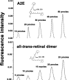
Figure 2.
The fluorescence intensity of RPE lipofuscin bisretinoids increased as the abundance of the pigments was augmented. A2E and all-_trans_-retinal dimer were injected into a UPLC (ultra-performance liquid chromatography) system, with reversed-phase column, at the indicated amounts in a 5-μL volume, and the samples were monitored with a fluorescence detector. UV-visible absorbances were monitored but are not shown. Insets: structures, and absorbance maxima (λmax) of A2E and all-_trans_-retinal dimer.
Figure 3.
Fluorescence intensity of RPE lipofuscin bisretinoids was increased after photooxidation on the short arm of the molecule. Samples of A2E were irradiated at 430 nm to generate photooxidation products (oxo-A2E 1, 2, and 3) and then analyzed by reversed-phase UPLC (ultra-performance liquid chromatography) with online monitoring of absorbance (black trace) and fluorescence (red trace). Fluorescence efficiency per absorbed photon, calculated as fluorescence peak height/absorbance peak height, was 83.6 for oxo-A2E 1, 36.1 for oxo-A2E 3, and 6.7 for A2E. Note that oxo-A2E 2 exhibited little or no fluorescence.
Figure 4.
Bisretinoid formation in impaired photoreceptors can greatly exceed that generated in healthy photoreceptor cell outer segments. (A) HPLC quantitation of all-_trans_-retinal (retinoid precursor of RPE lipofuscin) and two bisretinoids (all-_trans_-retinal dimer and A2PE) that form in photoreceptor cells via the lipofuscin biosynthetic pathway. Eyecups of RCS and control (RCS rdy+) albino rats, age 1 month, included RPE and neural retina. Under normal conditions, phospholipase D-mediated phosphate hydrolysis of A2PE (dashed line in structure) in RPE cell lysosomes releases A2E, and the latter then accumulates in RPE. However, in the RCS rat, because of the failure to phagocytose, most of the pigment generated within the A2PE/A2E pathway remains as A2PE. (B) Fluorescence emission spectra of A2E and A2PE recorded at an excitation of 488 nm. The slightly greater fluorescence intensity of A2PE probably reflects an excitation maximum (∼449 nm) that is closer to 488 nm than the excitation maximum of A2E (∼439 nm).
Similar articles
- Fundus autofluorescence and the bisretinoids of retina.
Sparrow JR, Wu Y, Nagasaki T, Yoon KD, Yamamoto K, Zhou J. Sparrow JR, et al. Photochem Photobiol Sci. 2010 Nov;9(11):1480-9. doi: 10.1039/c0pp00207k. Epub 2010 Sep 23. Photochem Photobiol Sci. 2010. PMID: 20862444 Free PMC article. - Fundus autofluorescence and photoreceptor cell rosettes in mouse models.
Flynn E, Ueda K, Auran E, Sullivan JM, Sparrow JR. Flynn E, et al. Invest Ophthalmol Vis Sci. 2014 Jul 11;55(9):5643-52. doi: 10.1167/iovs.14-14136. Invest Ophthalmol Vis Sci. 2014. PMID: 25015357 Free PMC article. - Lipofuscin-associated photo-oxidative stress during fundus autofluorescence imaging.
Teussink MM, Lambertus S, de Mul FF, Rozanowska MB, Hoyng CB, Klevering BJ, Theelen T. Teussink MM, et al. PLoS One. 2017 Feb 24;12(2):e0172635. doi: 10.1371/journal.pone.0172635. eCollection 2017. PLoS One. 2017. PMID: 28235055 Free PMC article. - The bisretinoids of retinal pigment epithelium.
Sparrow JR, Gregory-Roberts E, Yamamoto K, Blonska A, Ghosh SK, Ueda K, Zhou J. Sparrow JR, et al. Prog Retin Eye Res. 2012 Mar;31(2):121-35. doi: 10.1016/j.preteyeres.2011.12.001. Epub 2011 Dec 22. Prog Retin Eye Res. 2012. PMID: 22209824 Free PMC article. Review. - Bisretinoids of RPE lipofuscin: trigger for complement activation in age-related macular degeneration.
Sparrow JR. Sparrow JR. Adv Exp Med Biol. 2010;703:63-74. doi: 10.1007/978-1-4419-5635-4_5. Adv Exp Med Biol. 2010. PMID: 20711707 Review.
Cited by
- Fundus autofluorescence imaging patterns in central serous chorioretinopathy according to chronicity.
Lee WJ, Lee JH, Lee BR. Lee WJ, et al. Eye (Lond). 2016 Oct;30(10):1336-1342. doi: 10.1038/eye.2016.113. Epub 2016 Jun 10. Eye (Lond). 2016. PMID: 27285318 Free PMC article. - Influence of lens opacities and cataract severity on quantitative fundus autofluorescence as a secondary outcome of a randomized clinical trial.
Reiter GS, Schwarzenbacher L, Schartmüller D, Röggla V, Leydolt C, Menapace R, Schmidt-Erfurth U, Sacu S. Reiter GS, et al. Sci Rep. 2021 Jun 16;11(1):12685. doi: 10.1038/s41598-021-92309-6. Sci Rep. 2021. PMID: 34135449 Free PMC article. Clinical Trial. - An unusual pAIR: Anti-PKM2 antibody and occult pancreatic adenocarcinoma.
Spitz MP, Anderson DR, Vrabec TR. Spitz MP, et al. Am J Ophthalmol Case Rep. 2024 Sep 7;36:102166. doi: 10.1016/j.ajoc.2024.102166. eCollection 2024 Dec. Am J Ophthalmol Case Rep. 2024. PMID: 39351584 Free PMC article. - Multiple evanescent white dot syndrome (MEWDS): update on practical appraisal, diagnosis and clinicopathology; a review and an alternative comprehensive perspective.
Papasavvas I, Mantovani A, Tugal-Tutkun I, Herbort CP Jr. Papasavvas I, et al. J Ophthalmic Inflamm Infect. 2021 Dec 18;11(1):45. doi: 10.1186/s12348-021-00279-7. J Ophthalmic Inflamm Infect. 2021. PMID: 34921620 Free PMC article. Review. - Structures and biogenetic analysis of lipofuscin bis-retinoids.
Wu YL, Li J, Yao K. Wu YL, et al. J Zhejiang Univ Sci B. 2013 Sep;14(9):763-73. doi: 10.1631/jzus.B1300051. J Zhejiang Univ Sci B. 2013. PMID: 24009196 Free PMC article. Review.
References
- Delori FC, Dorey CK, Staurenghi G, Arend O, Goger DG, Weiter JJ. In vivo fluorescence of the ocular fundus exhibits retinal pigment epithelium lipofuscin characteristics. Invest Ophthalmol Vis Sci. 1995;36:718–729 - PubMed
- von Ruckmann A, Fitzke FW, Bird AC. In vivo fundus autofluorescence in macular dystrophies. Arch Ophthalmol. 1997;115:609–615 - PubMed
- Holz FG, Bellman C, Staudt S, Schutt F, Volcker HE. Fundus autofluorescence and development of geographic atrophy in age-related macular degeneration. Invest Ophthalmol Vis Sci. 2001;42:1051–1056 - PubMed
Publication types
MeSH terms
Substances
LinkOut - more resources
Full Text Sources
Other Literature Sources
Medical


