Membrane delivery to the yeast autophagosome from the Golgi-endosomal system - PubMed (original) (raw)
Membrane delivery to the yeast autophagosome from the Golgi-endosomal system
Yohei Ohashi et al. Mol Biol Cell. 2010.
Abstract
While many of the proteins required for autophagy have been identified, the source of the membrane of the autophagosome is still unresolved with the endoplasmic reticulum (ER), endosomes, and mitochondria all having been evoked. The integral membrane protein Atg9 is delivered to the autophagosome during starvation and in the related cytoplasm-to-vacuole (Cvt) pathway that occurs constitutively in yeast. We have examined the requirements for delivery of Atg9-containing membrane to the yeast autophagosome. Atg9 does not appear to originate from mitochondria, and Atg9 cannot reach the forming autophagosome directly from the ER or early Golgi. Components of traffic between Golgi and endosomes are known to be required for the Cvt pathway but do not appear required for autophagy in starved cells. However, we find that pairwise combinations of mutations in Golgi-endosomal traffic components apparently only required for the Cvt pathway can cause profound defects in Atg9 delivery and autophagy in starved cells. Thus it appears that membrane that contains Atg9 is delivered to the autophagosome from the Golgi-endosomal system rather than from the ER or mitochondria. This is underestimated by examination of single mutants, providing a possible explanation for discrepancies between yeast and mammalian studies on Atg9 localization and autophagosome formation.
Figures
Figure 1.
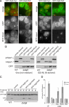
Atg9 cannot get directly from the ER to the forming autophagosome. (A) Fluorescence micrographs of yeast expressing GFP-Atg9 with a C-terminal myc-tag followed by nothing or an RXR motif [KLRRRRI (Michelsen et al., 2007)]. The fusions were expressed with a TPI1 promoter from a centromeric vector and the cells also express either RFP-Ape1, or the ER protein RFP-Scs2. The RXR motif increases the amount of Atg9 in the ER and reduces that present at aggregates of mRFP-Ape1. Scale bars = 2 μm. (B) Anti-Ape1 immunoblots of lysates from wild-type cells, or from an ATG9 deletion strain transformed with either an empty plasmid, or the same GFP-Atg9 plasmids shown in A, and also one with the C-terminal myc-tag followed by a KKXX motif (SKKSL). Cells were harvested after growth in rich medium or after four hours of nitrogen starvation to induce autophagy. Attachment of the RXR motif to Atg9 blocks its activity under both conditions. (C) Anti-GFP immunoblots of lysates from cells expressing GFP-Atg8 from the ATG8 promoter on a CEN plasmid. The yeast strains are as in A and B but with the ATG9 fusions integrated into the genome. The cells were grown to midlog phase and either harvested (0) or starved for nitrogen for 4 h. Deletion of Atg9 prevents delivery of GFP-Atg8 to the vacuole and release of free GFP. (D) Alkaline phosphatase activity in strains expressing a cytosolic form of Pho8 (Pho8Δ60) that lacks a transmembrane domain and so only becomes active upon delivery to the vacuole by bulk autophagy (Noda and Ohsumi, 1998). Strains are as in C but with PHO13 deleted and PHO8 truncated to _PHO8_Δ60 by integration of TDH3 promoter. Cells were grown and starved for 4 h as in C (error bars indicate SD of three independent experiments).
Figure 2.
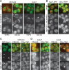
Intracellular distribution of Atg9-GFP. (A) Fluorescent micrographs comparing Atg9-GFP with the mitochondrial outer membrane marker Tom20-RFP in wild-type cells or cells lacking the mitochondrial fusion protein Ugo1 and grown in YEPD or starved for nitrogen for four hours [SD(-N)] as indicated. The loss of Ugo1 causes the mitochondria to form clumps of fragments (Sesaki and Jensen, 2001), but Atg9-GFP remains scattered, with colocalization only observed occasionally, usually in forming buds. (B) Fluorescence micrographs comparing Atg9-GFP to the late Golgi protein Sec7-RFP or the late endosomal protein Nhx1-RFP in wild-type cells in rich medium. (C) Fluorescence micrographs comparing Atg9-GFP to RFP-Ape1, the endocytic tracer FM4-64 (10 min pulse, 10 min chase), or mCherry-Tlg1 (Chry-Tlg1) in wild-type cells in rich medium. (D) As for C except that the cells are either lacking the autophagosome scaffold protein Atg11 which is required for membrane to accumulate at the autophagosome (Shintani and Klionsky, 2004), or lacking the endosomal protein Vps4, in which the endocytic compartment expands (Babst et al., 1997).
Figure 3.
Golgi SNAREs and COG subunits are involved in the Cvt pathway. (A) Ape1 immunoblot analysis in SNARE mutants grown in rich medium (YEPD) for three hours or under conditions of nitrogen starvation for four hours [SD(-N)]. Vam3 and Vam7 are involved in the fusion of vacuolar membranes and so are required for maturation of Ape1 in vegetative and starvation conditions. Deletion of Pep12 inhibits Ape1 maturation, which at least in part appears to be because of its known role in the delivery of vacuolar hydrolases (Becherer et al., 1996). In starved cells some GFP-Ape1 could be seen in undigested autophagosomes inside the vacuole (Supplemental Figure 1B), but in rich medium we also observed an accumulation of GFP-Ape1 aggregates adjacent to the vacuole and so there may also be defects in autophagosome formation in the Cvt pathway. (B) Localization of GFP-Ape1 by fluorescence microscopy in the indicated strains. Cells were grown in YEPD or in SD(-N) and labeled with FM4-64. In YEPD a puncta of GFP-Ape1 was observed in 4% of wild-type cells, but this rose in Δatg1 (31%), Δcog8 (29%), Δgos1 (12%), and Δtlg2 (24%). In SD(-N), GFP signals were mainly observed inside the vacuole in all the strains except Δatg1. (C) Ape1 immunoblot analysis in wild-type (WT) or the indicated deletion mutants. Mutants lacking COG lobe B subunits, or YPT6 or its exchange factor RIC1 show reduced Ape1 maturation only in vegetative phase (YEPD).
Figure 4.
Combinations of Golgi-endosomal mutations cause defects in autophagy. Ape1 maturation was analyzed both in YEPD and SD(-N) by immunoblotting in the indicated mutants. (A) Analysis of Δcog8 and two SNARE mutants, Δgos1 and Δtlg2. (B) Analysis of Δcog8 and single mutants for three related sorting nexins, Atg20, Atg24, and Snx41. (C) Analysis of two SNARE mutants (Δgos1 and Δtlg2) and two sorting nexin mutants.
Figure 5.
Combinations of Cvt pathway mutations cause defects in autophagy. Ape1 maturation was analyzed both in YEPD and SD(-N) by immunoblotting in the indicated mutants. (A) Analysis of mutants lacking GARP/VFT subunit Vps51, or Ypt6 that recruits GARP/VFT, and the sorting nexins (Δatg20 and Δatg24). (B) Analysis of mutants in Cvt component Atg21 and sorting nexins (Δatg20 and Δatg24). (C) Analysis of mutants in TRAPP subunit Gsg1 and sorting nexin Atg24.
Figure 6.
Combined mutants in Golgi-endosomal components show synergistic defects in starvation-induced autophagy. (A) Anti-GFP immunoblot of the indicated strains expressing GFP-Atg8. Cells were grown without uracil (rich medium) to midlog phase and either harvested or shifted to starvation conditions for four hours (SD-N). The positions of GFP-Atg8 and the free GFP that is released after autophagic delivery to the vacuole are indicated. (B) Alkaline phosphatase activity in the indicated strains expressing Pho8Δ60 that lacks a transmembrane domain and so is only activated after delivery to the vacuole by autophagy. Starvation and strains (with PHO13 deleted) are as in A, and error bars indicate the SD of three independent experiments. (C) Quantitation of the distribution of GFP-Atg8 in cells grown as in A. For each strain >150 cells were counted. The values show the proportion of the population with one, or more than one puncta of GFP-Atg8, and are based on a projections of six focal planes. The increases in frequency of cells with multiple GFP-Atg8 puncta in all the double mutants are statistically significant (χ2 test, two-tailed values p < 0.0001). (D) Representative single focal plane images of the cells quantified in C. Combination of SNARE and Atg24 mutations results in the appearance of multiple puncta of GFP-Atg8 consistent with defects in autophagosome formation.
Figure 7.
Atg9 does not reach Ape1-containing autophagosomes in double mutants for Golgi-endosomal trafficking components. (A) Fluorescence micrographs comparing the distribution of Atg9-GFP and RFP-Ape1 in the indicated representative strains in YEPD or SD(-N). Arrows indicate RFP-Ape1 dots which are not colocalized with Atg9-GFP. (B) Quantification of Atg9-GFP and RFP-Ape1 colocalization in both YEPD (rich) and SD(-N). For each strain 50–100 RFP-Ape1 containing structures were examined.
Figure 8.
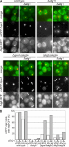
Combining loss of Atg24 with loss of different SNAREs affects different parts of the Atg9 recycling itinerary. (A) Fluorescence micrographs comparing the distribution of Atg9-GFP and RFP-Ape1 in the indicated strains with or without Atg1 deleted. Cells were grown in YEPD to midlog phase and shifted to starvation conditions for four hours (SD-N). Deletion of Atg1 blocks exit of Atg9 from the autophagosome that forms around Ape1 aggregates (Reggiori et al., 2004a). The autophagosome does not form in the absence of Atg11, and so this serves as a negative control. In Δgos1Δatg24 the majority of forming autophagosomes show an accumulation of Atg9 in the absence of Atg1 (arrows), whereas in Δtlg2Δatg24, the forming autophagosomes tend to lack Atg9 (arrows), indicating that delivery rather than retrieval is defective. (B) Quantitation of the experiments in A in which the presence of Atg9 in forming autophagosomes was quantified by counting how many Ape1 aggregates had associated Atg9-GFP. 100–200 mRFP-Ape1 puncta were analyzed for each condition.
Similar articles
- Atg9 vesicles are an important membrane source during early steps of autophagosome formation.
Yamamoto H, Kakuta S, Watanabe TM, Kitamura A, Sekito T, Kondo-Kakuta C, Ichikawa R, Kinjo M, Ohsumi Y. Yamamoto H, et al. J Cell Biol. 2012 Jul 23;198(2):219-33. doi: 10.1083/jcb.201202061. J Cell Biol. 2012. PMID: 22826123 Free PMC article. - Atg27 is required for autophagy-dependent cycling of Atg9.
Yen WL, Legakis JE, Nair U, Klionsky DJ. Yen WL, et al. Mol Biol Cell. 2007 Feb;18(2):581-93. doi: 10.1091/mbc.e06-07-0612. Epub 2006 Nov 29. Mol Biol Cell. 2007. PMID: 17135291 Free PMC article. - TRAPPIII is responsible for vesicular transport from early endosomes to Golgi, facilitating Atg9 cycling in autophagy.
Shirahama-Noda K, Kira S, Yoshimori T, Noda T. Shirahama-Noda K, et al. J Cell Sci. 2013 Nov 1;126(Pt 21):4963-73. doi: 10.1242/jcs.131318. Epub 2013 Aug 28. J Cell Sci. 2013. PMID: 23986483 - Protein transport from the late Golgi to the vacuole in the yeast Saccharomyces cerevisiae.
Bowers K, Stevens TH. Bowers K, et al. Biochim Biophys Acta. 2005 Jul 10;1744(3):438-54. doi: 10.1016/j.bbamcr.2005.04.004. Biochim Biophys Acta. 2005. PMID: 15913810 Review. - Atg9 trafficking in the yeast Saccharomyces cerevisiae.
Mari M, Reggiori F. Mari M, et al. Autophagy. 2007 Mar-Apr;3(2):145-8. doi: 10.4161/auto.3608. Epub 2007 Mar 21. Autophagy. 2007. PMID: 17204846 Review.
Cited by
- A Genome Scale Screen for Mutants with Delayed Exit from Mitosis: Ire1-Independent Induction of Autophagy Integrates ER Homeostasis into Mitotic Lifespan.
Ghavidel A, Baxi K, Ignatchenko V, Prusinkiewicz M, Arnason TG, Kislinger T, Carvalho CE, Harkness TA. Ghavidel A, et al. PLoS Genet. 2015 Aug 6;11(8):e1005429. doi: 10.1371/journal.pgen.1005429. eCollection 2015 Aug. PLoS Genet. 2015. PMID: 26247883 Free PMC article. - SNAP23 regulates BAX-dependent adipocyte programmed cell death independently of canonical macroautophagy.
Feng D, Amgalan D, Singh R, Wei J, Wen J, Wei TP, McGraw TE, Kitsis RN, Pessin JE. Feng D, et al. J Clin Invest. 2018 Aug 31;128(9):3941-3956. doi: 10.1172/JCI99217. Epub 2018 Aug 13. J Clin Invest. 2018. PMID: 30102258 Free PMC article. - Deficiency of the exportomer components Pex1, Pex6, and Pex15 causes enhanced pexophagy in Saccharomyces cerevisiae.
Nuttall JM, Motley AM, Hettema EH. Nuttall JM, et al. Autophagy. 2014 May;10(5):835-45. doi: 10.4161/auto.28259. Epub 2014 Mar 18. Autophagy. 2014. PMID: 24657987 Free PMC article. - Live-cell imaging of Aspergillus nidulans autophagy: RAB1 dependence, Golgi independence and ER involvement.
Pinar M, Pantazopoulou A, Peñalva MA. Pinar M, et al. Autophagy. 2013 Jul;9(7):1024-43. doi: 10.4161/auto.24483. Epub 2013 Apr 8. Autophagy. 2013. PMID: 23722157 Free PMC article. - Therapeutic targeting of autophagy in neurodegenerative and infectious diseases.
Rubinsztein DC, Bento CF, Deretic V. Rubinsztein DC, et al. J Exp Med. 2015 Jun 29;212(7):979-90. doi: 10.1084/jem.20150956. Epub 2015 Jun 22. J Exp Med. 2015. PMID: 26101267 Free PMC article. Review.
References
- Axe E. L., Walker S. A., Manifava M., Chandra P., Roderick H. L., Habermann A., Griffiths G., Ktistakis N. T. Autophagosome formation from membrane compartments enriched in phosphatidylinositol 3-phosphate and dynamically connected to the endoplasmic reticulum. J. Cell. Biol. 2008;182:685–701. - PMC - PubMed
Publication types
MeSH terms
Substances
LinkOut - more resources
Full Text Sources
Other Literature Sources
Molecular Biology Databases


