Effect of high-frequency, low-magnitude vibration on bone and muscle in children with cerebral palsy - PubMed (original) (raw)
Randomized Controlled Trial
Effect of high-frequency, low-magnitude vibration on bone and muscle in children with cerebral palsy
Tishya A L Wren et al. J Pediatr Orthop. 2010 Oct-Nov.
Abstract
Background: Children with cerebral palsy (CP) have decreased strength, low bone mass, and an increased propensity to fracture. High-frequency, low-magnitude vibration might provide a noninvasive, nonpharmacologic, home-based treatment for these musculoskeletal deficits. The purpose of this study was to examine the effects of this intervention on bone and muscle in children with CP.
Methods: Thirty-one children with CP ages 6 to 12 years (mean 9.4, SD 1.4) stood on a vibrating platform (30 Hz, 0.3 g peak acceleration) at home for 10 min/d for 6 months and on the floor without the platform for another 6 months. The order of vibration and standing was randomized, and outcomes were measured at 0, 6, and 12 months. The outcome measures included computed tomography measurements of vertebral cancellous bone density (CBD) and cross-sectional area, CBD of the proximal tibia, geometric properties of the tibial diaphysis, and dynamometer measurements of plantarflexor strength. They were assessed using mixed model linear regression and Pearson correlation.
Results: The main difference between vibration and standing was that there was a greater increase in the cortical bone properties (cortical bone area and moments of inertia) during the vibration period (all P's ≤ 0.03). There was no difference in cancellous bone or muscle between vibration and standing (all P's > 0.10) and no correlation between compliance and outcome (all r's < 0.27; all P's > 0.15). The results did not depend on the order of treatment (P > 0.43) and were similar for children in gross motor function classification system (GMFCS) 1 to 2 and GMFCS 3 to 4.
Conclusions: The primary benefit of the vibration intervention in children with CP was to the cortical bone in the appendicular skeleton. Increased cortical bone area and the structural (strength) properties could translate into a decreased risk of long bone fractures in some patients. More research is needed to corroborate these findings, to elucidate the mechanisms of the intervention, and to determine the most effective age and duration of the treatment.
Level of evidence: Level II, prospective randomized cross-over study.
Figures
Figure 1
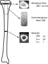
Location of CT scans from the tibia.
Figure 2
Percent change in tibia cortical bone area as a function of compliance.
Similar articles
- Site-Specific Transmission of a Floor-Based, High-Frequency, Low-Magnitude Vibration Stimulus in Children With Spastic Cerebral Palsy.
Singh H, Whitney DG, Knight CA, Miller F, Manal K, Kolm P, Modlesky CM. Singh H, et al. Arch Phys Med Rehabil. 2016 Feb;97(2):218-23. doi: 10.1016/j.apmr.2015.08.434. Epub 2015 Sep 21. Arch Phys Med Rehabil. 2016. PMID: 26392035 Free PMC article. - Bone density and size in ambulatory children with cerebral palsy.
Al Wren T, Lee DC, Kay RM, Dorey FJ, Gilsanz V. Al Wren T, et al. Dev Med Child Neurol. 2011 Feb;53(2):137-41. doi: 10.1111/j.1469-8749.2010.03852.x. Epub 2010 Dec 17. Dev Med Child Neurol. 2011. PMID: 21166671 Free PMC article. - Complicated Muscle-Bone Interactions in Children with Cerebral Palsy.
Modlesky CM, Zhang C. Modlesky CM, et al. Curr Osteoporos Rep. 2020 Feb;18(1):47-56. doi: 10.1007/s11914-020-00561-y. Curr Osteoporos Rep. 2020. PMID: 32060718 Free PMC article. Review. - Vibration therapy.
Rauch F. Rauch F. Dev Med Child Neurol. 2009 Oct;51 Suppl 4:166-8. doi: 10.1111/j.1469-8749.2009.03418.x. Dev Med Child Neurol. 2009. PMID: 19740225 Review.
Cited by
- The effects of whole body vibration on mobility and balance in children with cerebral palsy: a systematic review with meta-analysis.
Saquetto M, Carvalho V, Silva C, Conceição C, Gomes-Neto M. Saquetto M, et al. J Musculoskelet Neuronal Interact. 2015 Jun;15(2):137-44. J Musculoskelet Neuronal Interact. 2015. PMID: 26032205 Free PMC article. Review. - Effectiveness of whole-body vibration in patients with cerebral palsy: A systematic review and meta-analysis.
Han YG, Kim MK. Han YG, et al. Medicine (Baltimore). 2023 Dec 1;102(48):e36441. doi: 10.1097/MD.0000000000036441. Medicine (Baltimore). 2023. PMID: 38050249 Free PMC article. - Vibration therapy in patients with cerebral palsy: a systematic review.
Ritzmann R, Stark C, Krause A. Ritzmann R, et al. Neuropsychiatr Dis Treat. 2018 Jun 18;14:1607-1625. doi: 10.2147/NDT.S152543. eCollection 2018. Neuropsychiatr Dis Treat. 2018. PMID: 29950843 Free PMC article. Review. - Fractures in children with cerebral palsy.
Mughal MZ. Mughal MZ. Curr Osteoporos Rep. 2014 Sep;12(3):313-8. doi: 10.1007/s11914-014-0224-1. Curr Osteoporos Rep. 2014. PMID: 24964775 Review. - Site-Specific Transmission of a Floor-Based, High-Frequency, Low-Magnitude Vibration Stimulus in Children With Spastic Cerebral Palsy.
Singh H, Whitney DG, Knight CA, Miller F, Manal K, Kolm P, Modlesky CM. Singh H, et al. Arch Phys Med Rehabil. 2016 Feb;97(2):218-23. doi: 10.1016/j.apmr.2015.08.434. Epub 2015 Sep 21. Arch Phys Med Rehabil. 2016. PMID: 26392035 Free PMC article.
References
- Gilsanz V, Gibbens DT, Carlson M, et al. Peak trabecular vertebral density: a comparison of adolescent and adult females. Calcif Tissue Int. 1988;43:260–2. - PubMed
- Binkley T, Johnson J, Vogel L, et al. Bone measurements by peripheral quantitative computed tomography (pQCT) in children with cerebral palsy. J Pediatr. 2005;147:791–6. - PubMed
- Tasdemir HA, Buyukavci M, Akcay F, et al. Bone mineral density in children with cerebral palsy. Pediatr Int. 2001;43:157–60. - PubMed
Publication types
MeSH terms
LinkOut - more resources
Full Text Sources
Medical
Research Materials
Miscellaneous

