Computer control of microscopes using µManager - PubMed (original) (raw)
Computer control of microscopes using µManager
Arthur Edelstein et al. Curr Protoc Mol Biol. 2010 Oct.
Abstract
With the advent of digital cameras and motorization of mechanical components, computer control of microscopes has become increasingly important. Software for microscope image acquisition should not only be easy to use, but also enable and encourage novel approaches. The open-source software package µManager aims to fulfill those goals. This unit provides step-by-step protocols describing how to get started working with µManager, as well as some starting points for advanced use of the software.
© 2010 by John Wiley & Sons, Inc.
Figures
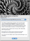
Figure 1
μManager’s “splash screen” window that appears when the application is started. Use this window to choose a configuration file that contains the hardware settings for your system. A drop down menu lets you select recent configuration files, and the button to its right (…) opens a file browser dialog window where you can choose a new configuration file.
Figure 2
The main μManager window, where the application’s primary controls are located.
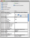
Figure 3
The property browser window. To reach it, choose Tools -> Device/Property Browser…. All major hardware properties and their values are included in this table.
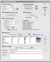
Figure 4
The Multi-Dimensional Acquisition window. Use the check-boxes to select which of the five dimensions you want to include in an acquisition: Time points, Multiple positions (XY), Z-stacks (slices), and Channels.
Figure 5
The 5D Image Viewer of μManager. Use the z and t sliders to view different slices and time points; use the boxes at the right to show and hide channels; use the buttons at bottom to save files, halt acquisition, examine metadata, adjust contrast, and play back time lapse sequences.
Figure 6
The Stage-position List window. Manage waypoints in 2 or 3 dimensions in the position list table. Each position can refer to one or more stage axes
Figure 7
The Image Metadata window. Experimental annotations can be entered in the Comment tab.
Figure 8
The Script Window. Scripts are written in Beanshell and allow fine-grained control of all aspects of μManager.
Similar articles
- Integrative Toolkit to Analyze Cellular Signals: Forces, Motion, Morphology, and Fluorescence.
Nguyen A, Battle K, Paudel SS, Xu N, Bell J, Ayers L, Chapman C, Singh AP, Palanki S, Rich T, Alvarez DF, Stevens T, Tambe DT. Nguyen A, et al. J Vis Exp. 2022 Mar 5;(181). doi: 10.3791/63095. J Vis Exp. 2022. PMID: 35311823 - Use of YouScope to implement systematic microscopy protocols.
Lang M, Rudolf F, Stelling J. Lang M, et al. Curr Protoc Mol Biol. 2012 Apr;Chapter 14:Unit 14.21.1-23. doi: 10.1002/0471142727.mb1421s98. Curr Protoc Mol Biol. 2012. PMID: 22470060 - Feedback regulation of microscopes by image processing.
Tsukada Y, Hashimoto K. Tsukada Y, et al. Dev Growth Differ. 2013 May;55(4):550-62. doi: 10.1111/dgd.12056. Epub 2013 Apr 18. Dev Growth Differ. 2013. PMID: 23594233 Review. - From image to data using common image-processing techniques.
Sysko LR, Davis MA. Sysko LR, et al. Curr Protoc Cytom. 2010 Oct;Chapter 12:Unit 12.21. doi: 10.1002/0471142956.cy1221s54. Curr Protoc Cytom. 2010. PMID: 20938916 - Digital autofocus methods for automated microscopy.
Shen F, Hodgson L, Hahn K. Shen F, et al. Methods Enzymol. 2006;414:620-32. doi: 10.1016/S0076-6879(06)14032-X. Methods Enzymol. 2006. PMID: 17110214 Review.
Cited by
- In Situ Structural Characterization of Cardiomyocyte Microenvironment by Multimodal STED Microscopy.
Zhang Z, Gao BZ, Ye T. Zhang Z, et al. Photonics. 2024 Jun;11(6):533. doi: 10.3390/photonics11060533. Epub 2024 Jun 3. Photonics. 2024. PMID: 39391533 Free PMC article. - Massive multiplexing of spatially resolved single neuron projections with axonal BARseq.
Yuan L, Chen X, Zhan H, Henry GL, Zador AM. Yuan L, et al. Nat Commun. 2024 Sep 27;15(1):8371. doi: 10.1038/s41467-024-52756-x. Nat Commun. 2024. PMID: 39333158 Free PMC article. - Protocol to study oxygen dynamics in the in vivo mouse brain using bioluminescence microscopy.
Asiminas A, Gomolka RS, Gregoriades S, Hirase H, Nedergaard M, Beinlich FRM. Asiminas A, et al. STAR Protoc. 2024 Sep 25;5(4):103334. doi: 10.1016/j.xpro.2024.103334. Online ahead of print. STAR Protoc. 2024. PMID: 39331498 Free PMC article. - Mechanism of barotaxis in marine zooplankton.
Bezares Calderón LA, Shahidi R, Jékely G. Bezares Calderón LA, et al. Elife. 2024 Sep 19;13:RP94306. doi: 10.7554/eLife.94306. Elife. 2024. PMID: 39298255 Free PMC article. - Neural Basis of Number Sense in Larval Zebrafish.
Luu P, Nadtochiy A, Zanon M, Moreno N, Messina A, Petrazzini MEM, Torres Perez JV, Keomanee-Dizon K, Jones M, Brennan CH, Vallortigara G, Fraser SE, Truong TV. Luu P, et al. bioRxiv [Preprint]. 2024 Sep 5:2024.08.30.610552. doi: 10.1101/2024.08.30.610552. bioRxiv. 2024. PMID: 39290349 Free PMC article. Preprint.
References
- μManager website. http://micro-manager.org. The μManager website contains further documentation, download links, and links to a μManager news group through which you can obtain community support.
- ImageJ website. http://rsbweb.nih.gov/ij/ ImageJ is a public domain cross-platform application for image analysis. Hundreds of plugins extend its capabilities. μManager runs as an ImageJ plugin.
- Beanshell Documentation. http://beanshell.org Beanshell is a java-like scripting language used in the μManager script panel.
Publication types
MeSH terms
Grants and funding
- HHMI/Howard Hughes Medical Institute/United States
- R01 EB007187/EB/NIBIB NIH HHS/United States
- R01 EB007187-04/EB/NIBIB NIH HHS/United States
- R01-EB007187/EB/NIBIB NIH HHS/United States
LinkOut - more resources
Full Text Sources
Other Literature Sources


