Evolved variants of the membrane protein can partially replace the envelope protein in murine coronavirus assembly - PubMed (original) (raw)
Evolved variants of the membrane protein can partially replace the envelope protein in murine coronavirus assembly
Lili Kuo et al. J Virol. 2010 Dec.
Abstract
The coronavirus small envelope (E) protein plays a crucial, but poorly defined, role in the assembly of virions. To investigate E protein function, we previously generated E gene point mutants of mouse hepatitis virus (MHV) that were defective in growth and assembled virions with anomalous morphologies. We subsequently constructed an E gene deletion (ΔE) mutant that was only minimally viable. The ΔE virus formed tiny plaques and reached optimal infectious titers many orders of magnitude below those of wild-type virus. We have now characterized highly aberrant viral transcription patterns that developed in some stocks of the ΔE mutant. Extensive analysis of three independent stocks revealed that, in each, a faster-growing virus harboring a genomic duplication had been selected. Remarkably, the net result of each duplication was the creation of a variant version of the membrane protein (M) gene that was situated upstream of the native copy of the M gene. Each different variant M gene encoded an expressed protein (M*) containing a truncated endodomain. Reconstruction of one variant M gene in a ΔE background showed that expression of the M* protein markedly enhanced the growth of the ΔE mutant and that the M* protein was incorporated into assembled virions. These findings suggest that M* proteins were repeatedly selected as surrogates for the E protein and that one role of E is to mediate interactions between transmembrane domains of M protein monomers. Our results provide a demonstration of the capability of coronaviruses to evolve new gene functions through recombination.
Figures
FIG. 1.
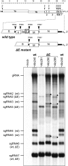
Production of unexpected viral RNA species by certain ΔE isolates of MHV. At the top is a schematic of the MHV genome, with expanded regions comparing the downstream ends of the wild-type (wt) and ΔE mutant genomes. The arrowheads denote TRSs. At the bottom is shown RNA synthesis in 17Cl1 cells that were mock infected or infected at a multiplicity of 0.125 PFU per cell. RNA was metabolically labeled with [33P]orthophosphate, purified, and electrophoretically separated, as described in Materials and Methods. gRNA, genomic RNA. The stars indicate additional RNA species from cells infected with ΔE isolate Alb290 or Alb289.
FIG. 2.
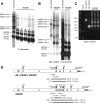
Genomic composition of ΔE mutant Alb290. (A) Northern blots of RNA from cells infected with stocks started from individual plaques of ΔE isolate Alb290 and detected with probes specific for the MHV 3′ genomic end (left) or the M gene (right). Control RNA samples were from mock-infected cells or from cells infected with the nonrearranged ΔE mutant Alb293 or the wild-type Alb240 (wt). The stars indicate the primary extra RNA band in Alb290 stocks. (B) RT-PCR analysis of the duplicated region of the Alb290 genome. Random-primed RT products obtained with RNA isolated from infected cells were amplified with primer pairs neighboring the region of the E gene deletion. PCR products were analyzed by agarose gel electrophoresis; the sizes (bp) of DNA markers are indicated on the left of each gel. (C) Schematic of the Alb290 genome compared with the ΔE genome. Beneath each genome are shown the loci of individual primers (arrows) or of RT-PCR products (bars); above each genome are shown the hybridizing regions of the probes used in the Northern blots. The M gene fragment in Alb290 is indicated by brackets; l denotes the embedded leader RNA.
FIG. 3.
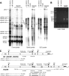
Genomic composition of ΔE mutant Alb295. (A and B) Northern blots of RNA from cells infected with stocks started from individual plaques of ΔE isolate Alb295 and detected with probes specific for the MHV 3′ genomic end or the 3′ end of the S gene. Control RNA samples were from cells infected with the wild-type Alb240 (wt). The stars indicate the primary extra RNA band in Alb295 stocks. In panel A, the wild-type control lane is a lower exposure than that shown for the other lanes. (C) RT-PCR analysis of the duplicated region of the Alb295 genome. Random-primed RT products obtained with RNA isolated from infected cells were amplified with a primer pair neighboring the region of the E gene deletion. The PCR products were analyzed by agarose gel electrophoresis; the sizes (bp) of DNA markers are indicated on the left of each gel. (D) Schematic of the Alb295 genome compared with the ΔE genome. Beneath each genome are shown the loci of individual primers (arrows) and of RT-PCR products (bars); above each genome are shown the hybridizing regions of the probes used in the Northern blots. The M and S gene fragments in Alb295 are indicated by brackets.
FIG. 4.
Genomic composition of ΔE mutant Alb294. (A) Northern blots of RNA from cells infected with stocks started from individual plaques of ΔE isolate Alb294 and detected with probes specific for the MHV 3′ genomic end (left), the middle of the S gene (middle), or the 3′ end of the S gene (right). Control RNA samples were from mock-infected cells or from cells infected with the nonrearranged ΔE mutant Alb293 or the wild type Alb240 (wt). The stars indicate the primary extra RNA band in Alb294 stocks. (B) RT-PCR analysis of the duplicated region of the Alb294 genome. Random-primed RT products obtained with RNA isolated from infected cells were amplified with a primer pair neighboring the region of the E gene deletion. The PCR products were analyzed by agarose gel electrophoresis; the sizes (bp) of DNA markers are indicated on the left of each gel. (C) Schematic of the Alb294 genome compared with the ΔE genome. Beneath each genome are shown the loci of individual primers (arrows) or of RT-PCR products (bars); the dashed lines indicate expected PCR products that we were unable to obtain. Note that PCR products obtained with primer LK99, which falls in the MHV leader sequence, originated from subgenomic templates. Above each genome are shown the hybridizing regions of the probes used in the Northern blots. The M and S gene fragments in Alb294 are indicated by brackets.
FIG. 5.
MHV variants evolved from ΔE mutants. (A) Summary of the 3′-end genomic structures of the five ΔE isolates that were analyzed in detail. Alb291 and Alb293 (top) were previously shown to contain only the predicted deletion between the S and M genes (31). Alb290, Alb295, and Alb294 each contain a duplication resulting from nonhomologous recombination, either between the ΔE genome and sgRNA6 (for Alb290) or between two ΔE genomes (for Alb295 and Alb294). In each case, the second component of the nonhomologous recombination event is shaded gray, and the crossover junction is indicated by an X. Fragments of genes are indicated by brackets; l denotes the leader sequence embedded in the Alb290 genome. The arrowheads denote TRSs. Schematics of the encoded wild-type M and variant (M*) proteins are shown below each genome. (B) Alignment of wild-type and M* protein sequences; for the latter, only those residues that differ from the wild type are shown. M*-290m is the M* protein encoded by a minor form of the Alb290 insert. The circles above the wild-type M sequence denote the three transmembrane domains, as defined by Rottier and coworkers (45); Δ, deletion of the F90 residue in M*-290. Following the crossover junction in M*-295, the extension of the M* ORF is in the −1 reading frame with respect to the S ORF; for M*-294, the extension of the M* ORF is in the +1 reading frame with respect to the S ORF. (C) Western blots of lysates from 17Cl1 cells infected at a multiplicity of 0.1 PFU per cell with wild-type MHV (Alb240) or the ΔE mutant Alb290, Alb294, or Alb295 and harvested at 18 h postinfection. The blot was probed with monoclonal antibody J.1.3, which is specific for the M ectodomain (13). mock, control lysate from mock-infected cells. Molecular mass standards (kDa) are indicated on the left of the panel.
FIG. 6.
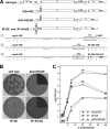
Expression of M*-290 from the gene 2 region of MHV. (A) Comparison of the downstream ends of the genomes of wild-type MHV and the mutants Δ(2a-HE), Δ(2a-HE)/ΔE, M*/ΔE, and M*-KO/ΔE. Beneath the schematics are shown details of the 1b-S or 1b-M* intergenic junction; SalI and AscI sites that were introduced into the parent transcription vectors are marked by dashed underlines; TRSs are boxed; indicates a nonfunctional TRS. Point mutations created to disrupt the TRS and the M* ORF in the M*-KO mutant are indicated in boldface with solid underlines. Deletion and substitution mutants were constructed by targeted RNA recombination, as described in Materials and Methods. (B) Plaques of mutants Δ(2a-HE)/ΔE, M*/ΔE, and M*-KO/ΔE compared with those of the wild type (Alb240). Plaque titrations were carried out on L2 cells at 37°C. Monolayers were stained with neutral red at 96 h postinfection and were photographed 18 h later. For the Δ(2a-HE)/ΔE and M*-KO/ΔE mutants, one quadrant of the photograph is shown with altered contrast to allow better visualization of the tiny plaques of these mutants. (C) Growth kinetics of the M*/ΔE mutant relative to those of its Δ(2a-HE) and Δ(2a-HE)/ΔE counterparts. Confluent monolayers of 17Cl1 cells were infected at a multiplicity of 0.1 PFU per cell. At the indicated times postinfection, aliquots of medium were removed, and infectious titers were determined by plaque assay on L2 cells. The open and shaded symbols represent results from two independent experiments.
FIG. 7.
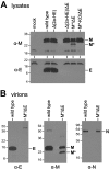
Expression and virion incorporation of M* protein. (A) Western blots of lysates from 17Cl1 cells infected at a multiplicity of 0.1 PFU per cell with wild-type MHV or with each of the mutants shown in Fig. 6A and harvested at 16 h postinfection. The blots were probed with anti-M monoclonal antibody J.1.3 (α-M) (top) or anti-E polyclonal antibody (α-E) (bottom). mock, control lysate from mock-infected cells. (B) Western blots of purified wild-type or M*/ΔE mutant virions probed with anti-E polyclonal antibody (left), anti-M monoclonal antibody J.1.3 (middle), or anti-N monoclonal antibody J.3.3 (α-N) (right). Molecular mass standards (kDa) are indicated on the left of each panel.
FIG. 8.
Mutations, other than M* duplications, that partially counteract the ΔE phenotype. (A) Summary of mutations in the M protein that were found to produce larger-plaque-size variants of E mutants. The solid boxes represent transmembrane (Tm) domains. The three M* proteins are also shown for comparison; the hatched boxes represent heterologous sequences resulting from frameshifts (Fig. 5). (B) Plaques of reconstructed mutants containing the ΔE mutation and one or more compensating M mutations compared to plaques of an original ΔE mutant (Alb291) and the wild type (Alb240). Plaque titrations were carried out on L2 cells at 37°C. The monolayers were stained with neutral red at 72 h postinfection and photographed 18 h later.
FIG. 9.
Model of M-M interactions based on cryo-electron microscopic (37, 38) and cryo-electron tomographic (4) reconstructions of coronaviruses. M multimers are depicted as dimers, but the same relationships would pertain for higher-order M oligomers. At the bottom is shown the domain structure of M protein in comparison to that of M* protein. Tm, transmembrane.
Similar articles
- The coronavirus E protein: assembly and beyond.
Ruch TR, Machamer CE. Ruch TR, et al. Viruses. 2012 Mar;4(3):363-82. doi: 10.3390/v4030363. Epub 2012 Mar 8. Viruses. 2012. PMID: 22590676 Free PMC article. Review. - Exceptional flexibility in the sequence requirements for coronavirus small envelope protein function.
Kuo L, Hurst KR, Masters PS. Kuo L, et al. J Virol. 2007 Mar;81(5):2249-62. doi: 10.1128/JVI.01577-06. Epub 2006 Dec 20. J Virol. 2007. PMID: 17182690 Free PMC article. - The small envelope protein E is not essential for murine coronavirus replication.
Kuo L, Masters PS. Kuo L, et al. J Virol. 2003 Apr;77(8):4597-608. doi: 10.1128/jvi.77.8.4597-4608.2003. J Virol. 2003. PMID: 12663766 Free PMC article. - Genetic analysis of determinants for spike glycoprotein assembly into murine coronavirus virions: distinct roles for charge-rich and cysteine-rich regions of the endodomain.
Ye R, Montalto-Morrison C, Masters PS. Ye R, et al. J Virol. 2004 Sep;78(18):9904-17. doi: 10.1128/JVI.78.18.9904-9917.2004. J Virol. 2004. PMID: 15331724 Free PMC article. - Coronavirus spike glycoprotein, extended at the carboxy terminus with green fluorescent protein, is assembly competent.
Bosch BJ, de Haan CA, Rottier PJ. Bosch BJ, et al. J Virol. 2004 Jul;78(14):7369-78. doi: 10.1128/JVI.78.14.7369-7378.2004. J Virol. 2004. PMID: 15220410 Free PMC article.
Cited by
- Functional analysis of the murine coronavirus genomic RNA packaging signal.
Kuo L, Masters PS. Kuo L, et al. J Virol. 2013 May;87(9):5182-92. doi: 10.1128/JVI.00100-13. Epub 2013 Feb 28. J Virol. 2013. PMID: 23449786 Free PMC article. - Characterization of an Immunodominant Epitope in the Endodomain of the Coronavirus Membrane Protein.
Dong H, Zhang X, Shi H, Chen J, Shi D, Zhu Y, Feng L. Dong H, et al. Viruses. 2016 Dec 10;8(12):327. doi: 10.3390/v8120327. Viruses. 2016. PMID: 27973413 Free PMC article. - The coronavirus E protein: assembly and beyond.
Ruch TR, Machamer CE. Ruch TR, et al. Viruses. 2012 Mar;4(3):363-82. doi: 10.3390/v4030363. Epub 2012 Mar 8. Viruses. 2012. PMID: 22590676 Free PMC article. Review. - Thermodynamic insight into viral infections 2: empirical formulas, molecular compositions and thermodynamic properties of SARS, MERS and SARS-CoV-2 (COVID-19) viruses.
Popovic M, Minceva M. Popovic M, et al. Heliyon. 2020 Sep;6(9):e04943. doi: 10.1016/j.heliyon.2020.e04943. Epub 2020 Sep 14. Heliyon. 2020. PMID: 32954038 Free PMC article. - Structure of a conserved Golgi complex-targeting signal in coronavirus envelope proteins.
Li Y, Surya W, Claudine S, Torres J. Li Y, et al. J Biol Chem. 2014 May 2;289(18):12535-49. doi: 10.1074/jbc.M114.560094. Epub 2014 Mar 25. J Biol Chem. 2014. PMID: 24668816 Free PMC article.
References
Publication types
MeSH terms
Substances
LinkOut - more resources
Full Text Sources
