Oestrogens ameliorate mitochondrial dysfunction in Leber's hereditary optic neuropathy - PubMed (original) (raw)
. 2011 Jan;134(Pt 1):220-34.
doi: 10.1093/brain/awq276. Epub 2010 Oct 13.
Monica Montopoli, Elena Perli, Maurizia Orlandi, Marianna Fantin, Fred N Ross-Cisneros, Laura Caparrotta, Andrea Martinuzzi, Eugenio Ragazzi, Anna Ghelli, Alfredo A Sadun, Giulia d'Amati, Valerio Carelli
Affiliations
- PMID: 20943885
- PMCID: PMC3025718
- DOI: 10.1093/brain/awq276
Oestrogens ameliorate mitochondrial dysfunction in Leber's hereditary optic neuropathy
Carla Giordano et al. Brain. 2011 Jan.
Abstract
Leber's hereditary optic neuropathy, the most frequent mitochondrial disease due to mitochondrial DNA point mutations in complex I, is characterized by the selective degeneration of retinal ganglion cells, leading to optic atrophy and loss of central vision prevalently in young males. The current study investigated the reasons for the higher prevalence of Leber's hereditary optic neuropathy in males, exploring the potential compensatory effects of oestrogens on mutant cell metabolism. Control and Leber's hereditary optic neuropathy osteosarcoma-derived cybrids (11778/ND4, 3460/ND1 and 14484/ND6) were grown in glucose or glucose-free, galactose-supplemented medium. After having shown the nuclear and mitochondrial localization of oestrogen receptors in cybrids, experiments were carried out by adding 100 nM of 17β-oestradiol. In a set of experiments, cells were pre-incubated with the oestrogen receptor antagonist ICI 182780. Leber's hereditary optic neuropathy cybrids in galactose medium presented overproduction of reactive oxygen species, which led to decrease in mitochondrial membrane potential, increased apoptotic rate, loss of cell viability and hyper-fragmented mitochondrial morphology compared with control cybrids. Treatment with 17β-oestradiol significantly rescued these pathological features and led to the activation of the antioxidant enzyme superoxide dismutase 2. In addition, 17β-oestradiol induced a general activation of mitochondrial biogenesis and a small although significant improvement in energetic competence. All these effects were oestrogen receptor mediated. Finally, we showed that the oestrogen receptor β localizes to the mitochondrial network of human retinal ganglion cells. Our results strongly support a metabolic basis for the unexplained male prevalence in Leber's hereditary optic neuropathy and hold promises for a therapeutic use for oestrogen-like molecules.
Figures
Figure 1
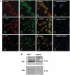
Oestrogen receptor localization to osteosarcoma-derived (143B. TK-) cybrid cell lines. (A) Fluorescence microscopy localization of oestrogen receptors to the nucleus and mitochondria of cybrid cells. Oestrogen receptor β is stained red by oestrogen receptor β BIO1974 antibody (a) and oestrogen receptor β H150 antibody (e); oestrogen receptor α is stained red by H-184 antibody (i); mitochondria are stained green using anti-mitochondria extract antibody (b, f and l); merged image of oestrogen receptors and mitochondria (c, g and m); negative controls loaded with 4',6-diamidino-2-phenylindole (blue) (d, h and n). Overlap is seen in bright green (original magnification ×40). (B) Western blot analysis of mitochondrial preparation from 143B.TK-derived wild-type cybrids and MCF7 oestrogen–dependent breast cancer cells used as positive control. The oestrogen receptor β (ERβ) protein (identified with oestrogen receptor β 485–503 antibody) is constitutively over-expressed in the mitochondrial fraction of MCF7 cells, whereas oestrogen receptor α (ERα) is present at lower levels, confirming previous data (Pedram et al., 2006). In wild-type cybrids, oestrogen receptor β is less abundant than in MCF7 cells, whereas oestrogen receptor α is not expressed.
Figure 2
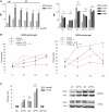
17β-Oestradiol decreases levels of reactive oxygen species and induces SOD2 activity. (A) Left: Control and 11778/ND4 cybrids were incubated for 1 h in glucose (glu) or galactose (gal) medium ± 10–200 nM 17β-oestradiol (E2), then the intracellular steady-state levels of reactive oxygen species was evaluated. Right: In a further experiment control and LHON cybrids were incubated for 1 h in glucose (glu) or galactose (gal) medium ± 100 nM 17β-oestradiol (E2). The oestrogen receptor antagonist ICI 182780 (I) was added 30 min before 17β-oestradiol. Untreated cells were maintained at the same final ethanol concentration. Data are expressed as mean fluorescence intensity (MFI) (± SEM) and are from three experiments. °P < 0.05; °°P < 0.01; °°°P < 0.001 LHON versus control; †P < 0.05 for glu + E2 versus glu; *P < 0.05, ***P < 0.001 for gal versus glu; +P < 0.05; ++P < 0.01 for gal + E2 versus gal and versus gal + E2+ I (see
Supplementary Fig. 1
). (B) Time course of SOD2 activity of control and 11778/ND4 (HPE9) cybrids incubated in glucose or galactose medium ± 100 nM 17β-oestradiol (E2). Incubation in galactose medium induced a significant increase of SOD2 activity in control cybrids (P < 0.001) that was not observed in LHON. SOD2 activity is expressed as unit per milligram of protein. Each data point is the mean of quadruplicate replicates. °°°P < 0.001 for LHON versus control; +++P < 0.001 for E2 versus ethanol. (C) Left: The relative expression of mitochondrial SOD2 gene was evaluated by real-time PCR in control (HGA and HP27) and 11778/ND4 (HPE9 and HFF3) cybrids incubated for 6 h in glucose (glu) or galactose (gal) medium ± 100 nM 17β-oestradiol (E2). The oestrogen receptor antagonist ICI 182780 was added 30 min before 17β-oestradiol. Data represent mean arbitrary units (± SEM) normalized to control values in glucose medium and are from three experiments. †P < 0.05 and ††P < 0.01 for glu + E2 versus glu; ***P < 0.001 for gal versus glu; +++P < 0.001 for gal+E2 versus gal. Right: Western blot analysis of SOD2 protein performed on extract from control (HGA) and 11778/ND4 (HPE9) cybrids incubated for 24 h in glucose or galactose medium ± 100 nM 17β-oestradiol. A representative blot from three is shown.
Figure 3
17β-Oestradiol ameliorates cell viability in galactose medium by reducing apoptosis. (A) Growth curves of control and LHON cybrids maintained in glucose (glu) or galactose (gal) medium ± 100 nM 17β-oestradiol (E2). ***P < 0.001 for gal versus glu; +P < 0.05, ++P < 0.01, +++P < 0.001 for gal + E2 versus gal. Data are expressed as % of untreated cell number in glucose medium, and are mean ± SEM from four different experiments in duplicate. Growth curves for control and 11778/ND4 are from HP27 and HFF3 clones. Similar results were obtained with HPE9 and HGA clones (data not shown). (B) Mitochondrial membrane potential (mtΔφ) of control and LHON cybrids incubated for 24 h in glucose (glu) or galactose (gal) medium ± 100 nM 17β-oestradiol (E2). In a subset of experiments, cells were pre-incubated with ICI 182780 (I). Data are expressed as mean fluorescence intensity (MFI; ± SEM) (% of values of untreated cells in glucose medium) and are from three independent experiments. Control and 11778/ND4 values are the mean from two clones. *P < 0.05, ***P < 0.001 for gal versus glu; +P < 0.05, +++P < 0.001 for gal + E2 versus gal and versus gal + E2+ I**.** (C) Percentages of apoptotic cells in control and LHON cybrids incubated in glucose (glu) or galactose (gal) medium ± 100 nM 17β-oestradiol (E2), as evaluated by labelling the cells with annexin V. Data are the mean ± SEM of the percent number of apoptotic cells from three repeated experiments. Control and 11778/ND4 values are the mean values from two clones. °°°P < 0.001 LHON versus control; ***P < 0.001 for gal versus glu; ++P < 0.01 for gal + E2 versus gal (see
Supplementary Fig. 2
).
Figure 4
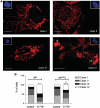
17β-Oestradiol reduces mitochondrial network fragmentation. (A) Representative images of the four classes of cells (see ‘Results’ section) as observed in 11778/ND4 cybrids incubated for 24 h in galactose medium, loaded with MitoTracker Orange and counterstained with 4',6-diamidino-2-phenylindole. The inset shows the corresponding nuclear morphology. (B) Bar graphs showing quantification of the four categories by blind test. Cybrids analysed were from 30 photos obtained from two controls (HGA, HP27) and two 11778/ND4 (HFF3, HPE9) cybrid cell lines grown in galactose medium (gal) ± 100 nM 17β-oestradiol (E2). °°°P < 0.001, °°P < 0.05 for LHON versus WT; +++P < 0.001 for gal + E2 versus gal.
Figure 5
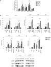
17β-Oestradiol induces mitochondrial biogenesis. (A) Amount of mitochondrial DNA in control and LHON cybrids maintained for 3 h in glucose (glu) or galactose (gal) medium ± 100 nM 17β-oestradiol (E2). In a subset of experiments, cells were pre-incubated with ICI 182780 (I). Bar graph represents the mean plus SEM from three experiments. †††P < 0.001 for glu + E2 versus glu and versus glu + E2+ I; **P < 0.01 for gal versus glu; +++P < 0.001 for gal + E2 versus gal (see
Supplementary Fig. 3
). (B) Control and LHON cybrids were incubated for 6 h in glucose (glu) or galactose (gal) medium ± 100 nM 17β-oestradiol (E2). In a subset of experiments, cells were pre-incubated with ICI 182780 (I). The relative expression of the following genes was evaluated by real-time PCR analysis: PGC1-α and its homologue PGC-1β, NRF1, NRF2, Tfam, MTCOI, MTND5. (C) In a subsequent experiment, cells were incubated for 24 h in glucose (glu) or galactose (gal) medium ± 100 nM 17β-oestradiol (E2), and the relative gene and protein expression of the nuclear encoded respiratory chain subunits COIV evaluated by real-time PCR (top) and western blot analysis of mitochondrial fraction (bottom) along with the protein expression of the mitochondrial encoded complex I subunit ND6. Data represent mean arbitrary units (± SEM) normalized to control values in glucose medium and are from three experiments. A representative blot out of three is shown. †††P < 0.001, ††P < 0.01, †P < 0.05 for glu + E2 versus glu and versus glu + E2+ I; ***P < 0.001, **P < 0.01, *P < 0.05 for gal versus glu; +++P < 0.001 for gal + E2 versus gal.
Figure 6
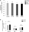
17β-Oestradiol ameliorates energetic competence of cybrid cells. (A) Control, 11778/ND4, 3460/ND1 and 14484/ND6 cybrids were incubated for 24 h in glucose (glu) ± 100 nM 17β-oestradiol (E2) and the rate of oxygen consumption measured. Data are mean ± SEM from three to four separate experiments. For each clone, the rate of oxygen consumption in glucose medium supplemented with 17β-oestradiol has been normalized to the rate of oxygen consumption in glucose medium plus ethanol. *P < 0.03; **P < 0.003 for glu + E2 versus glu. (B) Control and 11778/ND4 LHON cybrids were incubated for 24 h in glucose (glu) ± 100 nM 17β-oestradiol and the total ATP cellular content measured by luciferin/luciferase assay. Data are mean ± SEM from four separate experiments. For each clone, values are normalized for the total ATP content in glucose medium. +++P < 0.001 for E2 versus ethanol; *** for galactose (gal) versus glucose.
Figure 7
Oestrogen receptor β localizes to retinal ganglion cells. (A) Immunoperoxidase stain with anti-oestrogen receptor β antibodies (ERβ-H150) on horizontal retinal sections from a normal individual (left: male, 59-years-old) and a patient with LHON with the 11778/ND4 mutation (Middle: male, 52-years-old). In the normal individual, a positive stain is observed in the somata of retinal ganglion cells (between arrows) and the unmyelinated portion of the axons in the retinal nerve fibre layer, as well as in the inner and outer plexiform layers. Residual retinal ganglion cells in the patient with LHON (arrows) show a similar expression pattern. Similar results were obtained with the antibody ERβ 485-503 both on male and female control individuals (not shown). (B) Higher magnification showing a typical mitochondrial punctuate pattern of oestrogen receptor β in the somata of retinal ganglion cells. (C) Immunoperoxidase stain with anti-oestrogen receptor β antibodies (ERβ-H150) on a formalin fixed, paraffin embedded section of breast cancer used as a positive control. Note the faint nuclear and the strong cytoplasm staining, similar to that reported in BRC7 breast cancer cells (Pedram et al., 2006). (D) Double immunofluorescence stain with anti-mitochondrial (green) and anti-oestrogen receptor β (red) antibodies. The negative control (right) shows a non-specific background fluorescence induced by fixation media, yet on top of this background is the evident co-localization of mitochondria and oestrogen receptor β in the somata of retinal ganglion cells. Overlap is seen in yellow. NFL = nerve fiber layer; IPL = inner plexiform layer; OPL = outer plexiform layer.
Similar articles
- Inhibition of angiogenesis by the secretome from iPSC-derived retinal ganglion cells with Leber's hereditary optic neuropathy-like phenotypes.
Peng SY, Chen CY, Chen H, Yang YP, Wang ML, Tsai FT, Chien CS, Weng PY, Tsai ET, Wang IC, Hsu CC, Lin TC, Hwang DK, Chen SJ, Chiou SH, Chiao CC, Chien Y. Peng SY, et al. Biomed Pharmacother. 2024 Sep;178:117270. doi: 10.1016/j.biopha.2024.117270. Epub 2024 Aug 11. Biomed Pharmacother. 2024. PMID: 39126773 - The metabolomic signature of Leber's hereditary optic neuropathy reveals endoplasmic reticulum stress.
Chao de la Barca JM, Simard G, Amati-Bonneau P, Safiedeen Z, Prunier-Mirebeau D, Chupin S, Gadras C, Tessier L, Gueguen N, Chevrollier A, Desquiret-Dumas V, Ferré M, Bris C, Kouassi Nzoughet J, Bocca C, Leruez S, Verny C, Miléa D, Bonneau D, Lenaers G, Martinez MC, Procaccio V, Reynier P. Chao de la Barca JM, et al. Brain. 2016 Nov 1;139(11):2864-2876. doi: 10.1093/brain/aww222. Brain. 2016. PMID: 27633772 - Antioxidant defences in cybrids harboring mtDNA mutations associated with Leber's hereditary optic neuropathy.
Floreani M, Napoli E, Martinuzzi A, Pantano G, De Riva V, Trevisan R, Bisetto E, Valente L, Carelli V, Dabbeni-Sala F. Floreani M, et al. FEBS J. 2005 Mar;272(5):1124-35. doi: 10.1111/j.1742-4658.2004.04542.x. FEBS J. 2005. PMID: 15720387 - Bioenergetics shapes cellular death pathways in Leber's hereditary optic neuropathy: a model of mitochondrial neurodegeneration.
Carelli V, Rugolo M, Sgarbi G, Ghelli A, Zanna C, Baracca A, Lenaz G, Napoli E, Martinuzzi A, Solaini G. Carelli V, et al. Biochim Biophys Acta. 2004 Jul 23;1658(1-2):172-9. doi: 10.1016/j.bbabio.2004.05.009. Biochim Biophys Acta. 2004. PMID: 15282189 Review. - [Past, present, and future in Leber's hereditary optic neuropathy].
Oguchi Y. Oguchi Y. Nippon Ganka Gakkai Zasshi. 2001 Dec;105(12):809-27. Nippon Ganka Gakkai Zasshi. 2001. PMID: 11802455 Review. Japanese.
Cited by
- Surgical Menopause Impairs Retinal Conductivity and Worsens Prognosis in an Acute Model of Rat Optic Neuropathy.
Olakowska E, Rodak P, Pacwa A, Machowicz J, Machna B, Lewin-Kowalik J, Smedowski A. Olakowska E, et al. Cells. 2022 Sep 29;11(19):3062. doi: 10.3390/cells11193062. Cells. 2022. PMID: 36231022 Free PMC article. - Idebenone: A Review in Leber's Hereditary Optic Neuropathy.
Lyseng-Williamson KA. Lyseng-Williamson KA. Drugs. 2016 May;76(7):805-13. doi: 10.1007/s40265-016-0574-3. Drugs. 2016. PMID: 27071925 - Sex-specific regulation of mitochondrial DNA levels: genome-wide linkage analysis to identify quantitative trait loci.
López S, Buil A, Souto JC, Casademont J, Blangero J, Martinez-Perez A, Fontcuberta J, Lathrop M, Almasy L, Soria JM. López S, et al. PLoS One. 2012;7(8):e42711. doi: 10.1371/journal.pone.0042711. Epub 2012 Aug 20. PLoS One. 2012. PMID: 22916149 Free PMC article. - Mitochondrial optic neuropathies - disease mechanisms and therapeutic strategies.
Yu-Wai-Man P, Griffiths PG, Chinnery PF. Yu-Wai-Man P, et al. Prog Retin Eye Res. 2011 Mar;30(2):81-114. doi: 10.1016/j.preteyeres.2010.11.002. Epub 2010 Nov 26. Prog Retin Eye Res. 2011. PMID: 21112411 Free PMC article. Review. - Mitochondrial signal transduction.
Picard M, Shirihai OS. Picard M, et al. Cell Metab. 2022 Nov 1;34(11):1620-1653. doi: 10.1016/j.cmet.2022.10.008. Cell Metab. 2022. PMID: 36323233 Free PMC article. Review.
References
- Baracca A, Solaini G, Sgarbi G, Lenaz G, Baruzzi A, Schapira AH, et al. Severe impairment of complex I-driven adenosine triphosphate synthesis in Leber hereditary optic neuropathy cybrids. Arch Neurol. 2005;62:730–6. - PubMed
- Bernard G, Bellance N, James D, Parrone P, Fernandez H, Letellier T, et al. Mitochondrial bioenergetics and structural network organization. Journal of Cell Science. 2007;120:838–48. - PubMed
- Beretta S, Mattavelli L, Sala G, Tremolizzo L, Schapira AH, Martinuzzi A, et al. Leber hereditary optic neuropathy mtDNA mutations disrupt glutamate transport in cybrid cell lines. Brain. 2004;127:2183–92. - PubMed
- Borrás C, Gambini J, Gómez-Cabrera MC, Sastre J, Pallardó FV, Mann GE, et al. 17beta-oestradiol up-regulates longevity-related antioxidant enzyme expression via the ERK1 and ERK2[MAPK]/NFkappaB cascade. Aging Cell. 2005;4:113–18. - PubMed
Publication types
MeSH terms
Substances
LinkOut - more resources
Full Text Sources
Other Literature Sources
