Nuclear size is regulated by importin α and Ntf2 in Xenopus - PubMed (original) (raw)
Nuclear size is regulated by importin α and Ntf2 in Xenopus
Daniel L Levy et al. Cell. 2010.
Abstract
The size of the nucleus varies among different cell types, species, and disease states, but mechanisms of nuclear size regulation are poorly understood. We investigated nuclear scaling in the pseudotetraploid frog Xenopus laevis and its smaller diploid relative Xenopus tropicalis, which contains smaller cells and nuclei. Nuclear scaling was recapitulated in vitro using egg extracts, demonstrating that titratable cytoplasmic factors determine nuclear size to a greater extent than DNA content. Nuclear import rates correlated with nuclear size, and varying the concentrations of two transport factors, importin α and Ntf2, was sufficient to account for nuclear scaling between the two species. Both factors modulated lamin B3 import, with importin α increasing overall import rates and Ntf2 reducing import based on cargo size. Importin α also contributes to nuclear size changes during early X. laevis development. Thus, nuclear transport mechanisms are physiological regulators of both interspecies and developmental nuclear scaling.
Copyright © 2010 Elsevier Inc. All rights reserved.
Figures
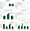
Figure 1. Nuclear Size and Import Scale Between X. Laevis and X. Tropicalis
(A) Nuclei were assembled in X. laevis or X. tropicalis egg extract with X. laevis sperm and visualized by immunofluorescence using mAb414 that recognizes the NPC. Scale bar, 20 μm. (B) NE surface area was quantified from images like those in (A) for at least 50 nuclei at each time point. Best-fit linear regression lines are displayed for six X. laevis and five X. tropicalis egg extracts, and the average difference between the two extracts was statistically significant by Student’s t-test (P < 0.001). R2 values range from 0.96 - 0.99 for X. laevis and 0.94 - 0.98 for X. tropicalis. Error bars represent standard deviation (SD). (C) X. laevis and X. tropicalis extracts were mixed as indicated, and nuclear size was measured at 90 min. One representative experiment of three is shown, and error bars represent SD. (D) Nuclei were assembled using the indicated source of extract and sperm, and nuclear size was measured at 90 min. One representative experiment of three is shown, and error bars represent SD. (E) GFP-NLS was added to nuclei at 30 min, and images were acquired live at 30 sec intervals with the same exposure time. Nuclear GFP-NLS fluorescence intensity per unit area was measured at each time point, averaged for five nuclei from each extract, and normalized to 1.0 (arbitrary units). Error bars represent SD. Representative images are at 70 min. Scale bar, 20 μm. (F) IBB-coated Qdots were added to nuclei at 30 min, and images were acquired live at the indicated time points for at least 30 nuclei with the same exposure time. Nuclear Qdot fluorescence intensity per unit area was calculated, averaged, and normalized to 1.0 (arbitrary units). Error bars represent SD. One representative experiment of three is shown. Representative images are at 75 min. Scale bar, 20 μm. See also Figure S1, Movie S1.
Figure 2. Importin α2 and Ntf2 Levels Differ in X. Laevis and X. Tropicalis
(A) 25 μg of protein from three different X. laevis and X. tropicalis egg extracts were separated by SDS-PAGE, transferred to nitrocellulose, and probed with antibodies against the indicated proteins. Values below each set of three lanes represent relative protein amounts (mean ± SD, n=3) quantified by infrared fluorescence. Absolute concentrations were determined by comparing band intensities to known concentrations of recombinant importin α2 or Ntf2 on the same blot. Two different antibodies against importin α2 and Ntf2 showed similar differences between the two species. (B) Nuclei at 80 min were processed for immunofluorescence using the same antibodies as in (A) and representative images are shown. For a given antibody, images were acquired with the same exposure time and scaled identically. Scale bar, 20 μm. (C) Quantification of nuclei displayed in (B). Nuclear fluorescence intensity per unit area was calculated for at least 50 nuclei per condition, averaged, and normalized to 1.0 (arbitrary units). Error bars represent SD. Two different antibodies against importin α2 and Ntf2 showed similar differences between the two species. See also Figure S2.
Figure 3. Importin α2 and Ntf2 Regulate Nuclear Size and Import
(A) Nuclei were assembled in X. tropicalis extract and at 40 min importin α-E was added at the indicated concentrations in addition to GFP-NLS. At 80 min images for at least 50 nuclei per condition were acquired with the same exposure time, and NE surface area was quantified, averaged, and normalized to the buffer control. Error bars represent standard error (SE). Scale bar, 20 μm. (B) Experiments were performed as in (A) with a fixed concentration (0.8 μM) of added importin α-E or a mutant version lacking the importin β binding domain (ΔIBB). Average fold change from the buffer control and SD are shown (n=4 extracts). The ΔIBB mutant did not have a strong dominant negative effect on import because it was added at a concentration below the endogenous importin α level. (C) Nuclei were assembled in X. laevis extract mock and partially immunodepleted of importin α2 (0.5-1 μM depleted). Kinetics of nuclear assembly were similar in the two extracts. At 40 min, indicated proteins were added at 1 μM as well as GFP-NLS. At 80 min images for at least 50 nuclei per condition were acquired with the same exposure time, and NE surface area and nuclear GFP-NLS fluorescence intensity were quantified. Average fold change from the mock depletion and SD are shown (n=4 extracts). (D) Recombinant Ntf2 was titrated into X. laevis extract prior to nuclear assembly. Initial kinetics of nuclear assembly were not altered by supplemental Ntf2. GFP-NLS or IBB-coated Qdots were added at 30 min. At 80 min nuclei were processed for immunofluorescence with an antibody against Ran, and images for at least 50 nuclei per condition were acquired with the same exposure time. NE surface area was quantified from Ran-stained nuclei, averaged, and normalized to the buffer control. Nuclear fluorescence intensities for Qdots, GFP-NLS, and Ran were similarly processed. Error bars represent SE. One representative experiment of three is shown. For each parameter, the difference between 0 and 1.6 μM added Ntf2 was statistically significant by Student’s t-test (P < 0.005). (E) Experiments similar to (D) were performed with a fixed Ntf2 concentration (1.6 μM) and over time. Nuclear Qdot or GFP-NLS fluorescence intensities for at least 50 nuclei per time point were averaged and normalized to 1.0 (arbitrary units). Error bars represent SE. At 95 min, the difference in Q dot import between 0 and 1.6 μM added Ntf2 was statistically significant by Student’s t-test (P < 0.001). (F) Nuclei were assembled in X. tropicalis extract supplemented with anti-Ntf2 or IgG antibodies (0.1 mg/ml). At 30 min, nuclear assembly was similar in the two conditions and Qdots or GFP-NLS was added. At 80 min immunofluorescence for Ran was performed, and nuclear parameters were quantified as in (D). Average fold change from the IgG control and SD are shown (n=6 extracts). See also Figure S3.
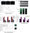
Figure 4. Importin α2 and Ntf2 are sufficient to Account for Interspecies Nuclear Scaling by Regulating LB3 Import
(A) X. tropicalis nuclei were assembled in the presence of anti-Ntf2 or IgG control antibodies (0.1 mg/ml) and 0.14 μM GFP-LB3 as indicated, and at 40 min 0.8 μM importin α-E was added to some reactions. LB3 was visualized in nuclei by immunofluorescence at 80 min and images for at least 50 nuclei per condition were acquired with the same exposure time. NE surface area and LB3 fluorescence were quantified. Average fold change from the buffer control and SD are shown (n=5 extracts). Scale bar, 20 μm. (B) Wild-type and mutant GFP-LB3 proteins, 1x Npl (nucleoplasmin), and GFP-NLS were added at 0.14 μM to X. tropicalis extract. For 5x Npl, 0.7 μM Npl was added. Nuclei were visualized at 75 min by immunofluorescence using an antibody against Ran. NE surface area was calculated for at least 50 nuclei. Average fold change from the buffer control and SD are shown (n=3 extracts). The K31Q mutant had a dominant negative effect on the structure of the lamina as nuclei were smaller and appeared crumpled, while the R385P mutant did not efficiently assemble into the lamina. (C) Nuclei were visualized by immunofluorescence with an antibody against Xenopus LB3. Images for at least 50 nuclei at each time point were acquired with the same exposure time. Fluorescence intensity was quantified, averaged, and normalized to 1.0 (arbitrary units). Error bars represent SD. One representative experiment of three is shown. The Western blot was performed as in Figure 2A using an antibody against Xenopus LB3. (D) Nuclei were assembled in X. tropicalis extract mock and immunodepleted of LB3 (0.1 μM depleted). Ntf2 antibodies, importin α-E, and GFP-LB3 were added to LB3-depleted extract in the same manner as in (A) with the exception that GFP-LB3 was added at 0.2 μM. At 80 min nuclei were stained for Ran by immunofluorescence, images for at least 50 nuclei per condition were acquired, and NE surface area was quantified. Average fold change from the mock depletion and SD are shown (n=4 extracts). Scale bar, 20 μm. See also Figure S4, Table S1.
Figure 5. Importin α2 Regulates X. Laevis Developmental Nuclear Scaling
(A) Different stage X. laevis embryos were arrested with cycloheximide for 60 min. Nuclei were isolated from embryo extracts and visualized by immunofluorescence using mAb414. Scale bar, 20 μm. For the graph, NE surface area was quantified for at least 50 nuclei from each stage. Error bars represent SD. (B) 25 μg of protein from different stage embryo extracts were analyzed by Western blot as in Figure 2A. (C) To assess nuclear import, GFP-NLS (1 μM) was added to embryo extracts and images of unfixed nuclei were acquired 30 min later with the same exposure time. Immunofluorescence was performed on fixed embryonic nuclei, and images were acquired with the same exposure time. Scale bar, 20 μm. (D) Quantification of (C). Nuclear fluorescence intensity per unit area was calculated for at least 50 nuclei per stage, averaged, and normalized to 1.0 (arbitrary units). Error bars represent SD. (E) Single-cell fertilized X. laevis embryos were injected with 1 ng importin α-E mRNA or water as control. Nuclei were isolated and quantified as in (A), except that an antibody against Xenopus LB3 was used for immunofluorescence. One representative experiment of two is shown for each stage, and error bars represent SD. (F) Experiments similar to (E) were performed except that embryos were arrested for different lengths of time in cycloheximide. Error bars represent SE. Representative stage 7 nuclei at 120 min are shown for control (bottom) and importin α-E (top) injected embryos. Scale bar, 20 μm. See also Figure S5.
Similar articles
- Altering the levels of nuclear import factors in early Xenopus laevis embryos affects later development.
Jevtić P, Mukherjee RN, Chen P, Levy DL. Jevtić P, et al. PLoS One. 2019 Apr 22;14(4):e0215740. doi: 10.1371/journal.pone.0215740. eCollection 2019. PLoS One. 2019. PMID: 31009515 Free PMC article. - Importin α Partitioning to the Plasma Membrane Regulates Intracellular Scaling.
Brownlee C, Heald R. Brownlee C, et al. Cell. 2019 Feb 7;176(4):805-815.e8. doi: 10.1016/j.cell.2018.12.001. Epub 2019 Jan 10. Cell. 2019. PMID: 30639102 Free PMC article. - Nuclear size is sensitive to NTF2 protein levels in a manner dependent on Ran binding.
Vuković LD, Jevtić P, Zhang Z, Stohr BA, Levy DL. Vuković LD, et al. J Cell Sci. 2016 Mar 15;129(6):1115-27. doi: 10.1242/jcs.181263. Epub 2016 Jan 28. J Cell Sci. 2016. PMID: 26823604 Free PMC article. - Importin α: functions as a nuclear transport factor and beyond.
Oka M, Yoneda Y. Oka M, et al. Proc Jpn Acad Ser B Phys Biol Sci. 2018;94(7):259-274. doi: 10.2183/pjab.94.018. Proc Jpn Acad Ser B Phys Biol Sci. 2018. PMID: 30078827 Free PMC article. Review. - Use of intact Xenopus oocytes in nucleocytoplasmic transport studies.
Panté N. Panté N. Methods Mol Biol. 2006;322:301-14. doi: 10.1007/978-1-59745-000-3_21. Methods Mol Biol. 2006. PMID: 16739732 Review.
Cited by
- Follicle dynamics and global organization in the intact mouse ovary.
Faire M, Skillern A, Arora R, Nguyen DH, Wang J, Chamberlain C, German MS, Fung JC, Laird DJ. Faire M, et al. Dev Biol. 2015 Jul 1;403(1):69-79. doi: 10.1016/j.ydbio.2015.04.006. Epub 2015 Apr 16. Dev Biol. 2015. PMID: 25889274 Free PMC article. - Colloid osmotic parameterization and measurement of subcellular crowding.
Mitchison TJ. Mitchison TJ. Mol Biol Cell. 2019 Jan 15;30(2):173-180. doi: 10.1091/mbc.E18-09-0549. Mol Biol Cell. 2019. PMID: 30640588 Free PMC article. Review. - Nuclear membrane protein Lem2 regulates nuclear size through membrane flow.
Kume K, Cantwell H, Burrell A, Nurse P. Kume K, et al. Nat Commun. 2019 Apr 23;10(1):1871. doi: 10.1038/s41467-019-09623-x. Nat Commun. 2019. PMID: 31015410 Free PMC article. - Nuclear F-actin and Lamin A antagonistically modulate nuclear shape.
Mishra S, Levy DL. Mishra S, et al. J Cell Sci. 2022 Jul 1;135(13):jcs259692. doi: 10.1242/jcs.259692. Epub 2022 Jul 4. J Cell Sci. 2022. PMID: 35665815 Free PMC article. - In vivo T-box transcription factor profiling reveals joint regulation of embryonic neuromesodermal bipotency.
Gentsch GE, Owens ND, Martin SR, Piccinelli P, Faial T, Trotter MW, Gilchrist MJ, Smith JC. Gentsch GE, et al. Cell Rep. 2013 Sep 26;4(6):1185-96. doi: 10.1016/j.celrep.2013.08.012. Epub 2013 Sep 19. Cell Rep. 2013. PMID: 24055059 Free PMC article.
References
- Aebi U, Cohn J, Buhle L, Gerace L. The nuclear lamina is a meshwork of intermediate-type filaments. Nature. 1986;323:560–564. - PubMed
- Clarkson WD, Kent HM, Stewart M. Separate binding sites on nuclear transport factor 2 (NTF2) for GDP-Ran and the phenylalanine-rich repeat regions of nucleoporins p62 and Nsp1p. J Mol Biol. 1996;263:517–524. - PubMed
- D’Angelo MA, Anderson DJ, Richard E, Hetzer MW. Nuclear pores form de novo from both sides of the nuclear envelope. Science. 2006;312:440–443. - PubMed
Publication types
MeSH terms
Substances
LinkOut - more resources
Full Text Sources
Other Literature Sources


