Biodistribution and radiation dosimetry of the hypoxia marker 18F-HX4 in monkeys and humans determined by using whole-body PET/CT - PubMed (original) (raw)
Clinical Trial
Biodistribution and radiation dosimetry of the hypoxia marker 18F-HX4 in monkeys and humans determined by using whole-body PET/CT
Mohan Doss et al. Nucl Med Commun. 2010 Dec.
Abstract
Objectives: F-HX4 is a novel positron emission tomography (PET) tracer for imaging hypoxia. The purpose of this study was to determine the biodistribution and estimate the radiation dose of F-HX4 using whole-body PET/computed tomography (CT) scans in monkeys and humans.
Methods: Successive whole-body PET/CT scans were done after the injection of F-HX4 in four healthy humans (422±142 MBq) and in three rhesus monkeys (189±3 MBq). Biodistribution was determined from PET images and organ doses were estimated using OLINDA/EXM software.
Results: The bladder, liver, and kidneys showed the highest percentage of the injected radioactivity for humans and monkeys. For humans, approximately 45% of the activity is eliminated by bladder voiding in 3.6 h, and for monkeys 60% is in the bladder content after 3 h. The critical organ is the urinary bladder wall with the highest absorbed radiation dose of 415±18 (monkeys) and 299±38 μGy/MBq (humans), in the 4.8-h bladder voiding interval model. The average value of effective dose for the adult male was estimated at 42±4.2 μSv/MBq from monkey data and 27±2 μSv/MBq from human data.
Conclusion: Bladder, kidneys, and liver have the highest uptake of injected F-HX4 activity for both monkeys and humans. The urinary bladder wall receives the highest dose of F-HX4 and is the critical organ. Thus, patients should be encouraged to maintain adequate hydration and void frequently. The effective dose of F-HX4 is comparable with that of other F-based imaging agents.
Trial registration: ClinicalTrials.gov NCT00606424.
Figures
Fig. 1
Synthesis of 18F-HX4 from Precursor
Fig. 2
Decay-corrected anterior maximum-intensity projections of PET at 17, 82, 120, 156, and 199 min (from left to right) after injection of 18F-HX4 in a female volunteer. There is rapid clearance of activity in kidneys, liver and bladder. Gallbladder activity peaks at 82 min then decreases with time
Fig. 3
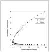
Mean percentage of injected activity and standard deviation (SD) for top 3 organs determined on the basis of 4 18F-HX4 PET emission scans in human volunteers, as a function of time after injection. Rapid clearance of activity is observed in the organs
Fig. 4
Decay-corrected anterior maximum-intensity projections of PET at 3, 13, 40, 77 and 187 min (from left to right) after injection of 18F-HX4 in a rhesus monkey. The liver and kidney activities decrease rapidly with time, and bladder accumulates activity with time (there is no voiding of bladder as the monkey is anesthetized)
Fig. 5
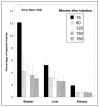
Mean percentage of injected activity and standard deviation (SD) for top three organs determined on the basis of three rhesus monkey 18F-HX4 PET emission scans, as a function of time after injection. Liver and kidney activities decrease rapidly with time, and bladder activity increases with time (there is no voiding as monkeys are anesthetized)
Similar articles
- Biodistribution and radiation dosimetry of the integrin marker 18F-RGD-K5 determined from whole-body PET/CT in monkeys and humans.
Doss M, Kolb HC, Zhang JJ, Bélanger MJ, Stubbs JB, Stabin MG, Hostetler ED, Alpaugh RK, von Mehren M, Walsh JC, Haka M, Mocharla VP, Yu JQ. Doss M, et al. J Nucl Med. 2012 May;53(5):787-95. doi: 10.2967/jnumed.111.088955. Epub 2012 Apr 12. J Nucl Med. 2012. PMID: 22499613 Free PMC article. Clinical Trial. - Biodistribution and radiation dosimetry of 18F-CP-18, a potential apoptosis imaging agent, as determined from PET/CT scans in healthy volunteers.
Doss M, Kolb HC, Walsh JC, Mocharla V, Fan H, Chaudhary A, Zhu Z, Alpaugh RK, Lango MN, Yu JQ. Doss M, et al. J Nucl Med. 2013 Dec;54(12):2087-92. doi: 10.2967/jnumed.113.119800. Epub 2013 Oct 17. J Nucl Med. 2013. PMID: 24136934 Free PMC article. Clinical Trial. - First Evaluation of PET-Based Human Biodistribution and Dosimetry of 18F-FAZA, a Tracer for Imaging Tumor Hypoxia.
Savi A, Incerti E, Fallanca F, Bettinardi V, Rossetti F, Monterisi C, Compierchio A, Negri G, Zannini P, Gianolli L, Picchio M. Savi A, et al. J Nucl Med. 2017 Aug;58(8):1224-1229. doi: 10.2967/jnumed.113.122671. Epub 2017 Feb 16. J Nucl Med. 2017. PMID: 28209906 - Biodistribution and Radiation Dosimetry for the Novel SV2A Radiotracer [(18)F]UCB-H: First-in-Human Study.
Bretin F, Bahri MA, Bernard C, Warnock G, Aerts J, Mestdagh N, Buchanan T, Otoul C, Koestler F, Mievis F, Giacomelli F, Degueldre C, Hustinx R, Luxen A, Seret A, Plenevaux A, Salmon E. Bretin F, et al. Mol Imaging Biol. 2015 Aug;17(4):557-64. doi: 10.1007/s11307-014-0820-6. Mol Imaging Biol. 2015. PMID: 25595813 - Noninvasive estimation of human radiation dosimetry of 18F-FDG by whole-body small animal PET imaging in rats.
Shidahara M, Funaki Y, Watabe H. Shidahara M, et al. Appl Radiat Isot. 2022 Mar;181:110071. doi: 10.1016/j.apradiso.2021.110071. Epub 2021 Dec 17. Appl Radiat Isot. 2022. PMID: 34952332 Review.
Cited by
- Detection of Hypoxia in Cancer Models: Significance, Challenges, and Advances.
Godet I, Doctorman S, Wu F, Gilkes DM. Godet I, et al. Cells. 2022 Feb 16;11(4):686. doi: 10.3390/cells11040686. Cells. 2022. PMID: 35203334 Free PMC article. Review. - Molecular imaging of hypoxia in non-small-cell lung cancer.
Yip C, Blower PJ, Goh V, Landau DB, Cook GJ. Yip C, et al. Eur J Nucl Med Mol Imaging. 2015 May;42(6):956-76. doi: 10.1007/s00259-015-3009-6. Epub 2015 Feb 21. Eur J Nucl Med Mol Imaging. 2015. PMID: 25701238 Review. - Radiation dosimetry and biodistribution in non-human primates of the sodium/iodide PET ligand [(18)F]-tetrafluoroborate.
Marti-Climent JM, Collantes M, Jauregui-Osoro M, Quincoces G, Prieto E, Bilbao I, Ecay M, Richter JA, Peñuelas I. Marti-Climent JM, et al. EJNMMI Res. 2015 Dec;5(1):70. doi: 10.1186/s13550-015-0148-5. Epub 2015 Dec 3. EJNMMI Res. 2015. PMID: 26635227 Free PMC article. - Pharmacokinetic modeling of a novel hypoxia PET tracer [18F]HX4 in patients with non-small cell lung cancer.
Verwer EE, Zegers CM, van Elmpt W, Wierts R, Windhorst AD, Mottaghy FM, Lambin P, Boellaard R. Verwer EE, et al. EJNMMI Phys. 2016 Dec;3(1):30. doi: 10.1186/s40658-016-0167-y. Epub 2016 Dec 12. EJNMMI Phys. 2016. PMID: 27957730 Free PMC article. - Evaluation of tumour hypoxia during radiotherapy using [18F]HX4 PET imaging and blood biomarkers in patients with head and neck cancer.
Zegers CM, Hoebers FJ, van Elmpt W, Bons JA, Öllers MC, Troost EG, Eekers D, Balmaekers L, Arts-Pechtold M, Mottaghy FM, Lambin P. Zegers CM, et al. Eur J Nucl Med Mol Imaging. 2016 Nov;43(12):2139-2146. doi: 10.1007/s00259-016-3429-y. Epub 2016 Jun 1. Eur J Nucl Med Mol Imaging. 2016. PMID: 27251643 Free PMC article. Clinical Trial.
References
- Lucignani G. PET imaging with hypoxia tracers: a must in radiation therapy. Eur J Nucl Med Mol Imaging. 2008;35:838–42. - PubMed
- Tatum JL, Kelloff GJ, Gillies RJ, Arbeit JM, Brown JM, Chao KS, et al. Hypoxia: importance in tumor biology, noninvasive measurement by imaging, and value of its measurement in the management of cancer therapy. Int J Radiat Biol. 2006;82:699–757. - PubMed
- Vaupel P. Hypoxia and aggressive tumor phenotype: implications for therapy and prognosis. Oncologist. 2008;13 3:21–6. - PubMed
- Zaffaroni N, Fiorentini G, De Giorgi U. Hyperthermia and hypoxia: new developments in anticancer chemotherapy. Eur J Surg Oncol. 2001;27:340–2. - PubMed
Publication types
MeSH terms
Substances
LinkOut - more resources
Full Text Sources
Other Literature Sources
Medical


