Preweaning Mn exposure leads to prolonged astrocyte activation and lasting effects on the dopaminergic system in adult male rats - PubMed (original) (raw)
Preweaning Mn exposure leads to prolonged astrocyte activation and lasting effects on the dopaminergic system in adult male rats
Cynthia H Kern et al. Synapse. 2011 Jun.
Abstract
Little is known about the effects of manganese (Mn) exposure over neurodevelopment and whether these early insults result in effects lasting into adulthood. To determine if early Mn exposure produces lasting neurobehavioral and neurochemical effects, we treated neonate rats with oral Mn (0, 25, or 50 mg Mn/kg/d over PND 1-21) and evaluated (1) behavioral performance in the open arena in the absence (PND 97) and presence (PND 98) of a d-amphetamine challenge, (2) brain dopamine D1 and D2-like receptors and dopamine transporter densities in the prefrontal cortex, striatum, and nucleus accumbens (PND 107), and (3) astrocyte marker glial fibrillary acidic protein (GFAP) levels in these same brain regions (PND 24 and 107). We found that preweaning Mn exposure did not alter locomotor activity or behavior disinhibition in adult rats, though Mn-exposed animals did exhibit an enhanced locomotor response to d-amphetamine challenge. Preweaning Mn exposure led to increased D1 and D2 receptor levels in the nucleus accumbens and prefrontal cortex, respectively, compared with controls. We also found increased GFAP expression in the prefrontal cortex in Mn-exposed PND 24 weanlings, and increased GFAP levels in prefrontal cortex, medial striatum and nucleus accumbens of adult (PND 107) rats exposed to preweaning Mn, indicating an effect of Mn exposure on astrogliosis that persisted and/or progressed to other brain regions in adult animals. These data show that preweaning Mn exposure leads to lasting molecular and functional impacts in multiple brain regions of adult animals, long after brain Mn levels returned to normal.
Copyright © 2010 Wiley-Liss, Inc.
Figures
Figure 1
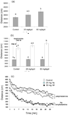
Pre-weaning Mn exposure increased open arena locomotor activity of PND 24 weanlings (panel ‘a’, ANOVA p=0.01, data from Kern et al., 2010), but not drug-naïve PND 97 adult male rats (panel ‘b’, solid bars, p=0.92). However, pre-weaning Mn exposure significantly increased the locomotor response to a d-amphetamine challenge (1.5 mg/kg) in PND 98 adults (panel ‘b’, open bars, p=0.03) (data are averages (±SE) of total distance traveled over 5–30 min). Superscripts denote significant differences between treatments based on Tukey's post-hoc analysis (p≤0.05). Panel ‘c’: Locomotor activity measured per minute over the entire 30 min test period for drug-naïve (no d-amphetamine, PND 97) and d-amphetamine-challenged (PND 98) adults; data show consistent increased activity over the entire test period after the d-amphetamine challenge (MANOVA p=0.03). Data are averages (error bars in ‘c’ omitted for clarity). N=15–20 rats per treatment. Activity was measured in 60 cm × 60 cm × 30 cm open enclosures using the SMART video tracking system (San Diego Instruments).
Figure 2
Pre-weaning Mn exposure did not alter center zone activity of drug-naïve (PND 97, solid bars, p=0.90) or 1.5 mg/kg d-amphetamine-challenged (PND 98, open bars, p=0.43) adult male rats, based on the ratio of center distance/total distance traveled over 5 – 30 min. Values are averages (±SE), n=15–20 rats per treatment. Activity was measured in 60 cm × 60 cm × 30 cm open enclosures using the SMART video tracking system (San Diego Instruments).
Figure 3
Representative immunohistochemistry photomicrographs showing that pre-weaning Mn exposure increased levels of D1 and D2 receptor proteins compared to controls in the nucleus accumbens (25 mg Mn/kg/d group) and prefrontal cortex (50 mg Mn/kg/d group) of PND 107 adult male rats (images reflect data summarized in Table II). Slides were prepared and stained with three animals/slide balanced by treatment and photographed at 20X magnification under defined illumination conditions (see text for details). Scale bar = 100 µm.
Figure 4
Representative immunohistochemistry (IHC) photomicrographs showing that pre-weaning Mn exposure increased levels of the astrocyte marker GFAP in prefrontal cortex of PND 24 rats, and the prefrontal cortex, medial striatum, and nucleus accumbens of PND 107 male rats. Panel ‘a’: GFAP expression in the prefrontal cortex of a representative 50 mg/kg/d Mn-exposed animal compared to control (40X magnification). Panel ‘b’: Diagram to indicate different striatal areas imaged for data shown in panel ‘c’ (Paxinos and Watson, 1998). Panel ‘c’: IHC fluorographs of data summarized in Table III (20× magnification). IHC slides were prepared and stained with three animals/slide balanced by treatment and photographed under defined illumination conditions (see text for details). Scale bar = 100 µm.
Similar articles
- Early postnatal manganese exposure causes arousal dysregulation and lasting hypofunctioning of the prefrontal cortex catecholaminergic systems.
Conley TE, Beaudin SA, Lasley SM, Fornal CA, Hartman J, Uribe W, Khan T, Strupp BJ, Smith DR. Conley TE, et al. J Neurochem. 2020 Jun;153(5):631-649. doi: 10.1111/jnc.14934. Epub 2020 Jan 10. J Neurochem. 2020. PMID: 31811785 Free PMC article. - Short-term manganese inhalation decreases brain dopamine transporter levels without disrupting motor skills in rats.
Saputra D, Chang J, Lee BJ, Yoon JH, Kim J, Lee K. Saputra D, et al. J Toxicol Sci. 2016;41(3):391-402. doi: 10.2131/jts.41.391. J Toxicol Sci. 2016. PMID: 27193731 - Effects of manganese on thyroid hormone homeostasis: potential links.
Soldin OP, Aschner M. Soldin OP, et al. Neurotoxicology. 2007 Sep;28(5):951-6. doi: 10.1016/j.neuro.2007.05.003. Epub 2007 May 13. Neurotoxicology. 2007. PMID: 17576015 Free PMC article. Review. - Role of Astrocytes in Manganese Neurotoxicity Revisited.
Ke T, Sidoryk-Wegrzynowicz M, Pajarillo E, Rizor A, Soares FAA, Lee E, Aschner M. Ke T, et al. Neurochem Res. 2019 Nov;44(11):2449-2459. doi: 10.1007/s11064-019-02881-7. Epub 2019 Sep 30. Neurochem Res. 2019. PMID: 31571097 Free PMC article. Review.
Cited by
- Postnatal manganese exposure does not alter dopamine autoreceptor sensitivity in adult and adolescent male rats.
McDougall SA, Mohd-Yusof A, Kaplan GJ, Abdulla ZI, Lee RJ, Crawford CA. McDougall SA, et al. Eur J Pharmacol. 2013 Apr 15;706(1-3):4-10. doi: 10.1016/j.ejphar.2013.02.030. Epub 2013 Feb 28. Eur J Pharmacol. 2013. PMID: 23458069 Free PMC article. - In vitro manganese exposure disrupts MAPK signaling pathways in striatal and hippocampal slices from immature rats.
Peres TV, Pedro DZ, de Cordova FM, Lopes MW, Gonçalves FM, Mendes-de-Aguiar CB, Walz R, Farina M, Aschner M, Leal RB. Peres TV, et al. Biomed Res Int. 2013;2013:769295. doi: 10.1155/2013/769295. Epub 2013 Nov 13. Biomed Res Int. 2013. PMID: 24324973 Free PMC article. - Impacts of a perinatal exposure to manganese coupled with maternal stress in rats: Tests of untrained behaviors.
McDaniel KL, Beasley TE, Oshiro WM, Huffstickler M, Moser VC, Herr DW. McDaniel KL, et al. Neurotoxicol Teratol. 2022 May-Jun;91:107088. doi: 10.1016/j.ntt.2022.107088. Epub 2022 Mar 10. Neurotoxicol Teratol. 2022. PMID: 35278630 Free PMC article. - Developmental manganese, lead, and barren cage exposure have adverse long-term neurocognitive, behavioral and monoamine effects in Sprague-Dawley rats.
Sprowles JLN, Amos-Kroohs RM, Braun AA, Sugimoto C, Vorhees CV, Williams MT. Sprowles JLN, et al. Neurotoxicol Teratol. 2018 May-Jun;67:50-64. doi: 10.1016/j.ntt.2018.04.001. Epub 2018 Apr 7. Neurotoxicol Teratol. 2018. PMID: 29631003 Free PMC article. - Evaluation of neurobehavioral and neuroinflammatory end-points in the post-exposure period in rats sub-acutely exposed to manganese.
Santos D, Batoréu MC, Tavares de Almeida I, Davis Randall L, Mateus ML, Andrade V, Ramos R, Torres E, Aschner M, Marreilha dos Santos AP. Santos D, et al. Toxicology. 2013 Dec 6;314(1):95-9. doi: 10.1016/j.tox.2013.09.008. Epub 2013 Sep 20. Toxicology. 2013. PMID: 24060432 Free PMC article.
References
- Aldskogius H, Kozlova EN. Central neuron-glial and glial-glial interactions following axon injury. Prog Neurobiol. 1998;55(1):1–26. - PubMed
- Antonopoulos J, Dori I, Dinopoulos A, Chiotelli M, Parnavelas JG. Postnatal development of the dopaminergic system of the striatum in the rat. Neuroscience. 2002;110(2):245–256. - PubMed
- Archer T, Fredriksson A. Functional changes implicating dopaminergic systems following perinatal treatments. Dev Pharmacol Ther. 1992;18(3–4):201–222. - PubMed
- Arcus-Arth A, Krowech G, Zeise L. Breast milk and lipid intake distributions for assessing cumulative exposure and risk. J Expo Anal Environ Epidemiol. 2005;15(4):357–365. - PubMed
- Arnsten AF. Fundamentals of attention-deficit/hyperactivity disorder: circuits and pathways. J Clin Psychiatry. 2006;67 Suppl 8:7–12. - PubMed
Publication types
MeSH terms
Substances
LinkOut - more resources
Full Text Sources
Miscellaneous


