Preparing synthetic Aβ in different aggregation states - PubMed (original) (raw)
Preparing synthetic Aβ in different aggregation states
W Blaine Stine et al. Methods Mol Biol. 2011.
Abstract
This chapter outlines protocols that produce homogenous preparations of oligomeric and fibrillar amyloid-β peptide (Aβ). While there are several isoforms of this peptide, the 42 amino acid form is the focus because of its genetic and pathological link to Alzheimer's disease (AD). Past decades of AD research highlight the dependence of Aβ42 function on its structural assembly state. Biochemical, cellular and in vivo studies of Aβ42 usually begin with purified peptide obtained by chemical synthesis or recombinant expression. The initial steps to solubilize and prepare these purified dry peptide stocks are critical to controlling the structural assembly of Aβ. To develop homogenous Aβ42 assemblies, we initially monomerize the peptide, erasing any "structural history" that could seed aggregation, by using a strong solvent. It is this starting material that has allowed us to define and optimize conditions that consistently produce homogenous solutions of soluble oligomeric and fibrillar Aβ42 assemblies. These preparations have been developed and characterized by using atomic force microscopy (AFM) to identify the structurally discrete species formed by Aβ42 under specific solution conditions. These preparations have been used extensively to demonstrate a variety of functional differences between oligomeric and fibrillar Aβ42. We also present a protocol for fluorescently labeling oligomeric Aβ42 that does not affect structure, as measured by AFM, or function, as measured by a cellular uptake assay. These reagents are critical experimental tools that allow for defining specific structure/function connections.
Figures
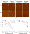
Fig. 1
Structure and neurotoxicity of oligomeric or fibrillar Aβ42 and Aβ40 assemblies. (a, b) Aβ42, but not Aβ40, forms oligomeric and fibrillar assemblies. 5 mM HFIP-treated Aβ42 (a) or Aβ40 (b) in DMSO was diluted to 100 μM in ice-cold F-12 culture media for oligomers, or 10 mM HCl for fibrils. Oligomer and fibril preparations were incubated for 24 h at 4°C and 37°C, respectively. Samples before (0 h) and after incubation (24 h) were mounted for AFM analysis at 10 μM. Representative 2 × 2 μm x–y, 10 nm total z-range AFM images. Inset images 200 × 200 nm x–y, 2 nm total z-range. Reprinted from Stine et al., JBC, 2003, with permission from ASBMB. (c, d) Oligomeric Aβ42, but not Aβ40, reduces neuronal viability significantly more than fibrillar and unaggregated species. Unaggregated, oligomeric, and fibrillar preparations of Aβ42 (c) or Aβ40 (d) were incubated with N2A cells for 20 h. Oligomeric and fibrillar preparations of Aβ were prepared as described above. For unaggregated peptide preparations, the 5 mM Aβ in DMSO was diluted directly into cell culture media. The MTT assay was used as an indicator of cell viability. Graph represents the mean ± SEM for n ≥ 10 from triplicate wells from at least three separate experiments using different Aβ preparations. * Significant difference between Aβassemblies prepared in oligomers and fibrils conditions (p < 0.001). ** Significantdifference between unaggregated and both Aβ assemblies prepared oligomers and fibrils conditions (p < 0.001). Reprinted from Dahlgren et al., JBC 2002, with permission from ASBMB.
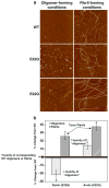
Fig. 2
Structure and neurotoxicity of oligomeric or fibrillar wild type (WT), Dutch (E22Q), and Arctic (E22G) Aβ42. (a) Oligomeric and fibrillar preparations of Aβ were prepared as described in Subheadings 3.3 and 3.4 and imaged at 10 μM. Both E22Q and E22G Aβ42 exhibit enhanced fibril formation, even under oligomer-forming conditions. Representative 2 × 2 μm, 10 nm total z-range AFM images of 100 μM Aβ. Reprinted from Dahlgren et al., JBC, 2002, with permission from ASBMB. (b) The “toxic fibrils” formed by E22Q and E22G are significantly more toxic than WT oligomers. Changes to structural assembly states of mutant Aβ42 observed by AFM (above) translate into changes in function as measured by cellular toxicity. N2A cells were treated for 20 h with 0.1 μM of WT Aβ42 oligomers and fibrils, or mutant E22Q Aβ42 or mutant E22G Aβ42 assemblies from oligomer and fibril-forming conditions. MTT assay was used as an indicator of cell viability. The data represent n ≥ 8 triplicate wells from at least two separate experiments using different Aβ preparations. * Significant difference between oligomers and fibrils (p < 0.01). Reprinted with modifications from Dahlgren et al., JBC, 2002, with permission from ASBMB.
Fig. 3
AFM analysis of Aβ42 solubilized in HFIP and H2O. Lyophilized synthetic Aβ42 was solubilized to 5 mM in 100% HFIP or deionized H2O. 5 mM stock solutions were incubated for 24 h at RT. Samples before (0 h) and after incubation (24 h) were mounted for AFM analysis at 10 μM. Representative 1 × 1 μm x–y, 5 nm total z-range AFM images. Inset image 390 × 390 nm x–y, 5 nm total z-range. Reprinted with modifications from Stine et al., JBC, 2003, with permission from ASBMB.
Fig. 4
Schematic diagram summarizing the solubilization and aggregation conditions developed for preparing oligomeric and fibrillar Aβ42. Synthetic Aβ42 is dissolved to 1 mM in 100% HFIP, HFIP is evaporated, and the dry peptide is stored at -20°C. For the aggregation protocols, the peptide is first resuspended in dry DMSO to 5 mM. For oligomeric conditions, F-12 (without phenol red) culture media is added to bring the peptide to a final concentration of 100 μM, and incubated at 4°C for 24 h. For fibrillar conditions, 10 mM HCl is added to bring the peptide to a final concentration of 100 μM, and incubated for 24 h at 37°C. Reprinted from Dahlgren et al., JBC, 2002, with permission from ASBMB.
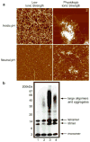
Fig. 5
Diverse Aβ42 assemblies imaged by AFM are not preserved by SDS-PAGE. (a) AFM images of Aβ42 fibrils, “plaque in a dish,” oligomers, and coalesced oligomers. 5 mM Aβ42 in DMSO was diluted to 100 μM in either 10 mM HCl (1, acidic pH, low ionic strength), 10 mM HCl + 150 mM NaCl (2, acidic pH, physiologic ionic strength), 10 mM Tris, pH 7.4 (3, neutral pH, low ionic strength), or 10 mM Tris, and pH 7.4 + 150 mM NaCl (4, neutral pH, physiologic ionic strength). Samples were prepared after a 2 h incubation at 37°C. Representative 2 × 2 μm x–y, 10 nm total z-range AFM images are shown, except for panel 2, which is scaled to 2 × 2μm x–y, 25 nm total z-range. Reprinted from Stine et al., JBC, 2003, with permission from ASBMB. (b) Western analysis of SDS-PAGE does not produce an immunoreactive pattern that correlates with AFM images in Panel A. Representative Western blots of Aβ42 assemblies prepared as described above, separated by SDS-PAGE on a 12% NuPAGE BisTRIS gel and probed with the monoclonal antibody 6E10 (recognizing residues 1–16 of Aβ). Samples were visualized by enhanced chemiluminescence. Lanes numbers correspond to panel numbers in (a): HCl (lane 1), HCl + NaCl (lane 2), Tris (lane 3), and Tris + NaCl (lane 4). Reprinted from Stine et al., JBC, 2003, with permission from ASBMB.
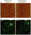
Fig. 6
Structure and neuronal uptake of Alexa Fluor® 488-labeled Aβ42 oligomers compared to unlabeled Aβ42 oligomers. (a) AFM analysis shows that oligomer assemblies are preserved after fluorophore-labeling. Aβ42 oligomers were prepared from unlabeled synthetic Aβ42 HFIP films (100 μM, PBS pH 7.4, 4°C) and analyzed by AFM (a1). Fluorophore-labeling of the oligomers with Alexa Fluor® 488 was performed using the Microscale Protein Labeling Kit and analyzed by AFM (a2). Unlabeled oligomers were diluted to 20 μM for analysis and Alexa-labeled oligomers were analyzed without dilution (estimated concentration of 25 μM). All AFM images shown are 2 × 2 μm x–y, 10 nm total z-range. (b) Following uptake, Alexa Fluor® 488-labeled oligomers (b2) appear as punctate fluorescence within the cell, similar to immunodetection of unlabeled Aβ42 oligomers (b1). Following 16 h treatment, Aβ uptake in N2A cells was analyzed using laser-scanning confocal microscopy. Panel B1 shows the image of cells treated with unlabeled Aβ42 oligomers, immunodetected with anti-Aβ monoclonal antibody 6E10 and Alexa488-rabbit-anti-mouse antibody. Panel B2 shows N2A cells treated for 16 h with 2 μM Alexa Fluor® 488-labeled Aβ42 oligomers. Scale bar = 44 μm. The insets show a single-cell magnification, scale bar = 12 μm. Reprinted from Jungbauer et al., Preparation of fluorescently labeled amyloid-beta peptide assemblies: the effect of fluorophore conjugation on structure and function, J. Mol. Recog., 2009, with permission from Wiley.
Similar articles
- Preparation of fluorescently-labeled amyloid-beta peptide assemblies: the effect of fluorophore conjugation on structure and function.
Jungbauer LM, Yu C, Laxton KJ, LaDu MJ. Jungbauer LM, et al. J Mol Recognit. 2009 Sep-Oct;22(5):403-13. doi: 10.1002/jmr.948. J Mol Recognit. 2009. PMID: 19343729 Free PMC article. - Dynamics of Inter-Molecular Interactions Between Single Aβ42 Oligomeric and Aggregate Species by High-Speed Atomic Force Microscopy.
Feng L, Watanabe H, Molino P, Wallace GG, Phung SL, Uchihashi T, Higgins MJ. Feng L, et al. J Mol Biol. 2019 Jul 12;431(15):2687-2699. doi: 10.1016/j.jmb.2019.04.044. Epub 2019 May 7. J Mol Biol. 2019. PMID: 31075274 - Physicochemical characteristics of soluble oligomeric Abeta and their pathologic role in Alzheimer's disease.
Watson D, Castaño E, Kokjohn TA, Kuo YM, Lyubchenko Y, Pinsky D, Connolly ES Jr, Esh C, Luehrs DC, Stine WB, Rowse LM, Emmerling MR, Roher AE. Watson D, et al. Neurol Res. 2005 Dec;27(8):869-81. doi: 10.1179/016164105X49436. Neurol Res. 2005. PMID: 16354549 Review. - Understanding amyloid fibril nucleation and aβ oligomer/drug interactions from computer simulations.
Nguyen P, Derreumaux P. Nguyen P, et al. Acc Chem Res. 2014 Feb 18;47(2):603-11. doi: 10.1021/ar4002075. Epub 2013 Dec 24. Acc Chem Res. 2014. PMID: 24368046 Review.
Cited by
- β-amyloid monomer scavenging by an anticalin protein prevents neuronal hyperactivity in mouse models of Alzheimer's Disease.
Zott B, Nästle L, Grienberger C, Unger F, Knauer MM, Wolf C, Keskin-Dargin A, Feuerbach A, Busche MA, Skerra A, Konnerth A. Zott B, et al. Nat Commun. 2024 Jul 10;15(1):5819. doi: 10.1038/s41467-024-50153-y. Nat Commun. 2024. PMID: 38987287 Free PMC article. - The Sigma-2 Receptor/TMEM97, PGRMC1, and LDL Receptor Complex Are Responsible for the Cellular Uptake of Aβ42 and Its Protein Aggregates.
Riad A, Lengyel-Zhand Z, Zeng C, Weng CC, Lee VM, Trojanowski JQ, Mach RH. Riad A, et al. Mol Neurobiol. 2020 Sep;57(9):3803-3813. doi: 10.1007/s12035-020-01988-1. Epub 2020 Jun 23. Mol Neurobiol. 2020. PMID: 32572762 Free PMC article. - BAG2 Is Repressed by NF-κB Signaling, and Its Overexpression Is Sufficient to Shift Aβ1-42 from Neurotrophic to Neurotoxic in Undifferentiated SH-SY5Y Neuroblastoma.
Santiago FE, Almeida MC, Carrettiero DC. Santiago FE, et al. J Mol Neurosci. 2015 Sep;57(1):83-9. doi: 10.1007/s12031-015-0579-5. Epub 2015 May 19. J Mol Neurosci. 2015. PMID: 25985852 - Early Aggregation of Amyloid-β(1-42) Studied by Fluorescence Correlation Spectroscopy.
Novo M, Pérez-González C, Freire S, Al-Soufi W. Novo M, et al. Methods Mol Biol. 2023;2551:1-14. doi: 10.1007/978-1-0716-2597-2_1. Methods Mol Biol. 2023. PMID: 36310192 - The amyloid-β degradation intermediate Aβ34 is pericyte-associated and reduced in brain capillaries of patients with Alzheimer's disease.
Kirabali T, Rigotti S, Siccoli A, Liebsch F, Shobo A, Hock C, Nitsch RM, Multhaup G, Kulic L. Kirabali T, et al. Acta Neuropathol Commun. 2019 Dec 3;7(1):194. doi: 10.1186/s40478-019-0846-8. Acta Neuropathol Commun. 2019. PMID: 31796114 Free PMC article.
References
- Mastrangelo IA, Ahmed M, Sato T, Liu W, Wang C, Hough P, Smith SO. High-resolution atomic force microscopy of soluble Abeta42 oligomers. J Mol Biol. 2006;358:106–19. - PubMed
- Huang TH, Yang DS, Plaskos NP, Go S, Yip CM, Fraser PE, Chakrabartty A. Structural studies of soluble oligomers of the Alzheimer beta-amyloid peptide. J Mol Biol. 2000;297:73–87. - PubMed
- Harper JD, Wong SS, Lieber CM, Lansbury PT. Observation of metastable Abeta amyloid protofibrils by atomic force microscopy. Chem Biol. 1997;4:119–25. - PubMed
Publication types
MeSH terms
Substances
Grants and funding
- P01 AG030128-03/AG/NIA NIH HHS/United States
- P01AG030128-A2/AG/NIA NIH HHS/United States
- P01AG021184/AG/NIA NIH HHS/United States
- P01 AG030128/AG/NIA NIH HHS/United States
- F32 AG030256/AG/NIA NIH HHS/United States
- R01 AG19121/AG/NIA NIH HHS/United States
- 1F32AG030256-01/AG/NIA NIH HHS/United States
- P01 AG021184/AG/NIA NIH HHS/United States
- R01 AG019121/AG/NIA NIH HHS/United States
LinkOut - more resources
Full Text Sources
Other Literature Sources
Miscellaneous


