Dermoscopic features of eccrine porocarcinoma arising from hidroacanthoma simplex - PubMed (original) (raw)
Case Reports
Dermoscopic features of eccrine porocarcinoma arising from hidroacanthoma simplex
Reiko Suzaki et al. Dermatol Res Pract. 2010.
Abstract
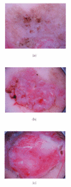
Eccrine porocarcinoma is a rare cutaneous neoplasm that mainly affects elderly people and grows slowly over a long period of time but often experiences an accelerated growth phase. Eccrine porocarcinoma may arise de novo or evolve from a pre-existing benign eccrine poroma. We reported a 86-year-old Japanese woman presenting with two reddish-colored pendulated lesions on a keratotic light brown plaque on the right thigh. Dermoscopic examination of the light-brown plaque demonstrated many whitish globular structures in a light-brown background. At the two reddish-colored pendulated lesions, polymorphous and prominent vessel proliferation was observed together with irregularly shaped whitish negative network. Immunohistochemical study demonstrated a positive CEA staining at ductal structures and atypical clear cells of reddish nodules. A diagnosis of eccrine porocarcinoma arising in a pigmented hidroacanthoma simplex was eventually established, and the dermoscopic features of eccrine porocarcinoma from hidroacanthoma simplex was described for the first time.
Figures
Figure 1
Two reddish-colored pendulated lesions at the peripheries of a keratotic light brown plaque.
Figure 2
(a) Dermoscopic examination of the light brown plaque demonstrated a lot of whitish globular structures on the light brown background. (b) In the left pendulated nodule, prominent polymorphous vessels were observed together with irregularly shaped whitish negative network. (c) Vessels were more conspicuous and irregular in the right nodule.
Figure 3
Histopathologic examination of the flat pigmented plaque disclosed many well-defined nests within the epidermis. (HE ×200).
Figure 4
(a) In the right red nodule, the epidermis was prominently acanthotic with intraepidermal proliferation of clear cells and squamoid cells. Dermal invasion of atypical squamoid cells was partially apparent (HE ×20). (b) In the right red nodule, the tumor cells were focally positive for carcinoembryonic antigen (CEA, ×20). (c) In the right red nodule, markedly atypical cells proliferated and extended throughout the entire thickness of the acanthotic epidermis (HE ×400). (d) The atypical clear cells expressed CEA (CEA ×400). (e) The ductal structures expressed CEA (CEA, ×400).
Figure 5
In the left red nodule, although the atypical basaloid cells proliferated in the epidermis, there were no visible invasions in the dermis (HE ×40).
Similar articles
- A case of porocarcinoma arising in pigmented hidroacanthoma simplex with multiple lymph node, liver and bone metastases.
Ishida M, Hotta M, Kushima R, Okabe H. Ishida M, et al. J Cutan Pathol. 2011 Feb;38(2):227-31. doi: 10.1111/j.1600-0560.2009.01440.x. J Cutan Pathol. 2011. PMID: 19788447 - Eccrine porocarcinoma shares dermoscopic characteristics with eccrine poroma: A report of three cases and review of the published work.
Edamitsu T, Minagawa A, Koga H, Uhara H, Okuyama R. Edamitsu T, et al. J Dermatol. 2016 Mar;43(3):332-5. doi: 10.1111/1346-8138.13082. Epub 2015 Sep 1. J Dermatol. 2016. PMID: 26333057 Review. - Porocarcinoma arising in pigmented hidroacanthoma simplex.
Ueo T, Kashima K, Daa T, Kondoh Y, Yanagi T, Yokoyama S. Ueo T, et al. Am J Dermatopathol. 2005 Dec;27(6):500-3. doi: 10.1097/01.dad.0000148874.20941.43. Am J Dermatopathol. 2005. PMID: 16314706 - [Eccrine porocarcinoma of the scalp].
Ekmekcı S, Lebe B. Ekmekcı S, et al. Turk Patoloji Derg. 2013;29(2):156-9. doi: 10.5146/tjpath.2013.01169. Turk Patoloji Derg. 2013. PMID: 23661356 Turkish. - [A case report of eccrine porocarcinoma].
Fumo G, Piras V, Pilloni L, Ferreli C, Pau M. Fumo G, et al. Clin Ter. 2015;166(4):e273-5. doi: 10.7417/T.2015.1873. Clin Ter. 2015. PMID: 26378762 Review. Italian.
Cited by
- Dermoscopy of non-pigmented eccrine poromas: study of Mexican cases.
Espinosa AE, Ortega BC, Venegas RQ, Ramírez RG. Espinosa AE, et al. Dermatol Pract Concept. 2013 Jan 31;3(1):25-8. doi: 10.5826/dpc.0301a07. Print 2013 Jan. Dermatol Pract Concept. 2013. PMID: 23785633 Free PMC article. - Dermoscopy findings of hidroacanthoma simplex.
Sato Y, Fujimura T, Tamabuchi E, Haga T, Aiba S. Sato Y, et al. Case Rep Dermatol. 2014 May 23;6(2):154-8. doi: 10.1159/000363369. eCollection 2014 May. Case Rep Dermatol. 2014. PMID: 24987351 Free PMC article. - Diagnosis and Management of Porocarcinoma.
Miyamoto K, Yanagi T, Maeda T, Ujiie H. Miyamoto K, et al. Cancers (Basel). 2022 Oct 25;14(21):5232. doi: 10.3390/cancers14215232. Cancers (Basel). 2022. PMID: 36358649 Free PMC article. Review. - Porocarcinoma; presentation and management, a meta-analysis of 453 cases.
Salih AM, Kakamad FH, Baba HO, Salih RQ, Hawbash MR, Mohammed SH, Othman S, Saeed YA, Habibullah IJ, Muhialdeen AS, Nawroly RO, Hammood ZD, Abdulkarim NH. Salih AM, et al. Ann Med Surg (Lond). 2017 Jun 20;20:74-79. doi: 10.1016/j.amsu.2017.06.027. eCollection 2017 Aug. Ann Med Surg (Lond). 2017. PMID: 28721214 Free PMC article. Review. - Hidroacanthoma simplex: dermoscopy and cryosurgery treatment.
Furlan KC, Kakizaki P, Chartuni JCN, Sittart JA, Valente NYS. Furlan KC, et al. An Bras Dermatol. 2017 Mar-Apr;92(2):253-255. doi: 10.1590/abd1806-4841.20174883. An Bras Dermatol. 2017. PMID: 28538891 Free PMC article.
References
- Brown CW, Jr., Dy LC. Eccrine porocarcinoma. Dermatologic Therapy. 2008;21(6):433–438. - PubMed
- Altamura D, Piccolo D, Lozzi GP, Peris K. Eccrine poroma in an unusual site: a clinical and dermoscopic simulator of amelanotic melanoma. Journal of the American Academy of Dermatology. 2005;53(3):539–541. - PubMed
- Nicolino R, Zalaudek I, Ferrara G, et al. Dermoscopy of eccrine poroma. Dermatology. 2007;215(2):160–163. - PubMed
- Kuo H-W, Ohara K. Pigmented eccrine poroma: a report of two cases and study with dermatoscopy. Dermatologic Surgery. 2003;29(10):1076–1079. - PubMed
- Ferrari A, Buccini P, Silipo V, et al. Eccrine poroma: a clinical-dermoscopic study of seven cases. Acta Dermato-Venereologica. 2009;89(2):160–164. - PubMed
Publication types
LinkOut - more resources
Full Text Sources


