Metabotropic glutamate receptors: from the workbench to the bedside - PubMed (original) (raw)
Review
Metabotropic glutamate receptors: from the workbench to the bedside
F Nicoletti et al. Neuropharmacology. 2011 Jun.
Abstract
Metabotropic glutamate (mGlu) receptors were discovered in the mid 1980s and originally described as glutamate receptors coupled to polyphosphoinositide hydrolysis. Almost 6500 articles have been published since then, and subtype-selective mGlu receptor ligands are now under clinical development for the treatment of a variety of disorders such as Fragile-X syndrome, schizophrenia, Parkinson's disease and L-DOPA-induced dyskinesias, generalized anxiety disorder, chronic pain, and gastroesophageal reflux disorder. Prof. Erminio Costa was linked to the early times of the mGlu receptor history, when a few research groups challenged the general belief that glutamate could only activate ionotropic receptors and all metabolic responses to glutamate were secondary to calcium entry. This review moves from those nostalgic times to the most recent advances in the physiology and pharmacology of mGlu receptors, and highlights the role of individual mGlu receptor subtypes in the pathophysiology of human disorders. This article is part of a Special Issue entitled 'Trends in neuropharmacology: in memory of Erminio Costa'.
Copyright © 2010 Elsevier Ltd. All rights reserved.
Figures
Fig. 1
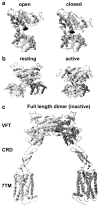
Modular structure of mGlu receptors. a) Ribbon view of the open (left) and closed (right) mGlu1 receptor Venus Fly Trap (VFT) bound with glutamate (back). Images were prepared using the coordinates of the glutamate-bound mGlu1 receptor VFT dimer (pdb 1EWK), in which one VFT is closed while the other is open. b) Side view of the mGlu1 receptor dimer bearing the VFT in its empty “resting” state (left) (pdb 1EWT), or agonist occupied “active” orientation (right) (pdb 1EWK). The front VFT is in light grey, while the one in the back is black. c) General organization of an mGlu receptor deduced from the structure of the dimeric mGlu3 extracellular domain (VFT + CRD) (pdb 1E4U) associated with two rhodopsinlike 7-TM domains.
Fig. 2
Protein–protein interactions involving group-I mGlu receptors in the post-synaptic densities. Long isoforms of Homer proteins allows the formation of multi-molecular complexes including mGlu1 and mGlu5 receptors. Interactions with NMDA receptors, TrpC ion channels, inositol-1,4,5-trisphosphate receptors (InsP3R), or PIKE-L are shown. Short Homer1a lacking the coiled-coil domain disrupts the formation of the multimolecular complex, thereby affecting mGlu1/5 receptor signalling.
Fig. 3
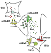
Synaptic distribution of group-II and group-III mGlu receptors. Note that presynaptic mGlu2/3 receptors are located in the pre-terminal regions of the axons, where they can be activated by glutamate released from astrocytes via the cystine/ glutamate antiporter. In contrast, presynaptic mGlu4/7/8 receptors are located near to the active zone of neurotransmitter release. Glial mGlu3 receptors induce the formation and secretion of TGF-β. The presence of mGlu5 receptors in glial cells is also shown.
Fig. 4
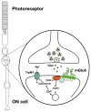
Localization and function of mGlu6 receptors in retinal ON-bipolar cells. mGlu6 receptors are present in the dendrites of the ON-bipolar cells of the retina, where their activation negatively modulates TrpM1 channels through a chain of events that include intracellular calcium release and activation of the protein phosphatase, calcineurin.
Similar articles
- Metabotropic glutamate receptors as therapeutic targets in Parkinson's disease: An update from the last 5 years of research.
Litim N, Morissette M, Di Paolo T. Litim N, et al. Neuropharmacology. 2017 Mar 15;115:166-179. doi: 10.1016/j.neuropharm.2016.03.036. Epub 2016 Apr 4. Neuropharmacology. 2017. PMID: 27055772 Review. - Group III metabotropic glutamate receptors: pharmacology, physiology and therapeutic potential.
Mercier MS, Lodge D. Mercier MS, et al. Neurochem Res. 2014 Oct;39(10):1876-94. doi: 10.1007/s11064-014-1415-y. Epub 2014 Aug 22. Neurochem Res. 2014. PMID: 25146900 Review. - The impact of metabotropic glutamate receptors into active neurodegenerative processes: A "dark side" in the development of new symptomatic treatments for neurologic and psychiatric disorders.
Bruno V, Caraci F, Copani A, Matrisciano F, Nicoletti F, Battaglia G. Bruno V, et al. Neuropharmacology. 2017 Mar 15;115:180-192. doi: 10.1016/j.neuropharm.2016.04.044. Epub 2016 Apr 30. Neuropharmacology. 2017. PMID: 27140693 Review. - Allosteric modulation of metabotropic glutamate receptors.
Sheffler DJ, Gregory KJ, Rook JM, Conn PJ. Sheffler DJ, et al. Adv Pharmacol. 2011;62:37-77. doi: 10.1016/B978-0-12-385952-5.00010-5. Adv Pharmacol. 2011. PMID: 21907906 Free PMC article. Review.
Cited by
- Neuronal autoantigens--pathogenesis, associated disorders and antibody testing.
Lancaster E, Dalmau J. Lancaster E, et al. Nat Rev Neurol. 2012 Jun 19;8(7):380-90. doi: 10.1038/nrneurol.2012.99. Nat Rev Neurol. 2012. PMID: 22710628 Free PMC article. Review. - Glial metabotropic glutamate receptor-4 increases maturation and survival of oligodendrocytes.
Spampinato SF, Merlo S, Chisari M, Nicoletti F, Sortino MA. Spampinato SF, et al. Front Cell Neurosci. 2015 Jan 14;8:462. doi: 10.3389/fncel.2014.00462. eCollection 2014. Front Cell Neurosci. 2015. PMID: 25642169 Free PMC article. - Amygdala pain mechanisms.
Neugebauer V. Neugebauer V. Handb Exp Pharmacol. 2015;227:261-84. doi: 10.1007/978-3-662-46450-2_13. Handb Exp Pharmacol. 2015. PMID: 25846623 Free PMC article. Review. - Role of presynaptic metabotropic glutamate receptors in the induction of long-term synaptic plasticity of vesicular release.
Upreti C, Zhang XL, Alford S, Stanton PK. Upreti C, et al. Neuropharmacology. 2013 Mar;66:31-9. doi: 10.1016/j.neuropharm.2012.05.004. Epub 2012 May 22. Neuropharmacology. 2013. PMID: 22626985 Free PMC article. Review. - Metabotropic Glutamate Receptor Subtype 7 in the Bed Nucleus of the Stria Terminalis is Essential for Intermale Aggression.
Masugi-Tokita M, Flor PJ, Kawata M. Masugi-Tokita M, et al. Neuropsychopharmacology. 2016 Feb;41(3):726-35. doi: 10.1038/npp.2015.198. Epub 2015 Jul 7. Neuropsychopharmacology. 2016. PMID: 26149357 Free PMC article.
References
- Abe T, Sugihara H, Nawa H, Shigemoto R, Mizuno N, Nakanishi S. Molecular characterization of a novel metabotropic glutamate receptor mGluR5 coupled to inositol phosphate/Ca2+ signal transduction. J Biol Chem. 1992;267:13361–13368. - PubMed
- Abraham WC. Metaplasticity: tuning synapses and networks for plasticity. Nat Rev Neurosci. 2008;9:387. - PubMed
- Adewale AS, Platt DM, Spealman RD. Pharmacological stimulation of group II metabotropic glutamate receptors reduces cocaine self-administration and cocaine-induced reinstatement of drug seeking in squirrel monkeys. J Pharmacol Exp Ther. 2006;318:922–931. - PubMed
- Aghajanian GK, Marek GJ. Serotonin, via 5-HT2A receptors, increases EPSCs in layer V pyramidal cells of prefrontal cortex by an asynchronous mode of glutamate release. Brain Res. 1999;825:161–171. - PubMed
Publication types
MeSH terms
Substances
Grants and funding
- G0601813/MRC_/Medical Research Council/United Kingdom
- R01 NS031373/NS/NINDS NIH HHS/United States
- R01 NS037436/NS/NINDS NIH HHS/United States
- R01 NS037436-08/NS/NINDS NIH HHS/United States
LinkOut - more resources
Full Text Sources
Other Literature Sources
Molecular Biology Databases


