Reducing excessive GABA-mediated tonic inhibition promotes functional recovery after stroke - PubMed (original) (raw)
Reducing excessive GABA-mediated tonic inhibition promotes functional recovery after stroke
Andrew N Clarkson et al. Nature. 2010.
Abstract
Stroke is a leading cause of disability, but no pharmacological therapy is currently available for promoting recovery. The brain region adjacent to stroke damage-the peri-infarct zone-is critical for rehabilitation, as it shows heightened neuroplasticity, allowing sensorimotor functions to re-map from damaged areas. Thus, understanding the neuronal properties constraining this plasticity is important for the development of new treatments. Here we show that after a stroke in mice, tonic neuronal inhibition is increased in the peri-infarct zone. This increased tonic inhibition is mediated by extrasynaptic GABA(A) receptors and is caused by an impairment in GABA (γ-aminobutyric acid) transporter (GAT-3/GAT-4) function. To counteract the heightened inhibition, we administered in vivo a benzodiazepine inverse agonist specific for α5-subunit-containing extrasynaptic GABA(A) receptors at a delay after stroke. This treatment produced an early and sustained recovery of motor function. Genetically lowering the number of α5- or δ-subunit-containing GABA(A) receptors responsible for tonic inhibition also proved beneficial for recovery after stroke, consistent with the therapeutic potential of diminishing extrasynaptic GABA(A) receptor function. Together, our results identify new pharmacological targets and provide the rationale for a novel strategy to promote recovery after stroke and possibly other brain injuries.
Figures
Figure 1. Elevated tonic inhibition in peri-infarct cortex
a, Images showing the peri-infarct recording site. Whole-cell patch-clamp recordings were made from post-stroke brain slices, within 200μm of infarct (top left), from layer-2/3 (top right) pyramidal neurons (bottom panels). b, Box-plot (boxes: 25–75%, whiskers:10–90%, lines: median) showing significantly elevated tonic inhibition in peri-infarct cortex (asterisk: P<0.05; see Supplementary Fig. 2 for additional analyses). **c,d,** Representative traces showing tonic inhibitory currents in control and peri-infarct neurons, respectively. Tonic currents were revealed by the shift in holding currents after blocking all GABAARs with gabazine (>100μM). Cells were voltage-clamped at +10mV.
Figure 2. Post-stroke impairment in GABA transport and effect of blocking α5-GABAARs
a, Blocking GAT-1 (NO-711) produced a higher % increase in _I_tonic after stroke; combined blockade of GAT-1 and GAT-3/4 (NO-711 + SNAP-5114) produced a substantial _I_tonic increase in controls but only an increase equivalent to blocking GAT-1 alone after stroke. b,c, _I_tonic in sequential drug applications. Note the lack of response to SNAP-5114 application in the post-stroke slice. d, L655,708 reduced _I_tonic. e, L655,708 significantly decreased post-stroke _I_tonic. f, Drug treatment reverted post-stroke _I_tonic to near-control level (asterisk: P<0.05; n.s.: no significance, bar graphs represent mean ± s.e.m.).
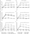
Figure 3. Behavioral recovery after stroke with L655,708 treatment and in Gabra5−/−and Gabrd−/− animals
a–c, L655,708 treatment starting from 3-days post-stroke resulted in a dose-dependent improvement in functional recovery post-stroke. d–f, Gabra5−/− and Gabrd−/− mice also showed decreased motor deficits post-stroke. Functional recovery was assessed with forelimb (a, d) and hindlimb foot-faults (b, e), and on the forelimb asymmetry (c, f). Low-dose L655,708 = 200μg/kg/day per animal; high-dose L655,708 = 400μg/kg/day per animal. Data are ± s.e.m. *** = _P_≤0.001 stroke + vehicle vs Sham; + = _P_≤0.05, ## = _P_≤0.01, # = _P_≤0.001 vs stroke + vehicle.
Figure 4. Inflection point in L655,708 treatment effect on infarct size
Representative Nissl stained sections at 7-days post-stroke from stroke + vehicle-treatment (a), stroke + L655,708-treatment starting at the time of stroke (b) and stroke + L655,708-treatment starting from 3-days post-insult (c). Quantification of the stroke volume is shown in panel (d). Data are mean ± s.e.m. for _n_=4 per group, * = _P_≤0.05.
Comment in
- Stroke: recovery inhibitors under attack.
Staley K. Staley K. Nature. 2010 Nov 11;468(7321):176-7. doi: 10.1038/468176a. Nature. 2010. PMID: 21068818 No abstract available. - Stroke: removing restraints on recovery.
Kingwell K. Kingwell K. Nat Rev Drug Discov. 2011 Jan;10(1):20-1. doi: 10.1038/nrd3341. Nat Rev Drug Discov. 2011. PMID: 21193863 No abstract available. - Neuroplasticity: Functional recovery after stroke.
Hutchinson E. Hutchinson E. Nat Rev Neurosci. 2011 Jan;12(1):4. doi: 10.1038/nrn2965. Nat Rev Neurosci. 2011. PMID: 21218570 No abstract available.
Similar articles
- Stroke: recovery inhibitors under attack.
Staley K. Staley K. Nature. 2010 Nov 11;468(7321):176-7. doi: 10.1038/468176a. Nature. 2010. PMID: 21068818 No abstract available. - Dissociation of nNOS from PSD-95 promotes functional recovery after cerebral ischaemia in mice through reducing excessive tonic GABA release from reactive astrocytes.
Lin YH, Liang HY, Xu K, Ni HY, Dong J, Xiao H, Chang L, Wu HY, Li F, Zhu DY, Luo CX. Lin YH, et al. J Pathol. 2018 Feb;244(2):176-188. doi: 10.1002/path.4999. Epub 2017 Dec 29. J Pathol. 2018. PMID: 29053192 - Environmental enrichment implies GAT-1 as a potential therapeutic target for stroke recovery.
Lin Y, Yao M, Wu H, Wu F, Cao S, Ni H, Dong J, Yang D, Sun Y, Kou X, Li J, Xiao H, Chang L, Wu J, Liu Y, Luo C, Zhu D. Lin Y, et al. Theranostics. 2021 Jan 27;11(8):3760-3780. doi: 10.7150/thno.53316. eCollection 2021. Theranostics. 2021. PMID: 33664860 Free PMC article. - Brain excitability in stroke: the yin and yang of stroke progression.
Carmichael ST. Carmichael ST. Arch Neurol. 2012 Feb;69(2):161-7. doi: 10.1001/archneurol.2011.1175. Epub 2011 Oct 10. Arch Neurol. 2012. PMID: 21987395 Free PMC article. Review. - Motor recovery after stroke: lessons from functional brain imaging.
Thirumala P, Hier DB, Patel P. Thirumala P, et al. Neurol Res. 2002 Jul;24(5):453-8. doi: 10.1179/016164102101200320. Neurol Res. 2002. PMID: 12117313 Review.
Cited by
- EEG Provides Insights Into Motor Control and Neuroplasticity During Stroke Recovery.
Delcamp C, Srinivasan R, Cramer SC. Delcamp C, et al. Stroke. 2024 Oct;55(10):2579-2583. doi: 10.1161/STROKEAHA.124.048458. Epub 2024 Aug 22. Stroke. 2024. PMID: 39171399 Review. - The neurophysiological effects of single-dose theophylline in patients with chronic stroke: A double-blind, placebo-controlled, randomized cross-over study.
Schambra HM, Martinez-Hernandez IE, Slane KJ, Boehme AK, Marshall RS, Lazar RM. Schambra HM, et al. Restor Neurol Neurosci. 2016 Sep 21;34(5):799-813. doi: 10.3233/RNN-160657. Restor Neurol Neurosci. 2016. PMID: 27567756 Free PMC article. Clinical Trial. - Activation of Meningeal Afferents Relevant to Trigeminal Headache Pain after Photothrombotic Stroke Lesion: A Pilot Study in Mice.
Krivoshein G, Bakreen A, van den Maagdenberg AMJM, Malm T, Giniatullin R, Jolkkonen J. Krivoshein G, et al. Int J Mol Sci. 2022 Oct 20;23(20):12590. doi: 10.3390/ijms232012590. Int J Mol Sci. 2022. PMID: 36293444 Free PMC article. - Are extrasynaptic GABAA receptors important targets for sedative/hypnotic drugs?
Houston CM, McGee TP, Mackenzie G, Troyano-Cuturi K, Rodriguez PM, Kutsarova E, Diamanti E, Hosie AM, Franks NP, Brickley SG. Houston CM, et al. J Neurosci. 2012 Mar 14;32(11):3887-97. doi: 10.1523/JNEUROSCI.5406-11.2012. J Neurosci. 2012. PMID: 22423109 Free PMC article. - A functional role for both -aminobutyric acid (GABA) transporter-1 and GABA transporter-3 in the modulation of extracellular GABA and GABAergic tonic conductances in the rat hippocampus.
Kersanté F, Rowley SC, Pavlov I, Gutièrrez-Mecinas M, Semyanov A, Reul JM, Walker MC, Linthorst AC. Kersanté F, et al. J Physiol. 2013 May 15;591(10):2429-41. doi: 10.1113/jphysiol.2012.246298. Epub 2013 Feb 4. J Physiol. 2013. PMID: 23381899 Free PMC article.
References
- Cramer SC. Repairing the human brain after stroke: I. Mechanisms of spontaneous recovery. Ann Neurol. 2008;63:272–287. - PubMed
- Brown CE, Aminoltejari K, Erb H, Winship IR, Murphy TH. In vivo voltage-sensitive dye imaging in adult mice reveals that somatosensory maps lost to stroke are replaced over weeks by new structural and functional circuits with prolonged modes of activation within both the peri-infarct zone and distant sites. J Neurosci. 2009;29:1719–1734. - PMC - PubMed
- Carmichael ST. Cellular and molecular mechanisms of neural repair after stroke: making waves. Ann Neurol. 2006;59:735–742. - PubMed
Methods References
- Barlow R. Cumulative frequency curves in population analysis. Trends Pharmacol Sci. 1990;11:404–406. - PubMed
- Baskin YK, Dietrich WD, Green EJ. Two effective behavioral tasks for evaluating sensorimotor dysfunction following traumatic brain injury in mice. J Neurosci Methods. 2003;129:87–93. - PubMed
- Schallert T, Fleming SM, Leasure JL, Tillerson JL, Bland ST. CNS plasticity and assessment of forelimb sensorimotor outcome in unilateral rat models of stroke, cortical ablation, parkinsonism and spinal cord injury. Neuropharmacology. 2000;39:777–787. - PubMed
Publication types
MeSH terms
Substances
Grants and funding
- R01 NS030549/NS/NINDS NIH HHS/United States
- R01 NS030549-18/NS/NINDS NIH HHS/United States
- R37 NS030549/NS/NINDS NIH HHS/United States
- NS30549/NS/NINDS NIH HHS/United States
LinkOut - more resources
Full Text Sources
Other Literature Sources
Medical
Molecular Biology Databases


