Capsaicin induces degeneration of cutaneous autonomic nerve fibers - PubMed (original) (raw)
Clinical Trial
Capsaicin induces degeneration of cutaneous autonomic nerve fibers
Christopher H Gibbons et al. Ann Neurol. 2010 Dec.
Abstract
Objective: To determine the effects of topical application of capsaicin on cutaneous autonomic nerves.
Methods: Thirty-two healthy subjects underwent occlusive application of 0.1% capsaicin cream (or placebo) for 48 hours. Subjects were followed for 6 months with serial assessments of sudomotor, vasomotor, pilomotor, and sensory function with simultaneous assessment of innervation through skin biopsies.
Results: There were reductions in sudomotor, vasomotor, pilomotor, and sensory function in capsaicin-treated subjects (p < 0.01 vs. placebo). Sensory function declined more rapidly than autonomic function, reaching a nadir by Day 6, whereas autonomic function reached a nadir by Day 16. There were reductions in sudomotor, vasomotor, pilomotor, and sensory nerve fiber densities in capsaicin-treated subjects (p < 0.01 vs. placebo). Intraepidermal nerve fiber density declined maximally by 6 days, whereas autonomic nerve fiber densities reached maximal degeneration by Day 16. Conversely, autonomic nerves generally regenerated more rapidly than sensory nerves, requiring 40-50 days to return to baseline levels, whereas sensory fibers required 140-150 days to return to baseline.
Interpretation: Topical capsaicin leads to degeneration of sudomotor, vasomotor, and pilomotor nerves accompanied by impairment of sudomotor, vasomotor, and pilomotor function. These results suggest the susceptibility and/or pathophysiologic mechanisms of nerve damage may differ between autonomic and sensory nerve fibers treated with capsaicin and enhances the capsaicin model for the study of disease-modifying agents. The data suggest caution should be taken when topical capsaicin is applied to skin surfaces at risk for ulceration, particularly in neuropathic conditions characterized by sensory and autonomic impairment.
Conflict of interest statement
Disclosure: The authors report no conflicts of interest.
Figures
Fig 1
Outline of the test paradigm on the forearm. The capsaicin treated area is divided into quadrants for structural and functional assessments: laser Doppler flowmetry to measure cutaneous vasomotor function, quantitative sensory testing (QST) to measure sensory function, silicone impressions to measure pilomotor function, quantitative sudomotor axon reflex testing (QSART) to measure sudomotor function and skin biopsies to quantify autonomic and sensory nerve fiber density.
Fig 2
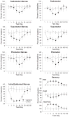
The change over time in cutaneous autonomic and sensory nerve fiber structure (Panels A, C, E, G) and function (Panels B, D, F, H) in capsaicin treated (closed black circles) and placebo treated subjects (open white circles). The results are expressed as percent of baseline for all panels. All results (A-H) were significant at _p<_0.01 compared to placebo treated subjects (ANOVA). A decrease in measures of structure and function is seen between days 3–16 after capsaicin treatment. There are no corresponding changes in the placebo-treated group. Increases in heat and heat-pain thresholds (Panel G) implies a greater temperature is required to reach threshold.
Fig 3
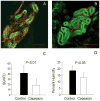
The effects of capsaicin on sudomotor nerves. Panels A and B: The sweat gland (green) and associated innervation (red) in a Day 16 control (A) and capsaicin-treated (B) biopsy. Fewer nerve fibers are visible in the capsaicin treated compared to the control treated biopsy. Red bar (A & B) = 100 μm. Panel C: The calculated sudomotor nerve fiber density in the control (black bar) and capsaicin (white bar) treated regions at day 16. There is a significant reduction in nerve fibers in the capsaicin treated area (p<0.01). Panel D: The maximum change in sudomotor function measured by QSART. There is a significant decrease in sweat output in the capsaicin treated region (_p<_0.01).
Fig 4
The effects of capsaicin on vasomotor nerves. Panels A and B: The cutaneous vasculature stained by CD 31 (green) and the associated innervation (red) stained by PGP 9.5 in a representative Day 16 control (A) and capsaicin treated (B) biopsy. The epithelial border is seen at the top of the image (white arrowheads). Fewer nerve fibers (yellow arrows) are visible in the Day 16 capsaicin-treated than control biopsy. Panel C: The vasomotor density for the control- (black bar) and capsaicin-treated (white bar) regions at day 16. There is a significant nerve fiber reduction in the capsaicin treated area (_p<_0.01). Panel D: Vasomotor function for the control- (black bar) and capsaicin-treated (white bar) regions at day 16. There is a significant axon reflex mediated flare response reduction in the capsaicin treated area (_p<_0.01). Scale bar (A & B) = 200 μm.
Fig 5
The effects of capsaicin on pilomotor nerves. Panels A and B: An arrector pili muscle is seen with nerve fibers extending parallel to the muscle in a control (A) and capsaicin-treated (B) biopsy stained with PGP 9.5. Fewer nerve fibers are visible in the capsaicin-treated compared to control on Day 16. Scale bar (A & B) = 200 μm. Panel C: Pilomotor nerve fiber density in the control regions (black bar) is significantly higher than the capsaicin treated regions (white bar) regions at day 16 (_p<_0.01). Panel D. Arrector pili, when electrically stimulated, cause piloerection (goose-bumps) that can be quantified with silicone impressions. The red circle surrounds a representative ‘goose-bump’ in a Day 16 control subject. Panel E: In a representative Day 16 capsaicin treated region, the stimulated arrector pili muscles form smaller impressions (red circle). Panel F: The mean area of piloerection is greater in control regions (black bar) compared to the capsaicin treated regions (white bar) (_p<_0.01).
Fig 6
The effects of capsaicin on sensory nerves. Panels A and B: The epidermal innervation stained by PGP 9.5 (green) penetrating the epidermal layer (yellow arrow) in a representative Day 16 control (A) and capsaicin-treated (B) biopsy. Fewer nerve fibers (green) are visible in the Day 16 capsaicin-treated than control biopsy. Scale bar (A & B) = 100 μm. Panel C: The intra-epidermal nerve fiber density (IENFD) for the control- (black bar) and capsaicin-treated (white bar) regions at day 16. There is a significant nerve fiber reduction in the capsaicin treated area (_p<_0.01). (D) The thermal and thermal-pain stimulation thresholds for control (black bar) and capsaicin (white bar) treated regions. There is a reduction in cold detection thresholds and an increase in heat and heat-pain (HP) detection thresholds in the capsaicin-treated regions at day 16 compared to control treated regions. *_p<_0.05.
Fig 7
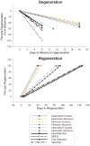
The rates of nerve fiber degeneration (A) and regeneration (B) by nerve fiber subtype. Panel A: The lines represent the percent change from baseline on day 1 to the day of maximal change. Sensory nerve fibers degenerate and sensory function decreases more rapidly than autonomic nerve fibers. Vasomotor function declines at a rate between that of sensory and autonomic function most likely in part due to physiologic desensitization and neuropeptide depletion. Panel B: The lines represent the time required for nerve fiber subtypes to return to baseline levels from the day of maximal degeneration (return to baseline levels defined as a non-significant change from baseline). Structural regeneration and functional improvement is more rapid in autonomic nerve fibers than in sensory nerve fibers. Vasomotor nerves, vasomotor function and cold detection thresholds return to baseline at a rate between sensory and other autonomic nerve fiber types.
Similar articles
- Pilomotor function is impaired in patients with Parkinson's disease: A study of the adrenergic axon-reflex response and autonomic functions.
Siepmann T, Frenz E, Penzlin AI, Goelz S, Zago W, Friehs I, Kubasch ML, Wienecke M, Löhle M, Schrempf W, Barlinn K, Siegert J, Storch A, Reichmann H, Illigens BM. Siepmann T, et al. Parkinsonism Relat Disord. 2016 Oct;31:129-134. doi: 10.1016/j.parkreldis.2016.08.001. Epub 2016 Aug 11. Parkinsonism Relat Disord. 2016. PMID: 27569843 - α-Synuclein in cutaneous autonomic nerves.
Wang N, Gibbons CH, Lafo J, Freeman R. Wang N, et al. Neurology. 2013 Oct 29;81(18):1604-10. doi: 10.1212/WNL.0b013e3182a9f449. Epub 2013 Oct 2. Neurology. 2013. PMID: 24089386 Free PMC article. - Assessment of cutaneous axon-reflex responses to evaluate functional integrity of autonomic small nerve fibers.
Hijazi MM, Buchmann SJ, Sedghi A, Illigens BM, Reichmann H, Schackert G, Siepmann T. Hijazi MM, et al. Neurol Sci. 2020 Jul;41(7):1685-1696. doi: 10.1007/s10072-020-04293-w. Epub 2020 Mar 3. Neurol Sci. 2020. PMID: 32125538 Free PMC article. Review. - Non-motor involvement in amyotrophic lateral sclerosis: new insight from nerve and vessel analysis in skin biopsy.
Nolano M, Provitera V, Manganelli F, Iodice R, Caporaso G, Stancanelli A, Marinou K, Lanzillo B, Santoro L, Mora G. Nolano M, et al. Neuropathol Appl Neurobiol. 2017 Feb;43(2):119-132. doi: 10.1111/nan.12332. Epub 2016 Jul 7. Neuropathol Appl Neurobiol. 2017. PMID: 27288647 - Contribution of Skin Biopsy in Peripheral Neuropathies.
Nolano M, Tozza S, Caporaso G, Provitera V. Nolano M, et al. Brain Sci. 2020 Dec 15;10(12):989. doi: 10.3390/brainsci10120989. Brain Sci. 2020. PMID: 33333929 Free PMC article. Review.
Cited by
- Objective evidence that small-fiber polyneuropathy underlies some illnesses currently labeled as fibromyalgia.
Oaklander AL, Herzog ZD, Downs HM, Klein MM. Oaklander AL, et al. Pain. 2013 Nov;154(11):2310-2316. doi: 10.1016/j.pain.2013.06.001. Epub 2013 Jun 5. Pain. 2013. PMID: 23748113 Free PMC article. - TRPV1: A Target for Rational Drug Design.
Carnevale V, Rohacs T. Carnevale V, et al. Pharmaceuticals (Basel). 2016 Aug 23;9(3):52. doi: 10.3390/ph9030052. Pharmaceuticals (Basel). 2016. PMID: 27563913 Free PMC article. Review. - Uses of skin biopsy for sensory and autonomic nerve assessment.
Myers MI, Peltier AC. Myers MI, et al. Curr Neurol Neurosci Rep. 2013 Jan;13(1):323. doi: 10.1007/s11910-012-0323-2. Curr Neurol Neurosci Rep. 2013. PMID: 23250768 Free PMC article. Review. - Neurovascular function and sudorimetry in health and disease.
Vinik AI, Nevoret M, Casellini C, Parson H. Vinik AI, et al. Curr Diab Rep. 2013 Aug;13(4):517-32. doi: 10.1007/s11892-013-0392-x. Curr Diab Rep. 2013. PMID: 23681491 Review. - Effect of diabetes type on long-term outcome of epidermal axon regeneration.
Khoshnoodi M, Truelove S, Polydefkis M. Khoshnoodi M, et al. Ann Clin Transl Neurol. 2019 Oct;6(10):2088-2096. doi: 10.1002/acn3.50904. Epub 2019 Sep 27. Ann Clin Transl Neurol. 2019. PMID: 31560176 Free PMC article.
References
- Caterina MJ. Vanilloid receptors take a TRP beyond the sensory afferent. Pain. 2003;105:5–9. - PubMed
- Pingle SC, Matta JA, Ahern GP. Capsaicin receptor: TRPV1 a promiscuous TRP channel. Handb Exp Pharmacol. 2007:155–171. - PubMed
Publication types
MeSH terms
Substances
LinkOut - more resources
Full Text Sources
Other Literature Sources


