Mitochondrial fission and cristae disruption increase the response of cell models of Huntington's disease to apoptotic stimuli - PubMed (original) (raw)
Mitochondrial fission and cristae disruption increase the response of cell models of Huntington's disease to apoptotic stimuli
Veronica Costa et al. EMBO Mol Med. 2010 Dec.
Abstract
Huntington's disease (HD), a genetic neurodegenerative disease caused by a polyglutamine expansion in the Huntingtin (Htt) protein, is accompanied by multiple mitochondrial alterations. Here, we show that mitochondrial fragmentation and cristae alterations characterize cellular models of HD and participate in their increased susceptibility to apoptosis. In HD cells, the increased basal activity of the phosphatase calcineurin dephosphorylates the pro-fission dynamin related protein 1 (Drp1), increasing its mitochondrial translocation and activation, and ultimately leading to fragmentation of the organelle. The fragmented HD mitochondria are characterized by cristae alterations that are aggravated by apoptotic stimulation. A genetic analysis indicates that correction of mitochondrial elongation is not sufficient to rescue the increased cytochrome c release and cell death observed in HD cells. Conversely, the increased apoptosis can be corrected by manoeuvres that prevent fission and cristae remodelling. In conclusion, the cristae remodelling of the fragmented HD mitochondria contributes to their hypersensitivity to apoptosis.
Figures
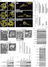
Figure 1. Mitochondrial fragmentation and cristae derangement in HD lymphoblasts and striatal precursors
- A. Lymphoblasts of the indicated genotype were transfected with mtYFP. Randomly selected confocal, 14 µm deep z axis stacks were acquired, stored, reconstructed and volume rendered. Scale bar: 5 µm.
- B. Striatal precursors of the indicated genotype were transfected with mtYFP. Confocal images of mtYFP from randomly selected cells. Scale bar: 20 µm.
- C. Morphometric analysis. Experiments were as in (A). Data represent mean ± SE of 4 independent experiments where 30 randomly selected, reconstructed and volume rendered z stack series were classified as described. Wt refers to gender-matched control of HD lymphoblasts. (p < 0.05 in a paired Student's _t_-test between HD samples and their relative control).
- D. Morphometric analysis of mitochondrial morphology. Experiments were as in (B). Data represent mean ± SE of 5 independent experiments where 50 randomly selected images of mtYFP fluorescence were classified as described. p < 0.05 in a paired Student's _t_-test between HD samples and their relative control.
- E. Representative electron micrographs of wt and Q111 striatal neurons. Cells were fixed and TEM images of randomly selected fields were acquired. Boxed areas represent a 2.3× magnification. Scale bar: 1 µm.
- F. Morphometric analysis. Experiments were performed as in (E). Data represent mean ± SE of 3 independent experiments (n = 50 mitochondria per condition from 15 different neurons of the indicated genotype). Data are normalized to the ratio of wt cells.
- G,H. Equal amounts of proteins (20 µg) from total cell lysates from lymphoblasts (G) and striatal precursors (H) of the indicated genotype were separated by SDS–PAGE and immunoblotted with the indicated antibodies. For MFN1, equal amounts of proteins (50 µg) from mitochondria isolated from lymphoblasts (G) and neurons (H) were separated by SDS–PAGE. For lymphoblasts, wt refers to gender-matched control.
- I,J. Equal amounts of proteins (30 µg) from mitochondria isolated from neurons (I) and lymphoblasts (J) of the indicated genotype were analysed by SDS–PAGE/immunoblotting using the indicated antibodies. Drp1/TOM20 levels in Q111/0 and Q111/1 mitochondria were 5.1 ± 1.03- and 3.05 ± 1.2-fold of wt (n = 3 independent experiments, p < 0.05 in a paired Student's _t_-test).
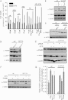
Figure 2. Hyperactivation of calcineurin and dephosphorylation of Drp1 in HD cells
- A. Phosphorylated and unphosphorylated proteins were separated from total cell lysates (2.5 mg) from cells of the indicated genotype. Equal amounts (20 µg) of total, phosphorylated and un-phosphorylated proteins were separated by SDS–PAGE and immunoblotted.
- B. Calcineurin enzyme activity measured in total cell extracts from cell lines of the indicated genotype. Data are mean ± SE of three independent experiments.
- C,D. Equal amounts of proteins (40 µg) from total cell lysates from cells of the indicated genotype were separated by SDS–PAGE and immunoblotted with the indicated antibodies. RCAN1L/actin levels (relative to their relative wt): 0.98 ± 0.05 in Q111/0; 0.82 ± 0.08 in Q111/1; 1.28 ± 0.21 in 48Q; 0.91 ± 0.04 in 70Q and 1.22 ± 0.02 in 45 + 47Q lymphoblasts.
- E. Representative traces of Fura-2 ratio of cytosolic Ca2+ ([Ca2+]i) following passive discharge of ER Ca2+ stores by CPA (100 µM) in cells of the indicated genotype.
- F. Quantification of peak and basal [Ca2+]i. Experiments were as in (E). Data represent mean ± SE of eight independent experiments (p < 0.002 by paired Student's _t_-test).
Figure 4. Correction of mitochondrial morphology in primary striatal YAC128 neurons
- Primary neurons of the indicated genotype were immunostained with anti-Tom20 (red) and anti-Tubulin III (green) antibodies. Randomly selected confocal, z axis stacks were acquired, stored, reconstructed and volume rendered. Scale bar: 20 µm.
- 3× magnification of the boxed areas in (A). The bottom panel was turned 90° counter-clockwise.
- Morphometric analysis of mitochondrial shape. Experiments were done as in (A). Data represent mean ± SE of three independent experiments (n = 70 stacks).
- Representative electron micrographs of wt and YAC128 primary striatal neurons. Cells were fixed and TEM images of randomly selected fields were acquired. Boxed areas are magnified 2.4×. Scale bar: 1 µm.
- Cells of the indicated genotype were electroporated with mtRFP and empty vector or the indicated plasmids. Samples were then immunostained with anti-Tubulin III antibody (magenta) and incubated with TUNEL reagent. Images were acquired exactly as in (A). Scale bar: 20 µm.
- Morphometric analysis of mitochondrial morphology. Experiments were performed as in (E). Data represent mean ± SE of three independent experiments (n = 40 stacks).
Figure 3. Correction of mitochondrial morphology in HD cells
- A,B. Lymphoblasts of the indicated genotype were cotransfected with mtYFP and the indicated plasmids. Experiments were performed exactly as in Fig 1. Scale bar: 5 µm.
- C. Cells of the indicated genotype were cotransfected with mtYFP and the indicated plasmids. When indicated, cells were treated with 1 µM FK506 for 1 h before acquisition of images. Images were acquired exactly as in Fig 1. Scale bar: 20 µm.
- D,E. Morphometric analysis of mitochondrial shape. Experiments were done as in (A) and (B), respectively. Data represent mean ± SE of four independent experiments (n = 30 stacks).
- F. Morphometric analysis of mitochondrial morphology. Experiments were performed as in (C). Data represent mean ± SE of five independent experiments (n = 50 cells).
Figure 5. Increased cytochrome c release and susceptibility to apoptosis in HD
- A-C. Lymphoblasts of the indicated genotype were treated with 2 µM staurosporine. At the indicated times, viability was determined cytofluorimetrically. Data represent mean ± SE of seven independent experiments.
- D. Cells of the indicated genotype were treated with 2 µM staurosporine. At the indicated times, cells were lysed and equal amounts of proteins (30 µg) were separated by SDS–PAGE and immunoblotted using the indicated antibodies.
- E. Densitometric analysis of uncleaved/cleaved PARP levels. Experiments were performed as in (D). Data are normalized to the ratio in untreated samples and represent mean ± SE of four independent experiments.
- F,H. Mitochondria isolated from lymphoblasts (F) and neurons (H) of the indicated genotype treated where indicated with 2 µM staurosporine for 4 h were treated with BMH where indicated. Equal amounts (40 µg) of mitochondrial protein were analysed by SDS–PAGE/immunoblotting using an anti-BAX antibody. Asterisks: BAX multimers.
- G. Mitochondria isolated from lymphoblasts of the indicated genotype were treated with cBID for the indicated time and with BMH where indicated. Equal amounts (40 µg) of mitochondrial protein were analysed by SDS–PAGE/immunoblotting using an anti-BAK antibody. Asterisks: BAK multimers.
- I. Mitochondria isolated from cells of the indicated genotype treated where indicated with 2 µM staurosporine for 4 h were incubated with BMH where indicated. Equal amounts (40 µg) of protein were analysed by SDS–PAGE/immunoblotting using an anti-BAK antibody. Asterisks: BAK multimers. In (F–I) immunoblots are representative of three independent experiments.
- J. Representative confocal images of striatal cells of the indicated genotype transfected with mtRFP and immunostained with anti-cytochrome c (green) antibody. When indicated, cells were treated for 2 h with 1 mM H2O2.
- K. Localization index of cytochrome c. Cells of the indicated genotype were treated where indicated for 2 h with 1 mM H2O2 or for 5 h with 0.75 µM staurosporine (STS). Localization index of cytochrome c was determined as described. Data represent mean ± SE of three independent experiments (n = 50 cells per condition in each experiment).
- L-N. Mitochondria isolated from lymphoblasts of the indicated genotype were treated for the indicated times with cBID. The amount of cytochrome c in supernatant and pellet was determined as described. Data represent mean ± SE of four independent experiments.
- O. Mitochondria from lymphoblasts of the indicated genotype were treated with cBID for the indicated times and then crosslinked with EDC. Equal amounts of proteins were analysed by SDS–PAGE/immunoblotting using anti-OPA1 antibody.
Figure 6. Increased susceptibility to apoptosis of striatal YAC128 primary neurons: correction by Opa1 and dominant-negative Drp1
- Representative confocal images of TUNEL assay of primary striatal neurons. Neurons of the indicated genotype were electroporated with mtRFP, treated where indicated with 500 nM staurosporine for 14 h, immunostained with anti-Tubulin III and processed for TUNEL. Arrowheads, TUNEL positive neurons. Scale bar: 20 µm.
- Quantification of TUNEL assay of primary neurons. Cells of the indicated genotype were transfected as indicated and treated as in (A). Cell death is represented as the percentage of TUNEL positive cells in the mtRPF–Tubulin III double positive population. Data represent mean ± SE of three independent experiments (n = 300 neurons per condition).
Figure 7. Apoptotic ultrastructural changes in HD mitochondria are faster and prevented by OPA1 and blockage of DRP1
- A. Representative EM fields of neurons of the indicated genotype. Cells were cotransfected with GFP and the indicated plasmids or siRNAs, sorted and GFP-positive cells were seeded and after 18 h treated when indicated with 2 µM staurosporine for 3 h, fixed and processed for EM. Scale bar: 1 µm.
- B-D. Morphometric analysis of cristae shape. Experiments were performed as in (A). Data represent mean ± SE of 3 independent experiments (n = 50 mitochondria from 30 different cells per each condition) and are normalized to the value in empty vector-transfected, untreated cells.
Figure 8. HD cells are more susceptible to apoptosis: correction by Opa1 and inhibition of Drp1
- A. Lymphoblasts of the indicated genotype transfected as indicated were treated with 2 µM staurosporine. At the indicated times, cell death was determined cytofluorimetrically. Data represent mean ± SE of five independent experiments.
- B-E. Cells of the indicated genotype were cotransfected with GFP and the indicated vectors or siRNAs. After 24 (B–D) or 30 h (E) cells were treated with 2 µM staurosporine for 3 h and 3 × 10⁵ GFP positive cells were sorted and lysed. Proteins were analysed by SDS–PAGE/immunoblotting using the indicated antibodies.
- F. Cells of the indicated genotype were treated where indicated with 1 µM FK506 and 2 µM staurosporine. Cells were lysed and equal amounts of proteins (30 µg) were separated by SDS–PAGE and immunoblotted using the indicated antibodies. (B–F) Western blots are representative of 3 independent experiments.
- G. Densitometric analysis of uncleaved/cleaved PARP levels. Experiments were performed as in (B,D,F). Data are normalized to the ratio in untreated samples and represent mean ± SE of four independent experiments.
Comment in
- Could successful (mitochondrial) networking help prevent Huntington's disease?
Oliveira JM, Lightowlers RN. Oliveira JM, et al. EMBO Mol Med. 2010 Dec;2(12):487-9. doi: 10.1002/emmm.201000104. EMBO Mol Med. 2010. PMID: 21117121 Free PMC article.
Similar articles
- Could successful (mitochondrial) networking help prevent Huntington's disease?
Oliveira JM, Lightowlers RN. Oliveira JM, et al. EMBO Mol Med. 2010 Dec;2(12):487-9. doi: 10.1002/emmm.201000104. EMBO Mol Med. 2010. PMID: 21117121 Free PMC article. - Effects of overexpression of huntingtin proteins on mitochondrial integrity.
Wang H, Lim PJ, Karbowski M, Monteiro MJ. Wang H, et al. Hum Mol Genet. 2009 Feb 15;18(4):737-52. doi: 10.1093/hmg/ddn404. Epub 2008 Nov 27. Hum Mol Genet. 2009. PMID: 19039036 Free PMC article. - S-nitrosylation of dynamin-related protein 1 mediates mutant huntingtin-induced mitochondrial fragmentation and neuronal injury in Huntington's disease.
Haun F, Nakamura T, Shiu AD, Cho DH, Tsunemi T, Holland EA, La Spada AR, Lipton SA. Haun F, et al. Antioxid Redox Signal. 2013 Oct 10;19(11):1173-84. doi: 10.1089/ars.2012.4928. Epub 2013 Jun 20. Antioxid Redox Signal. 2013. PMID: 23641925 Free PMC article. - Increased mitochondrial fission and neuronal dysfunction in Huntington's disease: implications for molecular inhibitors of excessive mitochondrial fission.
Reddy PH. Reddy PH. Drug Discov Today. 2014 Jul;19(7):951-5. doi: 10.1016/j.drudis.2014.03.020. Epub 2014 Mar 28. Drug Discov Today. 2014. PMID: 24681059 Free PMC article. Review. - Dynamin-related protein 1 and mitochondrial fragmentation in neurodegenerative diseases.
Reddy PH, Reddy TP, Manczak M, Calkins MJ, Shirendeb U, Mao P. Reddy PH, et al. Brain Res Rev. 2011 Jun 24;67(1-2):103-18. doi: 10.1016/j.brainresrev.2010.11.004. Epub 2010 Dec 8. Brain Res Rev. 2011. PMID: 21145355 Free PMC article. Review.
Cited by
- Huntington's disease affects mitochondrial network dynamics predisposing to pathogenic mitochondrial DNA mutations.
Neueder A, Kojer K, Gu Z, Wang Y, Hering T, Tabrizi S, Taanman JW, Orth M. Neueder A, et al. Brain. 2024 Jun 3;147(6):2009-2022. doi: 10.1093/brain/awae007. Brain. 2024. PMID: 38195181 Free PMC article. - Neuronal Ca(2+) dyshomeostasis in Huntington disease.
Giacomello M, Oliveros JC, Naranjo JR, Carafoli E. Giacomello M, et al. Prion. 2013 Jan-Feb;7(1):76-84. doi: 10.4161/pri.23581. Epub 2013 Jan 1. Prion. 2013. PMID: 23324594 Free PMC article. Review. - How Do Post-Translational Modifications Influence the Pathomechanistic Landscape of Huntington's Disease? A Comprehensive Review.
Lontay B, Kiss A, Virág L, Tar K. Lontay B, et al. Int J Mol Sci. 2020 Jun 16;21(12):4282. doi: 10.3390/ijms21124282. Int J Mol Sci. 2020. PMID: 32560122 Free PMC article. Review. - Raft-like microdomains play a key role in mitochondrial impairment in lymphoid cells from patients with Huntington's disease.
Ciarlo L, Manganelli V, Matarrese P, Garofalo T, Tinari A, Gambardella L, Marconi M, Grasso M, Misasi R, Sorice M, Malorni W. Ciarlo L, et al. J Lipid Res. 2012 Oct;53(10):2057-2068. doi: 10.1194/jlr.M026062. Epub 2012 Jul 6. J Lipid Res. 2012. PMID: 22773688 Free PMC article. - Targeting Mitochondrial Network Disorganization is Protective in C. elegans Models of Huntington's Disease.
Machiela E, Rudich PD, Traa A, Anglas U, Soo SK, Senchuk MM, Van Raamsdonk JM. Machiela E, et al. Aging Dis. 2021 Oct 1;12(7):1753-1772. doi: 10.14336/AD.2021.0404. eCollection 2021 Oct. Aging Dis. 2021. PMID: 34631219 Free PMC article.
References
- Almeida S, Domingues A, Rodrigues L, Oliveira CR, Rego AC. FK506 prevents mitochondrial-dependent apoptotic cell death induced by 3-nitropropionic acid in rat primary cortical cultures. Neurobiol Dis. 2004;17:435–444. - PubMed
- Bossy-Wetzel E, Barsoum MJ, Godzik A, Schwarzenbacher R, Lipton SA. Mitochondrial fission in apoptosis, neurodegeneration and aging. Curr Opin Cell Biol. 2003;15:706–716. - PubMed
- Chan DC. Mitochondrial dynamics in disease. N Engl J Med. 2007;356:1707–1709. - PubMed
Publication types
MeSH terms
Substances
LinkOut - more resources
Full Text Sources
Other Literature Sources
Medical
Research Materials
Miscellaneous


