Engraftment of human central memory-derived effector CD8+ T cells in immunodeficient mice - PubMed (original) (raw)
Engraftment of human central memory-derived effector CD8+ T cells in immunodeficient mice
Xiuli Wang et al. Blood. 2011.
Abstract
In clinical trials of adoptive T-cell therapy, the persistence of transferred cells correlates with therapeutic efficacy. However, properties of human T cells that enable their persistence in vivo are poorly understood, and model systems that enable investigation of the fate of human effector T cells (T(E)) have not been described. Here, we analyzed the engraftment of adoptively transferred human cytomegalovirus pp65-specific CD8(+) T(E) cells derived from purified CD45RO(+)CD62L(+) central memory (T(CM)) or CD45RO(+)CD62L(-) effector memory (T(EM)) precursors in an immunodeficient mouse model. The engraftment of T(CM)-derived effector cells (T(CM/E)) was dependent on human interleukin-15, and superior in magnitude and duration to T(EM)-derived effector cells (T(EM/E)). T-cell receptor Vβ analysis of persisting cells demonstrated that CD8(+) T(CM/E) engraftment was polyclonal, suggesting that the ability to engraft is a general feature of T(CM/E.) CD8(+) T(EM/E) proliferated extensively after transfer but underwent rapid apoptosis. In contrast, T(CM/E) were less prone to apoptosis and established a persistent reservoir of functional T cells in vivo characterized by higher CD28 expression. These studies predict that human CD8(+) effector T cells derived from T(CM) precursors may be preferred for adoptive therapy based on superior engraftment fitness.
Figures
Figure 1
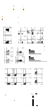
Frequency of CD8+ T-cell memory subsets in human peripheral blood. (A) Flow cytometric analysis of PBMCs from 4 different human donors gated on the lymphoid population by forward and side scatter (left panel) and analyzed for CD45RO, CD62L, and CD8 expression. CD45RO+ lymphocytes were then gated for CD8+CD62L+ TCM and CD8+CD62L− TEM (middle and right panels) and analyzed by multicolor flow cytometry with anti-CCR7, anti-perforin, or anti-granzyme A mAbs (B). Percentage of cells in each gate (red) is indicated, and mean percentage of the CD8+ TCM or TEM cells that were CCR7, perforin, or granzyme A positive (± SE; n = 3 donors) is indicated. *P < .05, CD8+ TCM versus TEM cells (unpaired Student t test). (C) Cytokine production profiles of the freshly isolated CD8+ TCM and TEM. Supernatants were collected after overnight coincubation with LCL-OKT3, and cytokine levels (mean ± SE of triplicate wells) were determined as described in “Cytokine production assays.” *P < .0001, cytokine levels of CD8+ TCM versus TEM cells (unpaired Student t test).
Figure 2
TE cells derived from TCM and TEM in vitro are similar in phenotype and function. (A) Schematic of methods for deriving CMV-specific TCM/E and TEM/E. Purified TCM, TEM, and pp65-expressing vAPCs were generated from the same CMV-seropositive donor's PBMCs. (B) CD45RO and CD62L staining of TCM (top) and TEM (bottom) after sorting from PBMCs. (C) CD8 and pp65-tetramer staining of gated PBMCs (left panels) and of CMV-specific TCM/E and TEM/E at day 7 (middle panels) and 21 (right panels) after stimulation with vAPCs. Histogram quadrants are based on staining with isotype and negative tetramer controls, and percentage of double-positive cells is indicated. (D) pp65 tetramer and intracellular IFN-γ staining of TCM/E and TEM/E before infusion, after overnight coincubation with LCL-pp65. (E) Fold expansion of pp65-tetramer+ cells was determined by multiplying the total number of cells by the percentage pp65tet+ (determined as shown in panel C) found at days 0, 7, 14, and 21 of vAPC stimulation and 14 days after the first and second anti-CD3 (REM) stimulations; these values were then normalized to the input cell number (day 0). (F) Expression of CD62L, CD127, CD28, CCR7, and CD8 on the TCM/E and TEM/E cell products. (G) Cytotoxic activity of TCM/E and TEM/E cell products against auto-LCLs loaded with either an HLA-A2-restricted control peptide (cLCL) or CMV pp65 peptide (pp65LCL). Mean percentage of 51Cr release (± SD) of triplicate wells is depicted. (H) Cytokine production by TCM/E and TEM/E. Supernatants were collected after coincubating T cells overnight with CMV pp65 peptide-loaded auto-LCLs, and mean (± SD of triplicate wells) cytokine levels were determined using cytometric bead array.
Figure 3
IL-15-dependent engraftment of CMV-specific TCM/E cells in NOG mice is greater than that of TEM/E. (A) Schematic of the experiment. (B) Mean percentage (± SE) of human T cells (CD45+ CD8+) in peripheral blood lymphocytes (PBLs) of mice engrafted with TCM/E (squares) or TEM/E (circles) was determined by flow cytometry (n = 5). *P < .05, TCM/E versus TEM/E cell engraftment in the presence of NS0-IL-15 cells (unpaired Student t test). (Inset) Mean levels of human IL-15 (± SE) in day 7 serum of NOG mice that had received 3 intraperitoneal injections of 1.5 × 10⁷ irradiated NS0-IL-15 cells (n = 6) or in control mice (n = 10). (C) Mean percentage of human T cells (CD45+ CD8+) plus or minus SE in mouse PBL, bone marrow, and spleen at day 21. *P < .05, TCM/E cell engraftment in each organ versus that of TEM/E in the presence of NS0-IL-15 cells. (D) TCR Vβ repertoire of the CMV-specific TCM/E and TEM/E before (Input) and after (d21) engraftment. Percentage of CD3+ cells (Input) or CD45+ CD3+ cells (d21) that were positive for the indicated TCR Vβ genes was determined by flow cytometry.
Figure 4
Human CD8+ TCM/E persist long-term (100 days) in huIL-15 NOG mice and remain functional. TCM/E and TEM/E (107) were injected intravenously at day 0, and irradiated NS0-IL-15 cells (1.5 × 10⁷) were administered 3 times a week starting at day 0, until mice were killed at day 100. (A) Mean percentage of human CD45+CD8+ cells (± SE) in mouse PBL, bone marrow, and spleen at day 100 was determined by flow cytometry (n = 5). (B) TCR Vβ repertoire of the input and long-term engrafted TCM/E and TEM/E. Bone marrow was pooled from mice, and human CD45+ cells were sorted and expanded by stimulation with anti-CD3. The percentage of CD3+ cells positive for the indicated TCR Vβ genes was determined by flow cytometry. (C) Bone marrow harvested at day 100 from mice engrafted with TCM/E and TEM/E was analyzed by flow cytometry for expression of human CD45, CD62L, CCR7, and CD28. Gating was based on staining with isotype control mAb, and the percentage of double-positive cells is indicated. (D) IL-2 production from CD45+ T cells derived from day 100 bone marrow of mice engrafted with TCM/E and TEM/E. Supernatants were collected after T cells were coincubated overnight with LCL-pp65, and IL-2 levels were determined using cytometric bead array. (E) Cytotoxic activity of human T cells derived from day 100 bone marrow of mice engrafted with TCM/E and TEM/E, and stimulated with anti-CD3 mAb. Target cells included OKT3-expressing LCLs, auto-LCLs or LCL-pp65. Mean percentage of 51Cr release (± SD) of triplicate wells. (F) Intracellular IFN-γ staining of human T cells derived from day 100 bone marrow of mice engrafted with TCM/E and TEM/E and coincubated overnight with LCL-pp65, LCL-OKT3, or auto-LCLs.
Figure 5
Differential cytokine receptor expression, IL-15–mediated proliferation, and caspase activity of TCM/E and TEM/E. (A) IL-15Rα, IL-2Rβ, or IL-2Rγ expression by TCM/E and TEM/E. Mean fluorescence intensity was normalized to that of isotype control staining in each case to determine ΔMFI. (B) Proliferation of TCM/E and TEM/E was determined after 48-hour incubation with different concentrations of rhIL-15 using a standard [3H]-thymidine incorporation assay. (C-D) CFSE-labeled TCM/E and TEM/E (107) were injected intravenously into mice at day 0, and irradiated NS0-IL-15 cells (1.5 × 10⁷) were administered 3 times a week starting at day 0, until mice were killed at either day 9 or day 12. (C) CFSE profiles of the input and engrafted TCM/E and TEM/E in day 9 PBL was assessed by flow cytometry. Percentage of CFSE-diluted cells that fall within the first log are indicated. (D) Engraftment of the CD45+ human T cells in the PBL, bone marrow, and spleen was assessed on days 9 and 12 by flow cytometry. (E) FL-1 profiles of CD45+ human T cells in the PBL were assessed on day 9 as a readout for cleavage of the caspase substrate D2R. The percentage of cells with cleaved D2R is depicted.
Figure 6
Adoptively transferred CMV-specific CD8+ TCM/E exhibit a better response to antigen challenge in vivo than TEM/E. (A) Schematic of in vivo antigenic stimulation of engrafted CMV-specific TCM/E and TEM/E. (B) Engraftment of CMV-specific TCM/E (squares) or TEM/E (circles) was carried out with (black) or without (white) administration of irradiated CMV pp65-expressing LCLs at days 3, 10, and17; and mean percentage (± SE) of human T cells (CD45+ CD8+) in mouse PBL was determined by flow cytometry (n = 6). *P < .05, engraftment of TCM/E alone versus pp65-driven TCM/E engraftment (unpaired Student t test). (C) On euthanasia at day 28, PBLs were harvested and analyzed by flow cytometry for percentage of Ki-67+ cells in the human T-cell population (left) and for the ability of CD45+ human T cells to cleave the caspase substrate D2R (right).
Figure 7
Adoptively transferred CMV-specific CD8+ TCM/E exhibit superior protection from tumor challenge. (A) Schematic of the in vivo tumor challenge experiment. (B) Engraftment of pp65+ ffLuc+ LCL in animals treated with or without CMV-specific TCM/E or TEM/E was determined by Xenogen imaging; and mean (± SE) of total flux levels of luciferase activity are shown for each group (n = 5). *P < .05, animals treated with TCM/E versus either untreated or TEM/E-treated animals (analysis of variance). (C) Mean percentage (± SE) of human CD45+CD8+ cells in day 10 mouse PBL was determined by flow cytometry. *P < .05, TCM/E versus TEM/E engraftment (unpaired Student t test).
Comment in
- Fitness without exhaustion.
Perreault C. Perreault C. Blood. 2011 Feb 10;117(6):1776. doi: 10.1182/blood-2010-12-324293. Blood. 2011. PMID: 21310933 No abstract available.
Similar articles
- Phenotypic and functional attributes of lentivirus-modified CD19-specific human CD8+ central memory T cells manufactured at clinical scale.
Wang X, Naranjo A, Brown CE, Bautista C, Wong CW, Chang WC, Aguilar B, Ostberg JR, Riddell SR, Forman SJ, Jensen MC. Wang X, et al. J Immunother. 2012 Nov-Dec;35(9):689-701. doi: 10.1097/CJI.0b013e318270dec7. J Immunother. 2012. PMID: 23090078 Free PMC article. - Tumor-Specific Effector CD8+ T Cells That Can Establish Immunological Memory in Humans after Adoptive Transfer Are Marked by Expression of IL7 Receptor and c-myc.
Chandran SS, Paria BC, Srivastava AK, Rothermel LD, Stephens DJ, Kammula US. Chandran SS, et al. Cancer Res. 2015 Aug 15;75(16):3216-26. doi: 10.1158/0008-5472.CAN-15-0584. Epub 2015 Jun 22. Cancer Res. 2015. PMID: 26100671 Free PMC article. - Proliferation-linked apoptosis of adoptively transferred T cells after IL-15 administration in macaques.
Berger C, Berger M, Beard BC, Kiem HP, Gooley TA, Riddell SR. Berger C, et al. PLoS One. 2013;8(2):e56268. doi: 10.1371/journal.pone.0056268. Epub 2013 Feb 13. PLoS One. 2013. PMID: 23418547 Free PMC article. - Uncoupling T-cell expansion from effector differentiation in cell-based immunotherapy.
Crompton JG, Sukumar M, Restifo NP. Crompton JG, et al. Immunol Rev. 2014 Jan;257(1):264-276. doi: 10.1111/imr.12135. Immunol Rev. 2014. PMID: 24329803 Free PMC article. Review. - Diversity in CD8(+) T cell differentiation.
Parish IA, Kaech SM. Parish IA, et al. Curr Opin Immunol. 2009 Jun;21(3):291-7. doi: 10.1016/j.coi.2009.05.008. Epub 2009 Jun 6. Curr Opin Immunol. 2009. PMID: 19497720 Free PMC article. Review.
Cited by
- A novel humanized mouse model to study the function of human cutaneous memory T cells in vivo in human skin.
Klicznik MM, Benedetti A, Gail LM, Varkhande SR, Holly R, Laimer M, Stoecklinger A, Sir A, Reitsamer R, Neuper T, Horejs-Hoeck J, Rosenblum MD, Campbell DJ, Murauer EM, Gratz IK. Klicznik MM, et al. Sci Rep. 2020 Jul 7;10(1):11164. doi: 10.1038/s41598-020-67430-7. Sci Rep. 2020. PMID: 32636404 Free PMC article. - Memory T Cells in the Immunoprevention of Cancer: A Switch from Therapeutic to Prophylactic Approaches.
Mittra S, Harding SM, Kaech SM. Mittra S, et al. J Immunol. 2023 Sep 15;211(6):907-916. doi: 10.4049/jimmunol.2300049. J Immunol. 2023. PMID: 37669503 Free PMC article. Review. - Development of T-cell immunotherapy for hematopoietic stem cell transplantation recipients at risk of leukemia relapse.
Dossa RG, Cunningham T, Sommermeyer D, Medina-Rodriguez I, Biernacki MA, Foster K, Bleakley M. Dossa RG, et al. Blood. 2018 Jan 4;131(1):108-120. doi: 10.1182/blood-2017-07-791608. Epub 2017 Oct 19. Blood. 2018. PMID: 29051183 Free PMC article. - Comprehensive Approach for Identifying the T Cell Subset Origin of CD3 and CD28 Antibody-Activated Chimeric Antigen Receptor-Modified T Cells.
Schmueck-Henneresse M, Omer B, Shum T, Tashiro H, Mamonkin M, Lapteva N, Sharma S, Rollins L, Dotti G, Reinke P, Volk HD, Rooney CM. Schmueck-Henneresse M, et al. J Immunol. 2017 Jul 1;199(1):348-362. doi: 10.4049/jimmunol.1601494. Epub 2017 May 26. J Immunol. 2017. PMID: 28550199 Free PMC article. - Applying a clinical lens to animal models of CAR-T cell therapies.
Duncan BB, Dunbar CE, Ishii K. Duncan BB, et al. Mol Ther Methods Clin Dev. 2022 Aug 30;27:17-31. doi: 10.1016/j.omtm.2022.08.008. eCollection 2022 Dec 8. Mol Ther Methods Clin Dev. 2022. PMID: 36156878 Free PMC article. Review.
References
- Mackinnon S, Thomson K, Verfuerth S, Peggs K, Lowdell M. Adoptive cellular therapy for cytomegalovirus infection following allogeneic stem cell transplantation using virus-specific T cells. Blood Cells Mol Dis. 2008;40(1):63–67. - PubMed
- Walter EA, Greenberg PD, Gilbert MJ, et al. Reconstitution of cellular immunity against cytomegalovirus in recipients of allogeneic bone marrow by transfer of T-cell clones from the donor. N Engl J Med. 1995;333(16):1038–1044. - PubMed
- Gottschalk S, Heslop HE, Rooney CM. Adoptive immunotherapy for EBV-associated malignancies. Leuk Lymphoma. 2005;46(1):1–10. - PubMed
Publication types
MeSH terms
Substances
Grants and funding
- R01 AI053193/AI/NIAID NIH HHS/United States
- R01 CA114536/CA/NCI NIH HHS/United States
- R01 CA136551/CA/NCI NIH HHS/United States
- AI053193/AI/NIAID NIH HHS/United States
- P01 CA030206/CA/NCI NIH HHS/United States
- P50 CA107399/CA/NCI NIH HHS/United States
LinkOut - more resources
Full Text Sources
Other Literature Sources
Research Materials


