Crystal structure of a prokaryotic homologue of the mammalian oligopeptide-proton symporters, PepT1 and PepT2 - PubMed (original) (raw)
. 2011 Jan 19;30(2):417-26.
doi: 10.1038/emboj.2010.309. Epub 2010 Dec 3.
David Drew, Alexander D Cameron, Vincent L G Postis, Xiaobing Xia, Philip W Fowler, Jean C Ingram, Elisabeth P Carpenter, Mark S P Sansom, Michael J McPherson, Stephen A Baldwin, So Iwata
Affiliations
- PMID: 21131908
- PMCID: PMC3025455
- DOI: 10.1038/emboj.2010.309
Crystal structure of a prokaryotic homologue of the mammalian oligopeptide-proton symporters, PepT1 and PepT2
Simon Newstead et al. EMBO J. 2011.
Abstract
PepT1 and PepT2 are major facilitator superfamily (MFS) transporters that utilize a proton gradient to drive the uptake of di- and tri-peptides in the small intestine and kidney, respectively. They are the major routes by which we absorb dietary nitrogen and many orally administered drugs. Here, we present the crystal structure of PepT(So), a functionally similar prokaryotic homologue of the mammalian peptide transporters from Shewanella oneidensis. This structure, refined using data up to 3.6 Å resolution, reveals a ligand-bound occluded state for the MFS and provides new insights into a general transport mechanism. We have located the peptide-binding site in a central hydrophilic cavity, which occludes a bound ligand from both sides of the membrane. Residues thought to be involved in proton coupling have also been identified near the extracellular gate of the cavity. Based on these findings and associated kinetic data, we propose that PepT(So) represents a sound model system for understanding mammalian peptide transport as catalysed by PepT1 and PepT2.
Conflict of interest statement
The authors declare that they have no conflict of interest.
Figures
Figure 1
Structure of PepTSo. (A) PepTSo topology. The central and extracellular cavities are shown as a closed diamond and open triangle, respectively. A bound ligand in the central cavity is represented as a black horizontal bar. Functionally important residues conserved between PepTSo and metazoan peptide transporters are highlighted by shapes in Supplementary Figure S1 and mapped onto the topology diagram. (B) PepTSo structure viewed in the plane of the membrane. The two hydrophilic cavities present in the structure are outlined in dashed lines. The hydrophobic core of the membrane (pale yellow) is distinguished from the interfacial region (light grey). N and C represent the N- and C-termini, respectively. Bound ligand is shown in black. Helices are labelled. (C) View from the extracellular side of the membrane.
Figure 2
Transport of peptides by PepTSo. (A). Concentration dependence of PepTSo-mediated glycylsarcosine (Gly-Sar) uptake in E. coli. Results shown, expressed per milligram of His-tagged PepTSo protein, are mean values±s.d. (_n_=4). (B) Effect on transport activity of mutating His61 to cysteine. Uptake of [3H]-glycylsarcosine (Gly-Sar) over a period of 10 min was measured in E. coli cells expressing the indicated forms of His-tagged PepTSo or in control cells lacking the transporter. Results shown are mean values±s.d. (_n_=3) and are expressed per milligram dry weight of bacteria. The inset shows western blots of equivalent samples from each culture, stained with a monoclonal antibody against oligohistidine. (C) Extracellular cavity viewed in the membrane plane. The central and extracellular cavities are isolated from each other by a putative extracellular gate. Residues in the central and extracellular cavities are highlighted in red and yellow, respectively. His61, part of the proposed proton–substrate coupling machinery is shown in green. Bound ligand is shown as a black CPK model of a di-alanine peptide. (D) Intracellular gate viewed in the membrane plane. Residues forming the gate are shown as stick models with transparent CPK surfaces. LacY helices (grey) are superposed onto PepTSo. Bound ligand is shown as a black CPK model as in C.
Figure 3
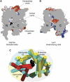
Comparison of PepTso and LacY structures. Electrostatic surface representation showing the location of the hydrophilic cavities in a section through the protein volumes of (A) PepTSo and (B) LacY. The N- and C-terminal six-helix bundles are labelled. (C) Superposed transmembrane helices of PepTSo and LacY viewed from the intracellular side of the membrane. PepTSo helices are labelled and shown in yellow except for helix H2 (green) and helices H7, H11 and H12 (red), which form sub-bundle C1. The N-terminal six-helix bundle and the C-terminal sub-bundles C1 and C2 are highlighted. LacY helices are shown in cyan. Bound ligand is shown as a black CPK model of a di-alanine peptide. Helices HA and HB have been omitted for clarity.
Figure 4
The peptide-binding site. Stereo view of the central cavity as viewed from above on the extracellular side of the membrane. Conserved residues between PepTSo and the mammalian peptide transporters are labelled and coloured according to side-chain type, Arg and Lys (blue), Glu and Ser (red), Tyr (green) and Trp, Phe and Leu (cyan). A di-peptide sized Cα baton (orange) is fitted as a size reference into the mFo-DFc electron density observed in the central cavity (blue mesh), contoured at 4 σ.
Figure 5
A possible mechanism for peptide–proton symport. (A) Outward-facing state: peptide (Pep) and proton (H+) can access respective binding sites through the outward-facing cavity that is open towards the extracellular side of the membrane. The peptide-binding site is made from the surfaces of both the N- and C-terminal helix bundles (indicated by + and − signs), whereas the proton-binding site is located in the area close to the extracellular gate. (B) Occluded state: both ends of the central cavity are closed with peptide occluded into the central cavity. The proton-binding site is still exposed to the extracellular side through the extracellular cavity. (C) Inward-facing state: peptide and proton are released on the intracellular side of the membrane through the inward-facing cavity. Note that the proton-binding site is exposed to the intracellular side in this conformation.
Similar articles
- Selectivity mechanism of a bacterial homolog of the human drug-peptide transporters PepT1 and PepT2.
Guettou F, Quistgaard EM, Raba M, Moberg P, Löw C, Nordlund P. Guettou F, et al. Nat Struct Mol Biol. 2014 Aug;21(8):728-31. doi: 10.1038/nsmb.2860. Epub 2014 Jul 27. Nat Struct Mol Biol. 2014. PMID: 25064511 - Crystal Structures of the Extracellular Domain from PepT1 and PepT2 Provide Novel Insights into Mammalian Peptide Transport.
Beale JH, Parker JL, Samsudin F, Barrett AL, Senan A, Bird LE, Scott D, Owens RJ, Sansom MSP, Tucker SJ, Meredith D, Fowler PW, Newstead S. Beale JH, et al. Structure. 2015 Oct 6;23(10):1889-1899. doi: 10.1016/j.str.2015.07.016. Epub 2015 Aug 27. Structure. 2015. PMID: 26320580 Free PMC article. - Expression of the peptide transporters PepT1, PepT2, and PHT1 in the embryonic and posthatch chick.
Zwarycz B, Wong EA. Zwarycz B, et al. Poult Sci. 2013 May;92(5):1314-21. doi: 10.3382/ps.2012-02826. Poult Sci. 2013. PMID: 23571341 - Recent advances in structural biology of peptide transporters.
Terada T, Inui K. Terada T, et al. Curr Top Membr. 2012;70:257-74. doi: 10.1016/B978-0-12-394316-3.00008-9. Curr Top Membr. 2012. PMID: 23177989 Review. - Towards a structural understanding of drug and peptide transport within the proton-dependent oligopeptide transporter (POT) family.
Newstead S. Newstead S. Biochem Soc Trans. 2011 Oct;39(5):1353-8. doi: 10.1042/BST0391353. Biochem Soc Trans. 2011. PMID: 21936814 Review.
Cited by
- The ABCs of membrane transporters in health and disease (SLC series): introduction.
Hediger MA, Clémençon B, Burrier RE, Bruford EA. Hediger MA, et al. Mol Aspects Med. 2013 Apr-Jun;34(2-3):95-107. doi: 10.1016/j.mam.2012.12.009. Mol Aspects Med. 2013. PMID: 23506860 Free PMC article. Review. - Structural basis for dynamic mechanism of proton-coupled symport by the peptide transporter POT.
Doki S, Kato HE, Solcan N, Iwaki M, Koyama M, Hattori M, Iwase N, Tsukazaki T, Sugita Y, Kandori H, Newstead S, Ishitani R, Nureki O. Doki S, et al. Proc Natl Acad Sci U S A. 2013 Jul 9;110(28):11343-8. doi: 10.1073/pnas.1301079110. Epub 2013 Jun 24. Proc Natl Acad Sci U S A. 2013. PMID: 23798427 Free PMC article. - Bacterial peptide transporters: Messengers of nutrition to virulence.
Garai P, Chandra K, Chakravortty D. Garai P, et al. Virulence. 2017 Apr 3;8(3):297-309. doi: 10.1080/21505594.2016.1221025. Epub 2016 Aug 9. Virulence. 2017. PMID: 27589415 Free PMC article. Review. - Di- and tripeptide transport in vertebrates: the contribution of teleost fish models.
Verri T, Barca A, Pisani P, Piccinni B, Storelli C, Romano A. Verri T, et al. J Comp Physiol B. 2017 Apr;187(3):395-462. doi: 10.1007/s00360-016-1044-7. Epub 2016 Nov 1. J Comp Physiol B. 2017. PMID: 27803975 Review. - Identification of the intracellular gate for a member of the equilibrative nucleoside transporter (ENT) family.
Valdés R, Elferich J, Shinde U, Landfear SM. Valdés R, et al. J Biol Chem. 2014 Mar 28;289(13):8799-809. doi: 10.1074/jbc.M113.546960. Epub 2014 Feb 4. J Biol Chem. 2014. PMID: 24497645 Free PMC article.
References
- Abramson J, Smirnova I, Kasho V, Verner G, Kaback HR, Iwata S (2003) Structure and mechanism of the lactose permease of Escherichia coli. Science 301: 610–615 - PubMed
- Adams PD, Grosse-Kunstleve RW, Hung LW, Ioerger TR, McCoy AJ, Moriarty NW, Read RJ, Sacchettini JC, Sauter NK, Terwilliger TC (2002) PHENIX: building new software for automated crystallographic structure determination. Acta Crystallogr D Biol Crystallogr 58(Pt 11): 1948–1954 - PubMed
- Bailey PD, Boyd CA, Bronk JR, Collier ID, Meredith D, Morgan KM, Temple CS (2000) How to make drugs orally active: a substrate template for peptide transporter PepT1. Angew Chem Int Ed Engl 39: 505–508 - PubMed
- Bapna A, Federici L, Venter H, Velamakanni S, Luisi B, Fan TP, van Veen HW (2007) Two proton translocation pathways in a secondary active multidrug transporter. J Mol Microbiol Biotechnol 12: 197–209 - PubMed
- Biegel A, Knütter I, Hartrodt B, Gebauer S, Theis S, Luckner P, Kottra G, Rastetter M, Zebisch K, Thondorf I, Daniel H, Neubert K, Brandsch M (2006) The renal type H+/peptide symporter PEPT2: structure-affinity relationships. Amino Acids 31: 137–156 - PubMed
Publication types
MeSH terms
Substances
Grants and funding
- WT_/Wellcome Trust/United Kingdom
- BB/G023425/1/BB_/Biotechnology and Biological Sciences Research Council/United Kingdom
- BBS/B/14418/BB_/Biotechnology and Biological Sciences Research Council/United Kingdom
- 062164/Z/00/Z/WT_/Wellcome Trust/United Kingdom
LinkOut - more resources
Full Text Sources
Research Materials


