Natural history of perihematomal edema after intracerebral hemorrhage measured by serial magnetic resonance imaging - PubMed (original) (raw)
Clinical Trial
Natural history of perihematomal edema after intracerebral hemorrhage measured by serial magnetic resonance imaging
Chitra Venkatasubramanian et al. Stroke. 2011 Jan.
Abstract
Background and purpose: knowledge on the natural history and clinical impact of perihematomal edema (PHE) associated with intracerebral hemorrhage is limited. We aimed to define the time course, predictors, and clinical significance of PHE measured by serial magnetic resonance imaging.
Methods: patients with primary supratentorial intracerebral hemorrhage ≥ 5 cm(3) underwent serial MRIs at prespecified intervals during the first month. Hematoma (H(v)) and PHE (E(v)) volumes were measured on fluid-attenuated inversion recovery images. Relative PHE was defined as E(v)/H(v). Neurologic assessments were performed at admission and with each MRI. Barthel Index, modified Rankin scale, and extended Glasgow Outcome scale scores were assigned at 3 months.
Results: twenty-seven patients with 88 MRIs were prospectively included. Median H(v) and E(v) on the first MRI were 39 and 46 cm(3), respectively. Median peak absolute E(v) was 88 cm(3). Larger hematomas produced a larger absolute E(v) (r(2)=0.6) and a smaller relative PHE (r(2)=0.7). Edema volume growth was fastest in the first 2 days but continued until 12 ± 3 days. In multivariate analysis, a higher admission hematocrit was associated with a greater delay in peak PHE (P=0.06). Higher admission partial thromboplastin time was associated with higher peak rPHE (P=0.02). Edema volume growth was correlated with a decline in neurologic status at 48 hours (81 vs 43 cm(3), P=0.03) but not with 3-month functional outcome.
Conclusions: PHE volume measured by MRI increases most rapidly in the first 2 days after symptom onset and peaks toward the end of the second week. The timing and magnitude of PHE volume are associated with hematologic factors. Its clinical significance deserves further study.
Figures
Figure 1
A representative slice from the FLAIR sequence at 48 hours (Panels A & B) and at 7 days (C) from a patient with a spontaneous left putaminal ICH demonstrating the method of outlining hematoma, perihematomal edema and total lesion volumes.
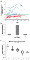
Figure 2. Temporal profile of perihematomal edema growth after spontaneous intracerebral hemorrhage
Panel A
: Growth curves (n=22 patients with 78 MRIs) of perihematomal edema in the first three weeks after ICH onset. Perihematomal edema peaks at a mean of 12 ± 3 days. Peak _r_PHE varies from 100-650%.
Panel B:
Timing of peak perihematomal edema (n=22 patients with 78 MRIs).
Panel C:
Boxplots of the rate and magnitude of perihematomal edema volume (Ev) growth in the first month after ICH. Edema growth is fastest in the first 48 hours after ICH onset.
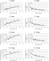
Figure 3. Relationship between hematoma and perihematomal edema volumes during the first month after spontaneous intracerebral hemorrhage
A larger hematoma volume is associated with larger perihematomal edema volume (Ev), but inversely correlated with relative perihematomal edema (_r_PHE).
Similar articles
- Peak Edema Extension Distance: An Edema Measure Independent from Hematoma Volume Associated with Functional Outcome in Intracerebral Hemorrhage.
Giede-Jeppe A, Gerner ST, Sembill JA, Kuramatsu JB, Lang S, Luecking H, Staykov D, Huttner HB, Volbers B. Giede-Jeppe A, et al. Neurocrit Care. 2024 Jun;40(3):1089-1098. doi: 10.1007/s12028-023-01886-z. Epub 2023 Nov 29. Neurocrit Care. 2024. PMID: 38030878 Free PMC article. - Fingolimod for the treatment of intracerebral hemorrhage: a 2-arm proof-of-concept study.
Fu Y, Hao J, Zhang N, Ren L, Sun N, Li YJ, Yan Y, Huang D, Yu C, Shi FD. Fu Y, et al. JAMA Neurol. 2014 Sep;71(9):1092-101. doi: 10.1001/jamaneurol.2014.1065. JAMA Neurol. 2014. PMID: 25003359 Clinical Trial. - Temporal pattern of cytotoxic edema in the perihematomal region after intracerebral hemorrhage: a serial magnetic resonance imaging study.
Li N, Worthmann H, Heeren M, Schuppner R, Deb M, Tryc AB, Bueltmann E, Lanfermann H, Donnerstag F, Weissenborn K, Raab P. Li N, et al. Stroke. 2013 Apr;44(4):1144-6. doi: 10.1161/STROKEAHA.111.000056. Epub 2013 Feb 7. Stroke. 2013. PMID: 23391767 - Perihematomal Edema After Intracerebral Hemorrhage: An Update on Pathogenesis, Risk Factors, and Therapeutic Advances.
Chen Y, Chen S, Chang J, Wei J, Feng M, Wang R. Chen Y, et al. Front Immunol. 2021 Oct 19;12:740632. doi: 10.3389/fimmu.2021.740632. eCollection 2021. Front Immunol. 2021. PMID: 34737745 Free PMC article. Review. - Treatment Strategies to Attenuate Perihematomal Edema in Patients With Intracerebral Hemorrhage.
Kim H, Edwards NJ, Choi HA, Chang TR, Jo KW, Lee K. Kim H, et al. World Neurosurg. 2016 Oct;94:32-41. doi: 10.1016/j.wneu.2016.06.093. Epub 2016 Jun 29. World Neurosurg. 2016. PMID: 27373415 Review.
Cited by
- Intraventricular hemorrhage expansion in patients with spontaneous intracerebral hemorrhage.
Witsch J, Bruce E, Meyers E, Velazquez A, Schmidt JM, Suwatcharangkoon S, Agarwal S, Park S, Falo MC, Connolly ES, Claassen J. Witsch J, et al. Neurology. 2015 Mar 10;84(10):989-94. doi: 10.1212/WNL.0000000000001344. Epub 2015 Feb 6. Neurology. 2015. PMID: 25663233 Free PMC article. - When the Blood Hits Your Brain: The Neurotoxicity of Extravasated Blood.
Stokum JA, Cannarsa GJ, Wessell AP, Shea P, Wenger N, Simard JM. Stokum JA, et al. Int J Mol Sci. 2021 May 12;22(10):5132. doi: 10.3390/ijms22105132. Int J Mol Sci. 2021. PMID: 34066240 Free PMC article. Review. - Hemorrhagic stroke: introduction.
Hanley DF, Awad IA, Vespa PM, Martin NA, Zuccarello M. Hanley DF, et al. Stroke. 2013 Jun;44(6 Suppl 1):S65-6. doi: 10.1161/STROKEAHA.113.000856. Stroke. 2013. PMID: 23709734 Free PMC article. No abstract available. - New avenues for treatment of intracranial hemorrhage.
Sonni S, Lioutas VA, Selim MH. Sonni S, et al. Curr Treat Options Cardiovasc Med. 2014 Jan;16(1):277. doi: 10.1007/s11936-013-0277-y. Curr Treat Options Cardiovasc Med. 2014. PMID: 24366522 Free PMC article. - Nomogram Prediction of Short-Term Outcome After Intracerebral Hemorrhage.
Kang H, Cai Q, Gong L, Wang Y. Kang H, et al. Int J Gen Med. 2021 Sep 7;14:5333-5343. doi: 10.2147/IJGM.S330742. eCollection 2021. Int J Gen Med. 2021. PMID: 34522130 Free PMC article.
References
- Xi G, Keep RF, Hoff JT. Mechanisms of brain injury after intracerebral haemorrhage. Lancet Neurol. 2006;5:53–63. - PubMed
- Mayer SA, Sacco RL, Shi T, Mohr JP. Neurologic deterioration in noncomatose patients with supratentorial intracerebral hemorrhage. Neurology. 1994;44:1379–1384. - PubMed
- Ladurner G, Sager WD, Iliff LD, Lechner H. A correlation of clinical findings and CT in ischaemic cerebrovascular disease. Eur Neurol. 1979;18:281–288. - PubMed
Publication types
MeSH terms
LinkOut - more resources
Full Text Sources
Medical


