Rapid expansion and long-term persistence of elevated NK cell numbers in humans infected with hantavirus - PubMed (original) (raw)
Rapid expansion and long-term persistence of elevated NK cell numbers in humans infected with hantavirus
Niklas K Björkström et al. J Exp Med. 2011.
Abstract
Natural killer (NK) cells are known to mount a rapid response to several virus infections. In experimental models of acute viral infection, this response has been characterized by prompt NK cell activation and expansion followed by rapid contraction. In contrast to experimental model systems, much less is known about NK cell responses to acute viral infections in humans. We demonstrate that NK cells can rapidly expand and persist at highly elevated levels for >60 d after human hantavirus infection. A large part of the expanding NK cells expressed the activating receptor NKG2C and were functional in terms of expressing a licensing inhibitory killer cell immunoglobulin-like receptor (KIR) and ability to respond to target cell stimulation. These results demonstrate that NK cells can expand and remain elevated in numbers for a prolonged period of time in humans after a virus infection. In time, this response extends far beyond what is considered normal for an innate immune response.
Figures
Figure 1.
Increase in CD56dim NK cells in human hantavirus infection. PBMCs from patients with acute hantavirus infection were analyzed by flow cytometry. (a and b) Absolute numbers of lymphocytes (a) and NK cells (b) at days 5 and 60 after symptom debut. (c) Relative changes of total lymphocytes and NK cells from days 5–60 after symptom debut (mean ± SEM). (d) Definition of CD56brightCD16− (i), CD56brightCD16+ (ii), CD56dimCD16+ (iii), and CD56−CD16+ (iv) NK cells by flow cytometry from one representative patient with acute hantavirus infection. Cells were gated on the CD3−CD4−CD14−CD19− population within the single cell lymphocyte gate. (e) Absolute numbers of the different NK cell subsets at days 5 and 60. For a–e, n = 16. *, P < 0.05; **, P < 0.01; ***, P < 0.0001, paired Student’s t test.
Figure 2.
Rapid proliferation and sustained elevation of CD56dim NK cell numbers. (a) Numbers of CD56dimCD16+ NK cells (solid lines) evaluated longitudinally from day 5 until day 450 after onset of clinical symptoms in infected patients (n = 16) and uninfected controls (n = 26; **, P < 0.01, Mann-Whitney test; mean ± SEM; the dashed lines represent upper and lower SEM intervals for mean of CD56dimCD16+ NK cells in the control individuals). (b–e) Expression of intracellular Ki67 used as an indicator of cells that are dividing or have recently divided. (b) Representative examples of Ki67 expression in CD56dim NK cells analyzed by flow cytometry at days 5, 11, and 60 after onset of symptoms in one hantavirus-infected patient. (c) Frequency of Ki67-positive cells in controls (n = 5) and patients 5 d after symptoms debut (n = 6; **, P = 0.0043, Mann-Whitney test; mean ± SEM). (d and e) Frequency (d) and absolute numbers (e) of Ki67+ CD56dim NK cells evaluated longitudinally in the infected patients (n = 8; mean ± SEM). (f) Plasma levels of IL-15 in the hantavirus-infected patients (n = 16) and noninfected controls (n = 24). IL-15 levels were significantly increased during acute infection compared with the convalescent stage (day 450; **, P < 0.01, Mann-Whitney test; mean ± SEM). (g and h) Expression of Bcl-2 in CD8 T cells and NK cells from patients with acute hantavirus infection analyzed by flow cytometry. Representative staining of Ki67 and Bcl-2 on CD8 T cells (g) and CD56dim NK cells (h) from one patient at day 5 after onset of symptoms, as well as the mean expression level (MFI) of Bcl-2 in Ki67+ and Ki67− CD8 T cells (g) and CD56dim NK cells (h) at days 5, 11, and 450 after onset of symptoms (n = 8; *, P < 0.05, Mann-Whitney test; horizontal bars represent mean).
Figure 3.
Hantavirus-infected endothelial cells up-regulate HLA-E. (a and b) Expression of ligands for NK cell receptors analyzed on hantavirus-infected primary human umbilical vein endothelial cells. (a) Immunofluorescence staining with patient serum on uninfected and infected cells 24 and 48 h after infection (hpi). Original magnification, 40×. Bars, 20 µm. (b) Kinetics of infection of endothelial cells. (c and d) Expression of ligands for NKG2D, NKG2A/C, DNAM-1, LFA-1, and KIRs assessed on infected (dashed lines) and uninfected (solid lines) endothelial cells 72 h after infection. Shaded histograms represent isotype control stainings. Co-culture of endothelial cells with UV-inactivated hantavirus for 72 h did not alter the expression of these ligands compared with uninfected cells (not depicted). Data in a–d are representative of three separate experiments.
Figure 4.
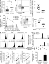
Expansion of NKG2C+ NK cells. (a) Frequency of NKG2C+ cells within the CD56dim NK cell population on day 5 after onset of symptoms in hantavirus-infected patients compared with noninfected controls (n = 16 infected, 59 uninfected; ***, P < 0.0001, unpaired Student’s t test; mean). (b) Representative example of costaining for NKG2A and NKG2C on CD56dim NK cells in hantavirus-infected patient at day 5 after onset of symptoms. (c) Co-expression of NKG2C and NKG2A evaluated at day 5 in infected (n = 6) and uninfected control (n = 9) individuals (horizontal bars represent mean). (d) Numbers of NKG2C+, NKG2A+, and NKG2C−NKG2A− CD56dimCD16+ NK cells evaluated longitudinally from day 5 until day 450 after onset of symptoms in infected patients (n = 6; mean ± SEM). Dashed lines represent the upper and lower SEM intervals for mean of NKG2C+ NK cell numbers in control individuals (n = 26). (e) Representative example of costaining for NKG2C and CD57 on CD56dim NK cells in one hantavirus-infected patient at day 60 after onset of symptoms. (f) Expression of CD57 evaluated at day 60 after symptom debut (n = 6) in NKG2C+ and NKG2C− CD56dim NK cells (**, P = 0.0022, Mann-Whitney test; mean). (g) Proliferation of NKG2C+ NK cells after stimulation with IL-15 and/or target cells measured by dilution of CellTrace violet. Purified NK cells were incubated for 7 d with or without irradiated K562 cells or K562*HLA-E cells, and in the absence (top) or presence (bottom) of IL-15. One representative experiment out of two is shown. (h) Expression pattern of the three major KIRs on NKG2C+ and NKG2C− NK cells. In one representative donor, a majority of the NKG2C+ NK cells were single positive for KIR2DL1/S1 (top), whereas the second representative donor shows a selective expression of KIR2DL2/S2/2DL3 (bottom) in the NKG2C+ population (see
Fig. S4
for separation between activation and inhibitory KIRs). Two representative donors out of five analyzed are shown. (i) Degranulation and cytokine production responses quantified in NKG2C+ CD56dim NK cells from patients in the convalescent phase of infection after triggering with K562-E cells with or without addition of an HLA-E stabilizing peptide (n = 5; *, P < 0.05, Mann-Whitney test). (j) Absolute numbers of NKG2C+ CD56dim NK cells in CMV IgG− and IgG+ infected individuals at day 5 (mean ± SEM).
Similar articles
- Artificial feeder cells expressing ligands for killer cell immunoglobulin-like receptors and CD94/NKG2A for expansion of functional primary natural killer cells with tolerance to self.
Michen S, Frosch J, Füssel M, Schackert G, Momburg F, Temme A. Michen S, et al. Cytotherapy. 2020 Jul;22(7):354-368. doi: 10.1016/j.jcyt.2020.02.004. Epub 2020 May 23. Cytotherapy. 2020. PMID: 32451262 - Adaptive reconfiguration of the human NK-cell compartment in response to cytomegalovirus: a different perspective of the host-pathogen interaction.
Muntasell A, Vilches C, Angulo A, López-Botet M. Muntasell A, et al. Eur J Immunol. 2013 May;43(5):1133-41. doi: 10.1002/eji.201243117. Eur J Immunol. 2013. PMID: 23552990 Review. - IL-12-producing monocytes and HLA-E control HCMV-driven NKG2C+ NK cell expansion.
Rölle A, Pollmann J, Ewen EM, Le VT, Halenius A, Hengel H, Cerwenka A. Rölle A, et al. J Clin Invest. 2014 Dec;124(12):5305-16. doi: 10.1172/JCI77440. Epub 2014 Nov 10. J Clin Invest. 2014. PMID: 25384219 Free PMC article. Clinical Trial. - NK cell activation in human hantavirus infection explained by virus-induced IL-15/IL15Rα expression.
Braun M, Björkström NK, Gupta S, Sundström K, Ahlm C, Klingström J, Ljunggren HG. Braun M, et al. PLoS Pathog. 2014 Nov 20;10(11):e1004521. doi: 10.1371/journal.ppat.1004521. eCollection 2014 Nov. PLoS Pathog. 2014. PMID: 25412359 Free PMC article. - The CD94/NKG2C+ NK-cell subset on the edge of innate and adaptive immunity to human cytomegalovirus infection.
López-Botet M, Muntasell A, Vilches C. López-Botet M, et al. Semin Immunol. 2014 Apr;26(2):145-51. doi: 10.1016/j.smim.2014.03.002. Epub 2014 Mar 22. Semin Immunol. 2014. PMID: 24666761 Review.
Cited by
- Influenza Vaccination Generates Cytokine-Induced Memory-like NK Cells: Impact of Human Cytomegalovirus Infection.
Goodier MR, Rodriguez-Galan A, Lusa C, Nielsen CM, Darboe A, Moldoveanu AL, White MJ, Behrens R, Riley EM. Goodier MR, et al. J Immunol. 2016 Jul 1;197(1):313-25. doi: 10.4049/jimmunol.1502049. Epub 2016 May 27. J Immunol. 2016. PMID: 27233958 Free PMC article. - Tissue-specific effector functions of innate lymphoid cells.
Björkström NK, Kekäläinen E, Mjösberg J. Björkström NK, et al. Immunology. 2013 Aug;139(4):416-27. doi: 10.1111/imm.12098. Immunology. 2013. PMID: 23489335 Free PMC article. Review. - NK cell frequencies, function and correlates to vaccine outcome in BNT162b2 mRNA anti-SARS-CoV-2 vaccinated healthy and immunocompromised individuals.
Cuapio A, Boulouis C, Filipovic I, Wullimann D, Kammann T, Parrot T, Chen P, Akber M, Gao Y, Hammer Q, Strunz B, Pérez Potti A, Rivera Ballesteros O, Lange J, Muvva JR, Bergman P, Blennow O, Hansson L, Mielke S, Nowak P, Söderdahl G, Österborg A, Smith CIE, Bogdanovic G, Muschiol S, Hellgren F, Loré K, Sobkowiak MJ, Gabarrini G, Healy K, Sällberg Chen M, Alici E, Björkström NK, Buggert M, Ljungman P, Sandberg JK, Aleman S, Ljunggren HG. Cuapio A, et al. Mol Med. 2022 Feb 8;28(1):20. doi: 10.1186/s10020-022-00443-2. Mol Med. 2022. PMID: 35135470 Free PMC article. - Natural Killer Cells Adapt to Cytomegalovirus Along a Functionally Static Phenotypic Spectrum in Human Immunodeficiency Virus Infection.
Holder KA, Lajoie J, Grant MD. Holder KA, et al. Front Immunol. 2018 Nov 12;9:2494. doi: 10.3389/fimmu.2018.02494. eCollection 2018. Front Immunol. 2018. PMID: 30483249 Free PMC article. - Is IFN expression by NK cells a hallmark of severe COVID-19?
Csordas BG, de Sousa Palmeira PH, Peixoto RF, Comberlang FCQDDS, de Medeiros IA, Azevedo FLAA, Veras RC, Janebro DI, Amaral IPG, Barbosa-Filho JM, Keesen TSL. Csordas BG, et al. Cytokine. 2022 Sep;157:155971. doi: 10.1016/j.cyto.2022.155971. Epub 2022 Jul 22. Cytokine. 2022. PMID: 35908408 Free PMC article.
References
Publication types
MeSH terms
Substances
LinkOut - more resources
Full Text Sources
Other Literature Sources
Medical


