Common neural mechanisms supporting spatial working memory, attention and motor intention - PubMed (original) (raw)
Review
Common neural mechanisms supporting spatial working memory, attention and motor intention
Akiko Ikkai et al. Neuropsychologia. 2011 May.
Abstract
The prefrontal cortex (PFC) and posterior parietal cortex (PPC) are critical neural substrates for working memory. Neural activity persists in these regions during the maintenance of a working memory representation. Persistent activity, therefore, may be the neural mechanism by which information is temporarily maintained. However, the nature of the representation or what is actually being represented by this persistent activity is not well understood. In this review, we summarize the recent functional magnetic resonance imaging (fMRI) studies conducted in our laboratory that test hypotheses about the nature of persistent activity during a variety of spatial cognition tasks. We find that the same areas in the PFC and PPC that show persistent activity during the maintenance of a working memory representation also show persistent activity during the maintenance of spatial attention and the maintenance of motor intention. Therefore, we conclude that persistent activity is not specific to working memory, but instead, carries information that can be used generally to support a variety of cognitions. Specifically, activity in topographically organized maps of prioritized space in PFC and PPC could be read out to guide attention allocation, spatial memory, and motor planning.
Copyright © 2010 Elsevier Ltd. All rights reserved.
Figures
Fig 1
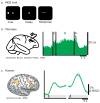
a. Trial schematic of a memory guided saccade (MGS) task. A subject must maintain the location of a briefly presented visual cue over a memory delay and then make a saccade to the past cue's location. b. A spike histogram from a neuron in the monkey PFC during a MGS task (Funahashi et al., 1989). c. BOLD signal time course from the human PFC during a MGS task (Srimal & Curtis, 2008). Notice that in both species neural activity persists during the memory delay period. C=cue, D=delay, and R=response.
Fig 2
Task and results summary from working memory (WM), attention, and intention studies. Trial schematics are shown for each task (a., d., and g.). Statistical parametric maps of significant delay period specific activity are projected onto the surface of a subject's cortical sheet for each task (b., e., and h.). Time courses (average, SEM) from the PFC (c., f., i. top panels) and PPC (bottom panels) are shown time locked to the presentation of the cue. Solid lines represent trials in which the memoranda (c.), locus of attention (f.), or direction of antisaccade (i.) was in hemifield contralateral to the cortical hemisphere and dashed lines represent ipsilateral trials. Notice that both PFC and PPC show delay period activation, that this activation persists throughout the delay period, and it shows a contralateral bias.
Fig 3
Delay period activity from subjects who participated in all three of the studies. (N=5). a. BOLD time-series from the exact same voxels in superior precentral sulcus (sPCS) across the three studies. Notice that delay period activity persists during the WM, attention, and intention delay periods and shows a contralateral bias. b. Cortex with significant delay period activity projected on an inflated cortical sheet of the right hemisphere. The color wheel is the legend for the delay period activity. Areas that show both activation for attention and intention would be depicted in magenta. Areas that show delay period activation during all three tasks are depicted in black and those areas are labeled.
Similar articles
- Maps of space in human frontoparietal cortex.
Jerde TA, Curtis CE. Jerde TA, et al. J Physiol Paris. 2013 Dec;107(6):510-6. doi: 10.1016/j.jphysparis.2013.04.002. Epub 2013 Apr 18. J Physiol Paris. 2013. PMID: 23603831 Free PMC article. Review. - Mnemonic Encoding and Cortical Organization in Parietal and Prefrontal Cortices.
Masse NY, Hodnefield JM, Freedman DJ. Masse NY, et al. J Neurosci. 2017 Jun 21;37(25):6098-6112. doi: 10.1523/JNEUROSCI.3903-16.2017. Epub 2017 May 24. J Neurosci. 2017. PMID: 28539423 Free PMC article. - Maintaining structured information: an investigation into functions of parietal and lateral prefrontal cortices.
Wendelken C, Bunge SA, Carter CS. Wendelken C, et al. Neuropsychologia. 2008 Jan 31;46(2):665-78. doi: 10.1016/j.neuropsychologia.2007.09.015. Epub 2007 Oct 6. Neuropsychologia. 2008. PMID: 18022652 - Working Memory and Decision-Making in a Frontoparietal Circuit Model.
Murray JD, Jaramillo J, Wang XJ. Murray JD, et al. J Neurosci. 2017 Dec 13;37(50):12167-12186. doi: 10.1523/JNEUROSCI.0343-17.2017. Epub 2017 Nov 7. J Neurosci. 2017. PMID: 29114071 Free PMC article. - Prefrontal and parietal contributions to spatial working memory.
Curtis CE. Curtis CE. Neuroscience. 2006 Apr 28;139(1):173-80. doi: 10.1016/j.neuroscience.2005.04.070. Epub 2005 Dec 2. Neuroscience. 2006. PMID: 16326021 Review.
Cited by
- Hemispheric lateralization of verbal and spatial working memory during adolescence.
Nagel BJ, Herting MM, Maxwell EC, Bruno R, Fair D. Nagel BJ, et al. Brain Cogn. 2013 Jun;82(1):58-68. doi: 10.1016/j.bandc.2013.02.007. Epub 2013 Mar 16. Brain Cogn. 2013. PMID: 23511846 Free PMC article. - The neural representation of objects formed through the spatiotemporal integration of visual transients.
Erlikhman G, Gurariy G, Mruczek REB, Caplovitz GP. Erlikhman G, et al. Neuroimage. 2016 Nov 15;142:67-78. doi: 10.1016/j.neuroimage.2016.03.044. Epub 2016 Mar 24. Neuroimage. 2016. PMID: 27033688 Free PMC article. - Oculomotor assessments of executive function in preterm children.
Loe IM, Luna B, Bledsoe IO, Yeom KW, Fritz BL, Feldman HM. Loe IM, et al. J Pediatr. 2012 Sep;161(3):427-433.e1. doi: 10.1016/j.jpeds.2012.02.037. Epub 2012 Apr 4. J Pediatr. 2012. PMID: 22480696 Free PMC article. Clinical Trial. - Virtual Reality Assessment of Classroom - Related Attention: An Ecologically Relevant Approach to Evaluating the Effectiveness of Working Memory Training.
Coleman B, Marion S, Rizzo A, Turnbull J, Nolty A. Coleman B, et al. Front Psychol. 2019 Aug 20;10:1851. doi: 10.3389/fpsyg.2019.01851. eCollection 2019. Front Psychol. 2019. PMID: 31481911 Free PMC article. - Sharpening Working Memory With Real-Time Electrophysiological Brain Signals: Which Neurofeedback Paradigms Work?
Jiang Y, Jessee W, Hoyng S, Borhani S, Liu Z, Zhao X, Price LK, High W, Suhl J, Cerel-Suhl S. Jiang Y, et al. Front Aging Neurosci. 2022 Mar 28;14:780817. doi: 10.3389/fnagi.2022.780817. eCollection 2022. Front Aging Neurosci. 2022. PMID: 35418848 Free PMC article. Review.
References
- Andersen RA, Buneo CA. Intentional maps in posterior parietal cortex. Annu Rev Neurosci. 2002;25:189–220. - PubMed
Publication types
MeSH terms
LinkOut - more resources
Full Text Sources
Miscellaneous


