ATP hydrolysis is required for relocating cohesin from sites occupied by its Scc2/4 loading complex - PubMed (original) (raw)
ATP hydrolysis is required for relocating cohesin from sites occupied by its Scc2/4 loading complex
Bin Hu et al. Curr Biol. 2011.
Abstract
Background: The Cohesin complex that holds sister chromatins together until anaphase is comprised of three core subunits: Smc1 and Smc3, two long-rod-shaped proteins with an ABC-like ATPase head (nucleotide-binding domain [NBD]) and a dimerization domain linked by a 50 nm long intramolecular antiparallel coiled-coil, and Scc1, an α-kleisin subunit interconnecting the NBD domains of Smc1 and Smc3. Cohesin's stable association with chromosomes is thought to involve entrapment of chromatin fibers by its tripartite Smc1-Smc3-Scc1 ring via a poorly understood mechanism dependent on a separate Scc2/4 loading complex. A key issue concerns where entrapment initially takes place: at sites where cohesin is found stably associated or at distinct "loading" sites from which it translocates.
Results: In this study, we find transition state mutant versions (Smc1E1158Q and SmcE1155Q) defective in disengagement of their nucleotide binding domains (NBDs), unlike functional cohesin, colocalize with Scc2/4 at core centromeres, sites that catalyze wild-type cohesin's recruitment to sequences 20 kb or more away. In addition to Scc2/4, the unstable association of transition state complexes with core centromeres requires Scc1's association with Smc1 and Smc3 NBDs, ATP-driven NBD engagement, cohesin's Scc3 subunit, and its hinge domain.
Conclusion: We propose that cohesin's association with chromosomes is driven by two key events. NBD engagement driven by ATP binding produces an unstable association with specific loading sites like core centromeres, whereas subsequent ATP hydrolysis triggers DNA entrapment, which permits translocation along chromatin fibers.
Copyright © 2011 Elsevier Ltd. All rights reserved.
Figures
Figure 1. Centromeres and Kinetochore Proteins Promote Recruitment of Scc2/4 and Cohesin
(A and B) An ectopic centromere promotes cohesin’s accumulation 20 kb away. The distribution of Scc1-PK9 was measured by ChIP-SEQ in exponentially grown cells from strain K16670 in which CEN14 was moved to a site between ADE12 and ALG9 (top panels) and compared to that of Scc1 in wild-type (WT) cells (K16586) (top and middle panels). The ratios of Scc1 ChIP-SEQ signals between K16670 and wild-type were mapped to chromosome XIV (bottom panels). Yellow bars indicate the ratio is more than 1.0 and gray bars indicate the ratio is less than 1.0. The Scc1 distribution within a 50 kb region along ectopic CEN14 (A) or endogenous CEN14 (B) is shown. See also Figure S1.
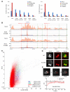
Figure 2. Unstable Association of Hydrolysis-Defective Cohesin Complexes with Centromeres
(A) Hydrolysis-defective cohesin is enriched at centromeres. Association with defined loci of wild-type and mutant Smc proteins tagged with myc9 (Smc1) or HA3 (Smc3) was measured by ChIP-qPCR. Chromatin was immunoprecipitated from extracts prepared from exponentially grown cells of strains K699, K11850, K11852, K11857, K11872, K13560, and K13561. The following abbreviations are used: Cen, centromere; inner pericen, inner pericentromere; outer pericen, outer pericentromere. Error bars represent standard deviation (SD); n = 3. (B) Genome-wide distribution of Smc3-HA3 and Smc3E1155Q-HA3. Crude extracts prepared from exponentially grown yeast cells (K16586 and K17458) were used for ChIP-SEQ. Red bars represent binding ratios within running 500 bp windows (50 bp step size) showing enrichment in the ChIP fraction. Positions of the centromere (CEN) and autonomously replicating sequences (ARSs) are shown. The horizontal axis represents kilobases along chromosome III. (C) Correlations between Smc3 and Smc3E1155Q. ChIP-SEQ signals were pooled from running 500 bp windows along each chromosome every 50 bp. Smc3E1155Q-HA3 signals from each window were plotted against those of Smc3-HA3. ChIP signals within 500 bp of tDNAs are marked as green dots, those within 5000 bp of centromeres are marked as blue dots, and the rest are marked as red dots. The correlation coefficients for centromeres, tDNA, the others, and the total are shown bottom right. (D) Localization of Smc3 and Smc3E1155Q in live diploid cells. Smc3 and Smc3E1155Q were fused with GFP (K18232 and K16715). Mtw1-RFP was used as a kinetochore marker. Smc3 forms pericentromeric barrels between sister kinetochore clusters at metaphase, whereas Smc3E1155Q colocalizes with Mtw1. (E) Association of Smc3E1155Q with centromeres is unstable. FRAP was measured in diploid yeast cells expressing Smc3E1155Q-GFP. One of two Smc3E1155Q fluorescent foci was bleached by exposing the area marked by a red circle to an argon laser for 200 ms. Relative fluorescence intensities of unbleached (black) and bleached (red) signals are plotted over time. Smc3E1155Q -GFP recovered with t1/2 = 3.4 s; n = 6; the error bars represent SD. The signal detected just after photobleaching (around 45% of that before photobleaching) was due to rapid turnover of soluble pool of Smc3E1158Q. See also Figure S2.
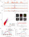
Figure 3. Hydrolysis-Defective Cohesin Colocalizes with and Depends on Scc2/4
(A) ChIP-SEQ distributions of Smc3-HA3, Smc3E1155Q-HA3, and Scc2-FLAG6 at selected regions of chromosome V. Yeast strains K13560, K13561, and K17458 were used. (B) Correlations of Smc3E1155Q with Scc2 performed as described in Figure 2C. (C) Localization of Scc2-GFP in live diploid cells (K16442). Kinetochores are marked by Mtw1-RFP. (D) Association of Scc2-GFP with centromeres is unstable. FRAP performed as in Figure 2E showed that fluorescence recovered after photobleaching with t1/2 = 0.8 s; n = 6. (E and F) Association of Smc1E1158Q with chromatin is Scc2-dependent. Exponential phase cultures of strains K16799, K16800, K16811, K16812, and K16331 growing in YEPraff at 25°C were arrested in G1 with α-factor. Degradation of td-Scc2 was triggered by shifting cultures to YEPgal, 20 μg/ml doxycycline, and 37°C for 1 hr before transferring cells to pheromone-free YEPgal media containing 20 μg/ml doxycycline. Chromatin was immunoprecipitated with myc9 tags, and association with indicated loci of myc9-tagged Smc1 and Smc1E1158Q was measured with ChIP-qPCR. See also Figure S3.
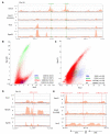
Figure 4. Scc2/4 and Hydrolysis-Defective Cohesin Accumulate on Highly Transcribed Genes
(A) Scc2 and Smc3E1155Q but not Smc3 colocalize with PolII subunit Rpo21 a PolII. Yeast cells expressing Rpo21-Flag3 (K17460) were used for ChIP-SEQ. The distributions of Smc3-HA3, Smc3E1155Q-HA3, and Scc2-FLAG6 are compared to Rpo21-FLAG3 within a selected region of chromosome VII. tDNAs are indicated with green lines. (B) Correlation of Rpc128 (K18393) with Scc2 performed as described in Figure 2C. (C) Correlation of Rpo21 (K18394) with Scc2 performed as described in Figure 2C. (D) The distributions of Scc2, Rpo21, and Spt15 within a selected region of chromosome IV. (E) The distributions of Smc3, Smc3E1155Q, and Scc2 at rDNA loci with transcription units of 35S and 5S shown at the bottom. See also Figure S4.
Figure 5. Cohesin Completely Blocked with NBDs Stably Engaged Associates with Centromeres
(A) ATP-dependent engagement of Smc1 and Smc3 NBDs. Equal amounts (3 nmol) of recombinant WT or ATP hydrolysis-defective Smc NBDs (Smc1 was associated with Scc1’s C-terminal fragment [F451–A566]) were subjected to size exclusion chromatography in the presence or absence of ATP. No dimer formation was observed with ATPγS or AMPPNP (data not shown). (B) Disruption of ATP-dependent engagement of Smc1 and Smc3 NBDs by mutation in signature motif. Equal amounts (3 nmol) of recombinant indicated Smc NBDs were subjected to size exclusion. (C) Cohesin incapable of hydrolyzing ATP associated with both NBDs also associates with centromeres. Exponential phase cells of strains K16331, K17240, K17241, and K17242 growing at 25°C were arrested in G1 with α-factor. Degradation of Smc3-td was induced and cells released from pheromone as in Figure 2E. The association of Smc1E1158Q-MYC9 with centromere was measured by ChIP-qPCR. See also Figure S5.
Figure 6. Ring Formation and Scc3 Are Required for Cohesin Loading
(A) Ring formation is required for cohesin loading. Wild-type or mutant Scc1-GFP was coexpressed with Smc1E1158Q (K16764, K16765, K16766, and K16767) and imaged in live cells. Wild-type Scc1-GFP and Scc1V81K-GFP colocalize with Mtw1-RFP foci and, in addition, form pericentric barrels in between. L75K and L89K mutations abolish both association with Mtw1 and barrel formation. (B) Scc3 is required for cohesin loading. Wild-type or mutant Scc1-GFP (K16915) was coexpressed with Smc1E1158Q and visualized by fluorescence microscopy. Wild-type Scc1-GFP colocalizes with Mtw1 foci as well as forming pericentric barrels in between. Scc1Δ319-327-GFP merely accumulates within nuclei. (C) Association of Smc1E1158Q with chromatin depends on Scc3 but not Pds5. Exponential phase cells of strains K16331, K16812, K17299, and K17300 were arrested at G1 phase with α-factor at 25°C. Degradation of Scc3-td or Pds5-td was induced and cells released from pheromone as in Figure 2E. Association of myc9-tagged Smc1E1158Q with centromeres was measured by ChIP-qPCR. (D) Smc1E1158Q-GFP was expressed in wild-type or rad61Δ strains (K16445 and K16796) and visualized together with Mtw1-RFP by fluorescence microscopy. See also Figure S6.
Figure 7. Smc1/3 Hinges Are Required for Cohesin Loading
(A and B) Hinge-substituted Smc1/3 heterodimers are not loaded onto chromosomes. (A) Association with CEN6 of myc9-tagged Smc1 or Smc1E1158Q with wild-type or p14/MP1 hinge domains measured by ChIP-qPCR in asynchronous yeast extract/peptone/dextrose (YPD) cultures of strains K699, K11587, K13585, K14133, and K16874. The error bars represent SD; n = 3. (B) Smc1E1158Q-GFP or Smc1P14/E1155Q-GFP was coexpressed with an extra copy of Smc3 or with Smc3MP1 (K16936 and K17070). GFP fusion proteins were visualized by fluorescence microscopy. Hinge substitution abolished association of Smc1E1158Q-GFP with Mtw1-RFP foci in metaphase cells. (C and D) The F584R Smc1 hinge mutation abolishes cohesin’s association with centromeres. (C) Association with CEN6 of myc9-tagged Smc1 or Smc1E1158Q proteins with wild-type or Smc1F584R hinge domains measured by ChIP-qPCR in asynchronous YPD cultures of strains K699, K11857, K14133, K14134, and K17000. The error bars represent SD; n = 3. (D) Smc1E1158Q-GFP or Smc1F584R/E1155Q-GFP (K16895 and K17070) was coexpressed with an extra copy of Smc3 and visualized together with Mtw1-RFP by fluorescence microscopy. See also Figure S7.
Similar articles
- Cohesin's ATPase activity is stimulated by the C-terminal Winged-Helix domain of its kleisin subunit.
Arumugam P, Nishino T, Haering CH, Gruber S, Nasmyth K. Arumugam P, et al. Curr Biol. 2006 Oct 24;16(20):1998-2008. doi: 10.1016/j.cub.2006.09.002. Curr Biol. 2006. PMID: 17055978 - ATP hydrolysis is required for cohesin's association with chromosomes.
Arumugam P, Gruber S, Tanaka K, Haering CH, Mechtler K, Nasmyth K. Arumugam P, et al. Curr Biol. 2003 Nov 11;13(22):1941-53. doi: 10.1016/j.cub.2003.10.036. Curr Biol. 2003. PMID: 14614819 - Folding of cohesin's coiled coil is important for Scc2/4-induced association with chromosomes.
Petela NJ, Gonzalez Llamazares A, Dixon S, Hu B, Lee BG, Metson J, Seo H, Ferrer-Harding A, Voulgaris M, Gligoris T, Collier J, Oh BH, Löwe J, Nasmyth KA. Petela NJ, et al. Elife. 2021 Jul 14;10:e67268. doi: 10.7554/eLife.67268. Elife. 2021. PMID: 34259632 Free PMC article. - The torments of the cohesin ring.
Chavda AP, Ang K, Ivanov D. Chavda AP, et al. Nucleus. 2017 May 4;8(3):261-267. doi: 10.1080/19491034.2017.1295200. Epub 2017 Feb 27. Nucleus. 2017. PMID: 28453390 Free PMC article. Review. - Chromosome organization by fine-tuning an ATPase.
Massari LF, Marston AL. Massari LF, et al. Genes Dev. 2023 Apr 1;37(7-8):259-260. doi: 10.1101/gad.350627.123. Epub 2023 Apr 12. Genes Dev. 2023. PMID: 37045607 Free PMC article. Review.
Cited by
- Structural evidence for Scc4-dependent localization of cohesin loading.
Hinshaw SM, Makrantoni V, Kerr A, Marston AL, Harrison SC. Hinshaw SM, et al. Elife. 2015 Jun 3;4:e06057. doi: 10.7554/eLife.06057. Elife. 2015. PMID: 26038942 Free PMC article. - Caenorhabditis elegans Dosage Compensation: Insights into Condensin-Mediated Gene Regulation.
Albritton SE, Ercan S. Albritton SE, et al. Trends Genet. 2018 Jan;34(1):41-53. doi: 10.1016/j.tig.2017.09.010. Epub 2017 Oct 13. Trends Genet. 2018. PMID: 29037439 Free PMC article. Review. - Evidence for cohesin sliding along budding yeast chromosomes.
Ocampo-Hafalla M, Muñoz S, Samora CP, Uhlmann F. Ocampo-Hafalla M, et al. Open Biol. 2016 Jun;6(6):150178. doi: 10.1098/rsob.150178. Open Biol. 2016. PMID: 27278645 Free PMC article. - The SMC Loader Scc2 Promotes ncRNA Biogenesis and Translational Fidelity.
Zakari M, Trimble Ross R, Peak A, Blanchette M, Seidel C, Gerton JL. Zakari M, et al. PLoS Genet. 2015 Jul 15;11(7):e1005308. doi: 10.1371/journal.pgen.1005308. eCollection 2015 Jul. PLoS Genet. 2015. PMID: 26176819 Free PMC article. - Topological in vitro loading of the budding yeast cohesin ring onto DNA.
Minamino M, Higashi TL, Bouchoux C, Uhlmann F. Minamino M, et al. Life Sci Alliance. 2018 Oct 26;1(5):e201800143. doi: 10.26508/lsa.201800143. Life Sci Alliance. 2018. PMID: 30381802 Free PMC article.
References
- Peters JM, Tedeschi A, Schmitz J. The cohesin complex and its roles in chromosome biology. Genes Dev. 2008;22:3089–3114. - PubMed
- Nasmyth K, Haering CH. Cohesin: Its roles and mechanisms. Annu. Rev. Genet. 2009;43:525–558. - PubMed
- Onn I, Heidinger-Pauli JM, Guacci V, Unal E, Koshland DE. Sister chromatid cohesion: A simple concept with a complex reality. Annu. Rev. Cell Dev. Biol. 2008;24:105–129. - PubMed
- Haering CH, Löwe J, Hochwagen A, Nasmyth K. Molecular architecture of SMC proteins and the yeast cohesin complex. Mol. Cell. 2002;9:773–788. - PubMed
- Gruber S, Haering CH, Nasmyth K. Chromosomal cohesin forms a ring. Cell. 2003;112:765–777. - PubMed
Publication types
MeSH terms
Substances
LinkOut - more resources
Full Text Sources
Molecular Biology Databases
Research Materials
Miscellaneous


