Analysing the eosinophil cationic protein--a clue to the function of the eosinophil granulocyte - PubMed (original) (raw)
Review
Analysing the eosinophil cationic protein--a clue to the function of the eosinophil granulocyte
Jonas Bystrom et al. Respir Res. 2011.
Abstract
Eosinophil granulocytes reside in respiratory mucosa including lungs, in the gastro-intestinal tract, and in lymphocyte associated organs, the thymus, lymph nodes and the spleen. In parasitic infections, atopic diseases such as atopic dermatitis and asthma, the numbers of the circulating eosinophils are frequently elevated. In conditions such as Hypereosinophilic Syndrome (HES) circulating eosinophil levels are even further raised. Although, eosinophils were identified more than hundred years ago, their roles in homeostasis and in disease still remain unclear. The most prominent feature of the eosinophils are their large secondary granules, each containing four basic proteins, the best known being the eosinophil cationic protein (ECP). This protein has been developed as a marker for eosinophilic disease and quantified in biological fluids including serum, bronchoalveolar lavage and nasal secretions. Elevated ECP levels are found in T helper lymphocyte type 2 (atopic) diseases such as allergic asthma and allergic rhinitis but also occasionally in other diseases such as bacterial sinusitis. ECP is a ribonuclease which has been attributed with cytotoxic, neurotoxic, fibrosis promoting and immune-regulatory functions. ECP regulates mucosal and immune cells and may directly act against helminth, bacterial and viral infections. The levels of ECP measured in disease in combination with the catalogue of known functions of the protein and its polymorphisms presented here will build a foundation for further speculations of the role of ECP, and ultimately the role of the eosinophil.
Figures
Figure 1
Identification of eosinophil granulocytes in peripheral blood by immunohistochemical detection of ECP. (A) Negative control (omission of primary antibody). Shown are peripheral leukocytes after fixation, incubation with alkaline phosphatase-anti-alkaline phosphatase (APAAP) with fast red substrate and counterstaining with Mayer's hematoxylin. The characteristic red immune-labelling reaction is absent. (B) Leukocytes are treated as in (A) but with addition of anti-ECP antibody. Peripheral leukocytes are visible but only the eosinophils have been stained for ECP. Original Magnification (X420).
Figure 2
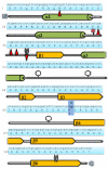
The RNASE3 (ECP) gene and ECP protein sequence with numbers referring to the amino acid sequence. Below the protein sequence is a schematic diagram of the peptide sequence where the beta sheet domains and the alpha helix domains are shown as red arrow and green barrel structures, respectively. Amino acids involved in RNase activity are represented by scissors. Amino acids involved in membrane interference, heparin binding and bactericidal activity are represented by red arrows. Glycosylated amino acids are represented with a glycomoiety while the letter N highlights the nitrated amino acid. A blue box shows the site of the amino acid altering polymorphism rs2073342.
Figure 3
Eosinophil granulocytes in the nasal mucosa. (A) Immunohistochemical staining of nasal biopsy specimens for ECP in (A) a healthy control and (B, C) a patient with perennial allergic rhinitis. In healthy controls (A), a few cells are staining weakly for ECP in the submucosa and epithelium. In patients with perennial allergic rhinitis cells staining intensely for ECP are present in the submucosa and epithelium. (original magnification, ×420). (C) Higher magnification highlighting eosinophil granules in the epithelium residing cells (original magnification ×1050); Mayer's hematoxylin.
Figure 4
Eosinophil granulocytes in the bronchial mucosa. Sections of bronchial biopsies from (A) a healthy control or (B) an individual with allergic asthma were stained with ECP antibody visualizing eosinophils in the mucosa. The figures show that only a few eosinophils are present in the tissue of the healthy control, but many eosinophils accumulate in areas of reduced epithelial integrity in a specimen from a patient with allergic asthma. Original magnification ×420; Mayer's haematoxylin.
Figure 5
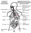
Known anatomical locations of eosinophil granulocytes and suggested activities of released ECP at these sites. On the left side are eosinophil granulocytes locations at homeostasis shown. On the right side are areas speculated to be affected by increased numbers of eosinophils and elevated levels of released ECP, in disease (pathology, P) and in physiological defense (function, F). This is a speculation by the authors of the review.
Figure 6
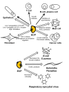
ECP's specific influences on various cell types and micro organisms in vitro. Alpha helixes in the protein are shown with green color and the location of the active site is marked with a red dot. Open arrow indicates moderate (1-5 μg/mL) to high (>5 μg/mL) concentrations of ECP used in the in vitro experiments. Filled arrow indicates low amounts of ECP used in the in vitro experiments (<1 μg/mL).
Similar articles
- Dissecting the role of eosinophil cationic protein in upper airway disease.
Bystrom J, Patel SY, Amin K, Bishop-Bailey D. Bystrom J, et al. Curr Opin Allergy Clin Immunol. 2012 Feb;12(1):18-23. doi: 10.1097/ACI.0b013e32834eccaf. Curr Opin Allergy Clin Immunol. 2012. PMID: 22157160 Review. - [Is the seric eosinophil cationic protein level a valuable tool of diagnosis in clinical practice?].
Moneret-Vautrin DA. Moneret-Vautrin DA. Rev Med Interne. 2006 Sep;27(9):679-83. doi: 10.1016/j.revmed.2006.02.007. Epub 2006 Mar 30. Rev Med Interne. 2006. PMID: 16647168 Review. French. - A (G->C) transversion in the 3' UTR of the human ECP (eosinophil cationic protein) gene correlates to the cellular content of ECP.
Jönsson UB, Byström J, Stålenheim G, Venge P. Jönsson UB, et al. J Leukoc Biol. 2006 Apr;79(4):846-51. doi: 10.1189/jlb.0904517. Epub 2006 Jan 24. J Leukoc Biol. 2006. PMID: 16434694 - Clara cell protein 16 and eosinophil cationic protein production in chronically inflamed sinonasal mucosa.
Špadijer-Mirković C, Perić A, Belić B, Vojvodić D. Špadijer-Mirković C, et al. Int Forum Allergy Rhinol. 2016 May;6(5):529-36. doi: 10.1002/alr.21710. Epub 2016 Feb 2. Int Forum Allergy Rhinol. 2016. PMID: 26833624 Clinical Trial. - Are sputum eosinophil cationic protein and eosinophils differently associated with clinical and functional findings of asthma?
Cianchetti S, Bacci E, Ruocco L, Pavia T, Bartoli ML, Cardini C, Costa F, Di Franco A, Malagrinò L, Novelli F, Vagaggini B, Celi A, Dente F, Paggiaro P. Cianchetti S, et al. Clin Exp Allergy. 2014;44(5):673-80. doi: 10.1111/cea.12236. Clin Exp Allergy. 2014. PMID: 24245689
Cited by
- Controlled human exposures: a review and comparison of the health effects of diesel exhaust and wood smoke.
Long E, Rider CF, Carlsten C. Long E, et al. Part Fibre Toxicol. 2024 Oct 23;21(1):44. doi: 10.1186/s12989-024-00603-8. Part Fibre Toxicol. 2024. PMID: 39444041 Free PMC article. Review. - Biomarkers of type 2 and non-type 2 inflammation in asthma exacerbations.
Ali KM, Jamal N, Wasman Smail S, Lauran M, Bystrom J, Janson C, Amin K. Ali KM, et al. Cent Eur J Immunol. 2024;49(2):203-213. doi: 10.5114/ceji.2024.141345. Epub 2024 Jul 12. Cent Eur J Immunol. 2024. PMID: 39381551 Free PMC article. - Evaluation of the Correlation Between Nasal Secretion ECP-MPO Test Papers and Immune Markers in Subcutaneous Immunotherapy of Dust Mites.
Xi Y, Deng YQ, Li HD, Jiao WE, Chen J, Chen JJ, Tao ZZ. Xi Y, et al. J Asthma Allergy. 2024 Sep 11;17:847-862. doi: 10.2147/JAA.S453414. eCollection 2024. J Asthma Allergy. 2024. PMID: 39281095 Free PMC article. - Probable drug-induced systemic reaction without blood eosinophilia and rash- utility of eosinophilic cationic protein for diagnosis.
Ziaka M, Liakoni E, Mani-Weber U, Exadaktylos A. Ziaka M, et al. Int J Immunopathol Pharmacol. 2024 Jan-Dec;38:3946320241271712. doi: 10.1177/03946320241271712. Int J Immunopathol Pharmacol. 2024. PMID: 39214525 Free PMC article. No abstract available. - Serum eosinophil-derived neurotoxin: a new promising biomarker for cow's milk allergy diagnosis.
Bahbah WA, Abo Hola AS, Bedair HM, Taha ET, El Zefzaf HMS. Bahbah WA, et al. Pediatr Res. 2024 May 27. doi: 10.1038/s41390-024-03260-x. Online ahead of print. Pediatr Res. 2024. PMID: 38802610
References
- Ehrlich P. Uber die specifischen Granulation des Blutes. Archiv fur Anatomie und Physiologie, Physiologische Abteilung. 1879.
- Wardlaw AJ, Moqbel R, Kay AB. Eosinophils: biology and role in disease. Adv Immunol. 1995;60:151–266. - PubMed
- Kurashima K, Numata M, Yachie A, Sai Y, Ishizaka N, Fujimura M, Matsuda T, Ohkuma S. The role of vacuolar H(+)-ATPase in the control of intragranular pH and exocytosis in eosinophils. Lab Invest. 1996;75(5):689–698. - PubMed
Publication types
MeSH terms
Substances
LinkOut - more resources
Full Text Sources
Other Literature Sources


