Exercise training increases size of hippocampus and improves memory - PubMed (original) (raw)
Randomized Controlled Trial
. 2011 Feb 15;108(7):3017-22.
doi: 10.1073/pnas.1015950108. Epub 2011 Jan 31.
Michelle W Voss, Ruchika Shaurya Prakash, Chandramallika Basak, Amanda Szabo, Laura Chaddock, Jennifer S Kim, Susie Heo, Heloisa Alves, Siobhan M White, Thomas R Wojcicki, Emily Mailey, Victoria J Vieira, Stephen A Martin, Brandt D Pence, Jeffrey A Woods, Edward McAuley, Arthur F Kramer
Affiliations
- PMID: 21282661
- PMCID: PMC3041121
- DOI: 10.1073/pnas.1015950108
Randomized Controlled Trial
Exercise training increases size of hippocampus and improves memory
Kirk I Erickson et al. Proc Natl Acad Sci U S A. 2011.
Abstract
The hippocampus shrinks in late adulthood, leading to impaired memory and increased risk for dementia. Hippocampal and medial temporal lobe volumes are larger in higher-fit adults, and physical activity training increases hippocampal perfusion, but the extent to which aerobic exercise training can modify hippocampal volume in late adulthood remains unknown. Here we show, in a randomized controlled trial with 120 older adults, that aerobic exercise training increases the size of the anterior hippocampus, leading to improvements in spatial memory. Exercise training increased hippocampal volume by 2%, effectively reversing age-related loss in volume by 1 to 2 y. We also demonstrate that increased hippocampal volume is associated with greater serum levels of BDNF, a mediator of neurogenesis in the dentate gyrus. Hippocampal volume declined in the control group, but higher preintervention fitness partially attenuated the decline, suggesting that fitness protects against volume loss. Caudate nucleus and thalamus volumes were unaffected by the intervention. These theoretically important findings indicate that aerobic exercise training is effective at reversing hippocampal volume loss in late adulthood, which is accompanied by improved memory function.
Conflict of interest statement
The authors declare no conflict of interest.
Figures
Fig. 1.
(A) Example of hippocampus segmentation and graphs demonstrating an increase in hippocampus volume for the aerobic exercise group and a decrease in volume for the stretching control group. The Time × Group interaction was significant (P < 0.001) for both left and right regions. (_B_) Example of caudate nucleus segmentation and graphs demonstrating the changes in volume for both groups. Although the exercise group showed an attenuation of decline, this did not reach significance (both _P_ > 0.10). (C) Example of thalamus segmentation and graph demonstrating the change in volume for both groups. None of the changes were significant for the thalamus. Error bars represent SEM.
Fig. 2.
The exercise group showed a selective increase in the anterior hippocampus and no change in the posterior hippocampus. See Table 2 for Means and SDs.
Fig. 3.
All scatterplots are of the aerobic exercise group only because it was the only group that showed an increase in volume across the intervention. (A and B) Scatterplots of the association between percent change in left and right hippocampus volume and percent change in aerobic fitness level from baseline to after intervention. (C and D) Scatterplots of percent change in left and right hippocampus volume and percent change in BDNF levels. (E and F) Scatterplots of percent change in left and right hippocampus and percent change in memory performance.
Fig. 4.
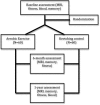
Flow diagram for the randomization and assessment sessions for both exercise and stretching control groups.
Fig. 5.
Display of the spatial memory task used in this study. The spatial memory task load was parametrically manipulated between one, two, or three items (two-item condition shown here). Participants were asked to remember the locations of one, two, or three black dots. After a brief delay, a red dot appeared, and participants were asked to respond whether the location of the red dot matched or did not match one of the locations of the previously shown black dots. This task was administered to all participants at baseline, after 6 mo, and again after completion of the intervention.
Comment in
- Failure to demonstrate that memory improvement is due either to aerobic exercise or increased hippocampal volume.
Coen RF, Lawlor BA, Kenny R. Coen RF, et al. Proc Natl Acad Sci U S A. 2011 May 3;108(18):E89; author reply E90. doi: 10.1073/pnas.1102593108. Epub 2011 Apr 19. Proc Natl Acad Sci U S A. 2011. PMID: 21504947 Free PMC article. No abstract available. - Both the body and brain benefit from exercise: potential win-win for Parkinson's disease patients.
Weintraub D, Morgan JC. Weintraub D, et al. Mov Disord. 2011 Mar;26(4):607. doi: 10.1002/mds.23726. Mov Disord. 2011. PMID: 21506145 No abstract available.
Similar articles
- Relationships of peripheral IGF-1, VEGF and BDNF levels to exercise-related changes in memory, hippocampal perfusion and volumes in older adults.
Maass A, Düzel S, Brigadski T, Goerke M, Becke A, Sobieray U, Neumann K, Lövdén M, Lindenberger U, Bäckman L, Braun-Dullaeus R, Ahrens D, Heinze HJ, Müller NG, Lessmann V, Sendtner M, Düzel E. Maass A, et al. Neuroimage. 2016 May 1;131:142-54. doi: 10.1016/j.neuroimage.2015.10.084. Epub 2015 Nov 3. Neuroimage. 2016. PMID: 26545456 Clinical Trial. - Brain-derived neurotrophic factor is associated with age-related decline in hippocampal volume.
Erickson KI, Prakash RS, Voss MW, Chaddock L, Heo S, McLaren M, Pence BD, Martin SA, Vieira VJ, Woods JA, McAuley E, Kramer AF. Erickson KI, et al. J Neurosci. 2010 Apr 14;30(15):5368-75. doi: 10.1523/JNEUROSCI.6251-09.2010. J Neurosci. 2010. PMID: 20392958 Free PMC article. - Aerobic fitness is associated with hippocampal volume in elderly humans.
Erickson KI, Prakash RS, Voss MW, Chaddock L, Hu L, Morris KS, White SM, Wójcicki TR, McAuley E, Kramer AF. Erickson KI, et al. Hippocampus. 2009 Oct;19(10):1030-9. doi: 10.1002/hipo.20547. Hippocampus. 2009. PMID: 19123237 Free PMC article. - The aging hippocampus: interactions between exercise, depression, and BDNF.
Erickson KI, Miller DL, Roecklein KA. Erickson KI, et al. Neuroscientist. 2012 Feb;18(1):82-97. doi: 10.1177/1073858410397054. Epub 2011 Apr 29. Neuroscientist. 2012. PMID: 21531985 Free PMC article. Review. - Physical exercise as a preventive or disease-modifying treatment of dementia and brain aging.
Ahlskog JE, Geda YE, Graff-Radford NR, Petersen RC. Ahlskog JE, et al. Mayo Clin Proc. 2011 Sep;86(9):876-84. doi: 10.4065/mcp.2011.0252. Mayo Clin Proc. 2011. PMID: 21878600 Free PMC article. Review.
Cited by
- Cognitive aging and reserve factors in the Metropolit 1953 Danish male cohort.
Mehdipour Ghazi M, Urdanibia-Centelles O, Bakhtiari A, Fagerlund B, Vestergaard MB, Larsson HBW, Mortensen EL, Osler M, Nielsen M, Benedek K, Lauritzen M. Mehdipour Ghazi M, et al. Geroscience. 2024 Nov 21. doi: 10.1007/s11357-024-01427-2. Online ahead of print. Geroscience. 2024. PMID: 39570569 - Acute and chronic effects of exercise intensity on cognitive functions of fastball athletes.
Kapur S, Joshi GM. Kapur S, et al. Cogn Neurodyn. 2024 Oct;18(5):2289-2298. doi: 10.1007/s11571-024-10083-3. Epub 2024 Mar 8. Cogn Neurodyn. 2024. PMID: 39555261 - The role of torso stiffness and prediction in the biomechanics of anxiety: a narrative review.
Chin S. Chin S. Front Sports Act Living. 2024 Nov 1;6:1487862. doi: 10.3389/fspor.2024.1487862. eCollection 2024. Front Sports Act Living. 2024. PMID: 39553377 Free PMC article. Review. - High-intensity interval training combined with cannabidiol supplementation improves cognitive impairment by regulating the expression of apolipoprotein E, presenilin-1, and glutamate proteins in a rat model of amyloid β-induced Alzheimer's disease.
Kordi MR, Khademi N, Zobeydi AM, Torabi S, Mahmoodifar E, Gaeini AA, Choobineh S, Pournemati P. Kordi MR, et al. Iran J Basic Med Sci. 2024;27(12):1583-1591. doi: 10.22038/ijbms.2024.79464.17210. Iran J Basic Med Sci. 2024. PMID: 39539449 Free PMC article. - The Effect of Two Somatic-Based Practices Dance and Martial Arts on Irisin, BDNF Levels and Cognitive and Physical Fitness in Older Adults: A Randomized Control Trial.
Hola V, Polanska H, Jandova T, Jaklová Dytrtová J, Weinerova J, Steffl M, Kramperova V, Dadova K, Durkalec-Michalski K, Bartos A. Hola V, et al. Clin Interv Aging. 2024 Nov 6;19:1829-1842. doi: 10.2147/CIA.S482479. eCollection 2024. Clin Interv Aging. 2024. PMID: 39525874 Free PMC article. Clinical Trial.
References
- Raz N, et al. Regional brain changes in aging healthy adults: General trends, individual differences and modifiers. Cereb Cortex. 2005;15:1676–1689. - PubMed
- Hillman CH, Erickson KI, Kramer AF. Be smart, exercise your heart: Exercise effects on brain and cognition. Nat Rev Neurosci. 2008;9:58–65. - PubMed
- Cotman CW, Berchtold NC. Exercise: A behavioral intervention to enhance brain health and plasticity. Trends Neurosci. 2002;25:295–301. - PubMed
Publication types
MeSH terms
Substances
Grants and funding
- P30 AG024827/AG/NIA NIH HHS/United States
- R37 AG025667/AG/NIA NIH HHS/United States
- R01 AG025032/AG/NIA NIH HHS/United States
- R01 AG25032/AG/NIA NIH HHS/United States
- P50 AG005133/AG/NIA NIH HHS/United States
- R01 AG25667/AG/NIA NIH HHS/United States
- R01 AG025667/AG/NIA NIH HHS/United States
LinkOut - more resources
Full Text Sources
Other Literature Sources
Medical


