Comparison of transplantation of bone marrow stromal cells (BMSC) and stem cell mobilization by granulocyte colony stimulating factor after traumatic brain injury in rat - PubMed (original) (raw)
Comparative Study
Comparison of transplantation of bone marrow stromal cells (BMSC) and stem cell mobilization by granulocyte colony stimulating factor after traumatic brain injury in rat
Mehrdad Bakhtiary et al. Iran Biomed J. 2010 Oct.
Abstract
Background: Recent clinical studies of treating traumatic brain injury (TBI) with autologous adult stem cells led us to compare effect of intravenous injection of bone marrow mesenchymal stem cells (BMSC) and bone marrow hematopoietic stem cell mobilization, induced by granulocyte colony stimulating factor (G-CSF), in rats with a cortical compact device.
Methods: Forty adult male Wistar rats were injured with controlled cortical impact device and divided randomly into four groups. The treatment groups were injected with 2 × 10⁶ intravenous bone marrow stromal stem cell (n = 10) and also with subcutaneous G-CSF (n = 10) and sham-operation group (n = 10) received PBS and "bromodeoxyuridine (Brdu)" alone, i.p. All injections were performed 1 day after injury into the tail veins of rats. All cells were labeled with Brdu before injection into the tail veins of rats. Functional neurological evaluation of animals was performed before and after injury using modified neurological severity scores (mNSS). Animals were sacrificed 42 days after TBI and brain sections were stained by Brdu immunohistochemistry.
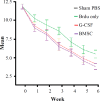
Results: Statistically, significant improvement in functional outcome was observed in treatment groups compared with control group (P<0.01). mNSS showed no significant difference between the BMSC and G-CSF-treated groups during the study period (end of the trial). Histological analyses showed that Brdu-labeled (MSC) were present in the lesion boundary zone at 42nd day in all injected animals.
Conclusion: In our study, we found that administration of a bone marrow-stimulating factor (G-CSF) and BMSC in a TBI model provides functional benefits.
Keywords: Stem cells; Injection; Traumatic brain injury (TBI).
Figures
Fig. 1
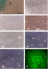
Stem cells from the bone marrow. (A-H)Appearance and growth of fibroblastoid cells or bone marrow stromal stem cell at primary culture (A) passage 1 on days 3, 7 (B and C), passage 2 (D and E) and passage 3 (F and G), respectively. Briefly, when BMSC initially grow outward from explants, two morphologically distinct populations of cells are present: spherical or flat mesenchymal cells. Bone marrow stromal stem cells express mesenchymal stem cell markers, integrin β1 (H).
Fig. 2
Results of behavioral functional tests (mNSS test) before and after TBI. Rats (10 in each group) were injured to TBI and were injected with PBS (Control), Brdu (control), G-CSF and BMSC one day after TBI. Significant functional recovery was detected in rats treated with BMSC and G-CSF treated group compared with control and sham. The data are presented as the mean ± SD. *P<0.01.
Fig. 3
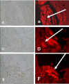
Brdu Immunohistochemistry. Forty two days after traumatic brain injury, 41 days after intravenous transplantation of BMSC and subcutaneous injection of G-CSF, cells derived from BMSC were identified by rhodamin conjugated secondary antibody (E and F) and G-CSF mobilized stem cell (C and D) and endogenous stem cell (Brdu alone) (A and B) distribute in the territory of the TBI (40×). Arrows show red spots.
Fig. 4
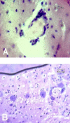
Degeneration of ipsilateral white matter of traumatic zone in rat brain revealed by cresyl violet staining at 7 days after TBI. (A) Nissle staining of traumatic brain site inflammatory cells (glial and microglial) is around traumatic zone. (B) Most of neurons are shrunk with condensed nuclei and sparse Nissl bodies.
Fig. 5
Comparition of Brdu positive cells. Data presented as mean ± SD, Brdu-labeled cells were increased in the BMSC and G-CSF injected group than alone Brdu group 41 days after injection (*_P<_0.01).
Similar articles
- Marrow stromal cell transplantation after traumatic brain injury promotes cellular proliferation within the brain.
Mahmood A, Lu D, Chopp M. Mahmood A, et al. Neurosurgery. 2004 Nov;55(5):1185-93. doi: 10.1227/01.neu.0000141042.14476.3c. Neurosurgery. 2004. PMID: 15509325 - Combination of stem cell mobilized by granulocyte-colony stimulating factor and human umbilical cord matrix stem cell: therapy of traumatic brain injury in rats.
Bakhtiary M, Marzban M, Mehdizadeh M, Joghataei MT, Khoei S, Tondar M, Mahabadi VP, Laribi B, Ebrahimi A, Hashemian SJ, Modiry N, Mehrabi S. Bakhtiary M, et al. Iran J Basic Med Sci. 2011 Jul;14(4):327-39. Iran J Basic Med Sci. 2011. PMID: 23492840 Free PMC article. - Human marrow stromal cell treatment provides long-lasting benefit after traumatic brain injury in rats.
Mahmood A, Lu D, Qu C, Goussev A, Chopp M. Mahmood A, et al. Neurosurgery. 2005 Nov;57(5):1026-31; discussion 1026-31. doi: 10.1227/01.neu.0000181369.76323.50. Neurosurgery. 2005. PMID: 16284572 Free PMC article. - Bone marrow stromal cells in traumatic brain injury (TBI) therapy: true perspective or false hope?
Opydo-Chanek M. Opydo-Chanek M. Acta Neurobiol Exp (Wars). 2007;67(2):187-95. doi: 10.55782/ane-2007-1647. Acta Neurobiol Exp (Wars). 2007. PMID: 17691227 Review.
Cited by
- Effects of bone marrow mesenchymal stromal cells-derived therapies for experimental traumatic brain injury: A meta-analysis.
Chen C, Peng C, Hu Z, Ge L. Chen C, et al. Heliyon. 2024 Jan 29;10(3):e25050. doi: 10.1016/j.heliyon.2024.e25050. eCollection 2024 Feb 15. Heliyon. 2024. PMID: 38322864 Free PMC article. - The effect of intrathecal delivery of bone marrow stromal cells on hippocampal neurons in rat model of Alzheimer's disease.
Eftekharzadeh M, Nobakht M, Alizadeh A, Soleimani M, Hajghasem M, Kordestani Shargh B, Karkuki Osguei N, Behnam B, Samadikuchaksaraei A. Eftekharzadeh M, et al. Iran J Basic Med Sci. 2015 May;18(5):520-5. Iran J Basic Med Sci. 2015. PMID: 26124940 Free PMC article. - Preclinical progenitor cell therapy in traumatic brain injury: a meta-analysis.
Jackson ML, Srivastava AK, Cox CS Jr. Jackson ML, et al. J Surg Res. 2017 Jun 15;214:38-48. doi: 10.1016/j.jss.2017.02.078. Epub 2017 Mar 8. J Surg Res. 2017. PMID: 28624058 Free PMC article. - Malignant transformation of bone marrow stromal cells induced by the brain glioma niche in rats.
He Q, Zou X, Duan D, Liu Y, Xu Q. He Q, et al. Mol Cell Biochem. 2016 Jan;412(1-2):1-10. doi: 10.1007/s11010-015-2602-0. Epub 2015 Nov 21. Mol Cell Biochem. 2016. PMID: 26590986 - The effects of BMSCs transplantation on autophagy by CX43 in the hippocampus following traumatic brain injury in rats.
Sun L, Gao J, Zhao M, Jing X, Cui Y, Xu X, Wang K, Zhang W, Cui J. Sun L, et al. Neurol Sci. 2014 May;35(5):677-82. doi: 10.1007/s10072-013-1575-6. Epub 2013 Nov 13. Neurol Sci. 2014. PMID: 24221859
References
- Rickard D.J., Sullivan T.A., Shenker B.J., Leboy P.S., Kazhdan I. Induction of rapid osteoblast differentiation in rat bone marrow stromal cell cultures by dexamethasone and BMP-2. Dev. Biol. 1994;161:218–228. - PubMed
- Bjorklund L.M., Sánchez-Pernaute R., Chung S., Andersson T., Chen I.Y., McNaught K.S., Brownell A.L., Jenkins B.G., Wahlestedt C., Kim K.S., Isacson O. Embryonic stem cells develop into functional dopaminergic neurons after transplantation in a Parkinson rat model. Proc. Natl. Acad. Sci. USA. 2002;99:2344–2349. - PMC - PubMed
Publication types
MeSH terms
Substances
LinkOut - more resources
Full Text Sources
Other Literature Sources
Medical


